Institution: University of New Mexico
Additional authors:Qian-Yun Zhang, David R. Czuchlewski, Nicholas D. Radovich
Session: AML with recurrent genetic abnormalities Part I
HISTORY
A 31-year-old woman with a history of diet-controlled diabetes, anxiety, and depression presented to the emergency room following a syncopal episode. She related a history of feeling weak, sluggish, and short of breath for approximately 3 weeks. Her physical exam was reportedly unremarkable. CBC was obtained. WBC: 3.9 K/mm3, Hgb 9.2 gm/dL, Hct 26.5%, Plt 20 K/mm3 WBC differential was reported as: Neutrophils 7%, Bands 1%, Lymphocytes 46%, monocytes 4%, metamyelocytes 1%, myelocytes 14%, promyelocytes 2%, blasts 25%.
DETAILS
Review of the patient’s bone marrow touch preparation and biopsy showed 90% immature cells with intermediate to large size, irregular nuclear contours with some folded nuclei, prominent nucleoli, and granular cytoplasm, including Auer rods, with some cells showing multiple Auer rods.
IMMUNOHISTOCHEMISTRY AND FLOW CYTOMETRY
Flow cytometry on the bone marrow showed 96% blasts, expressing CD33, dim HLA-DR, CD13, CD56, and CD117. The blasts were negative for CD3, CD7, CD10, CD19, CD20, and CD34. The cytochemical myeloperoxidase stain showed a pattern of strong staining, highlighting the Auer rods.
CYTOGENETIC FINDINGS
46,XX FISH for the RUNX1T1/RUNX1 fusion associated with the translocation t(8;21)(q22;q22), and rearrangement of the CBFB gene, associated with the inversion 16(p13q22) was negative. FISH for the PML/RARA fusion associated with the translocation t(15;17)(q22;q21) using the LSI PML/RARA dual color, dual fusion probe system was negative for the typical fusion as well as abnormal patterns.
MOLECULAR FINDINGS
t(15;17) Quantitative RT-PCR was positive for transcripts associated with the Bcr1 form PML-RARA translocation, with a PML-RARA/ABL1 ratio of 0.095
INTERESTING FEATURES
Cases of acute myeloid leukemia with normal cytogenetics and FISH which harbor a cryptic t(15;17) translocation identifiable by PCR are rare. In this case, the morphologic findings in the blasts and the flow immunophenotype were significantly worrisome for acute promyelocytic leukemia, and although the FISH study for t(15;17) was negative further workup with PCR was pursued. It has been documented that rare, extremely small insertions may not hybridize with available FISH probes, leading to a negative FISH study. RT-PCR offers a sensitive methodology and the ability to identify specific breakpoints. Next generation sequencing techniques have also been used to identify and classify these cryptic translocations. In this case, the distinctive blast morphology guided the diagnosis and motivated further testing despite negative FISH study and normal karyotype.
PROPOSED DIAGNOSIS
Acute promyelocytic leukemia with cryptic t(15;17) translocation identified by RT-PCR.
CONSENSUS DIAGNOSIS
Acute promyelocytic leukemia with cryptic t(15;17)(q22;q11); PML-RARA
| Cytospin of bone marrow aspirate showing immature cells with intermediate to large size, irregular nuclear contours with some folded nuclei, prominent nucleoli, and granular cytoplasm, including Auer rods. | 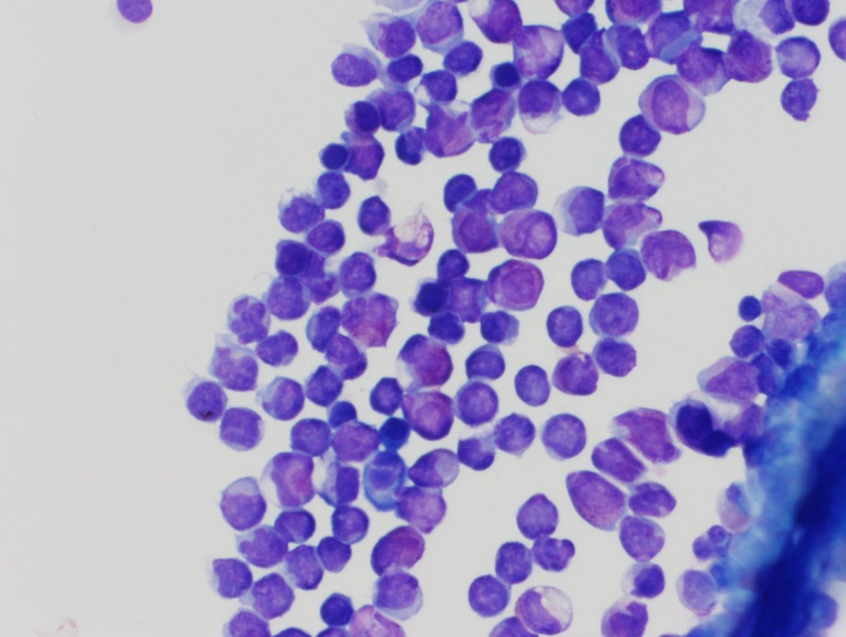 |
| Myeloperoxidase cytochemical stain showing strong positive staining in the immature cells. | 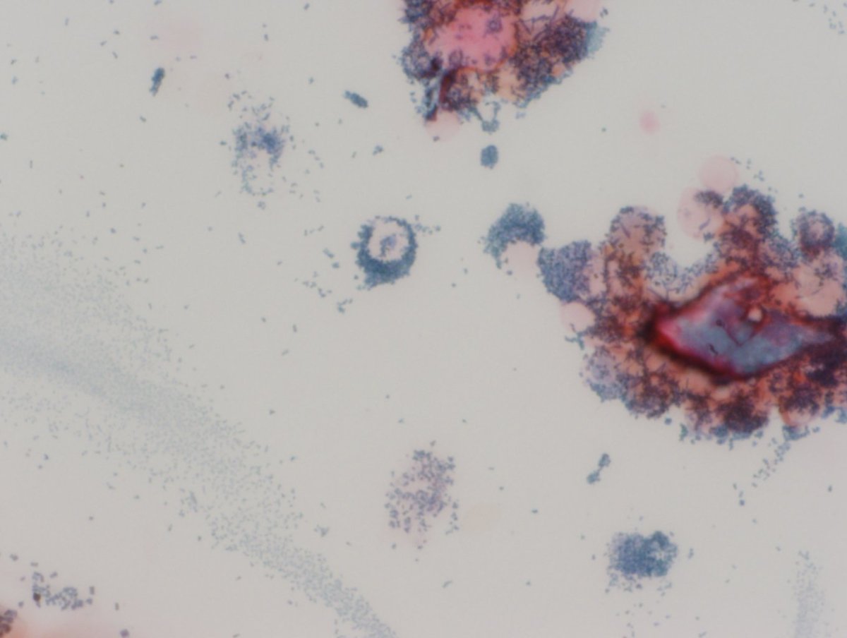 |
| Bone marrow biopsy showing sheets of immature cells. |  |
| Conventional cytogenetics showing normal karyotype: 46,XX | 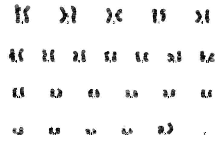 |
| FISH for the PML/RARA fusion associated with the translocation t(15;17)(q22;q21) was negative for the typical fusion as well as abnormal patterns. | 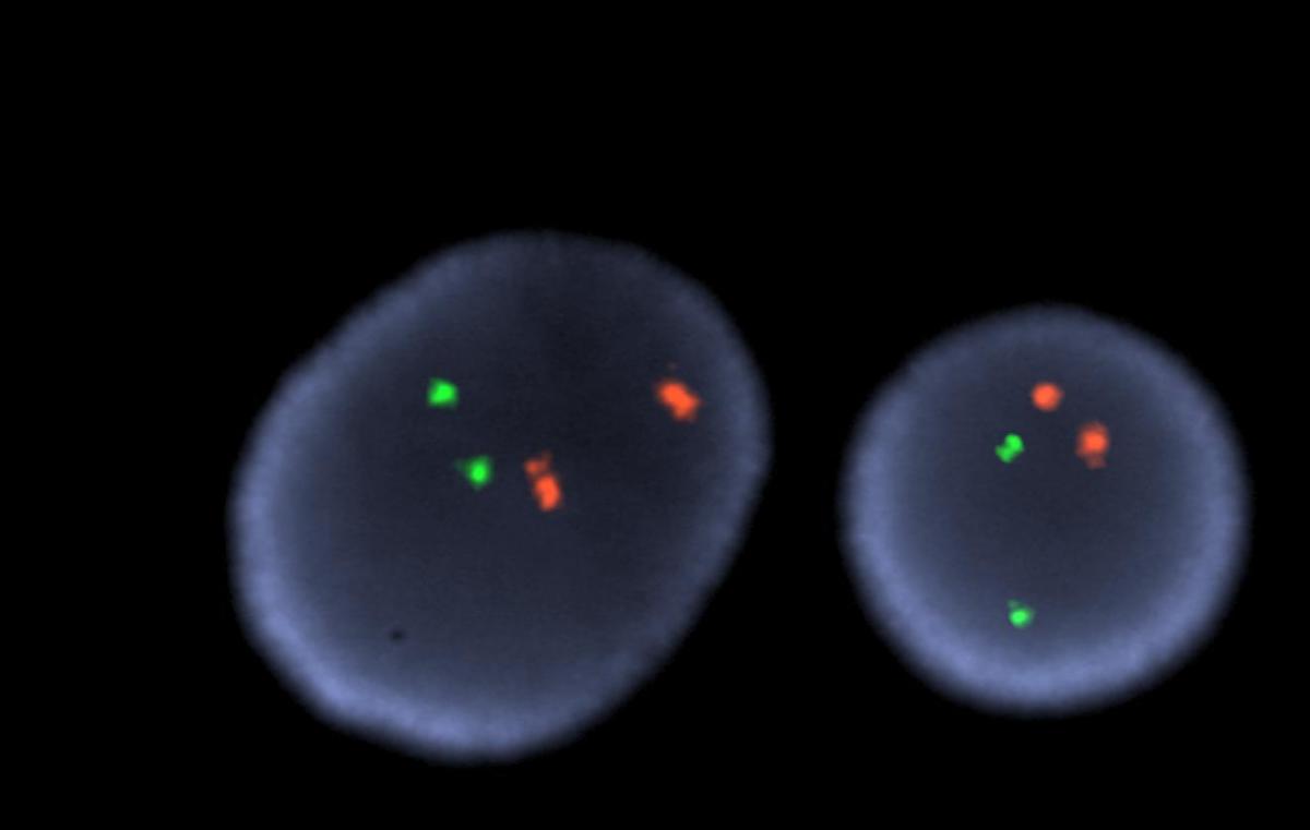 |
| RT-PCR positive for transcripts associated with the Bcr1 form PML-RARA translocation | 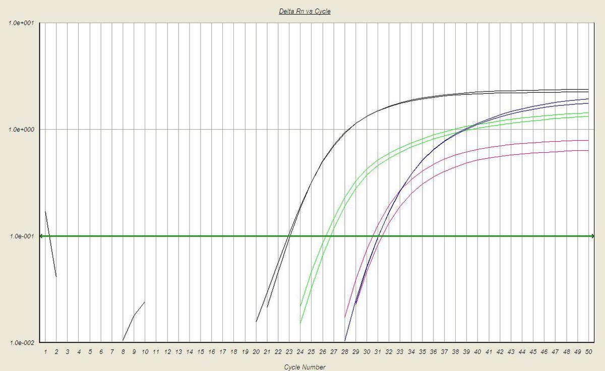 |