Institution: New York Presbyterian Hospital/Weill Cornell Medical Center
Additional authors:Susan Mathew, PhD Attilio Orazi, MD Wayne Tam, MD PhD
Session: AML with myelodysplasia-related changes
HISTORY
A 57 year old man presented with a one year history of unexplained anemia. During the most recent five months, he experienced a ten pound weight loss, dizziness, and fatigue and was found to be pancytopenic. Peripheral blood smear revealed circulating blasts (12%). He was then transferred to our institution for further evaluation and treatment. A bone marrow examination showed high grade myelodysplastic syndrome (see below). The patient was treated with decitabine for 6 cycles, with partial response. In September 2012, a bone marrow examination showed 8% blasts on aspirate smear. The patient then received 7+3 idarubicin/ara-c as salvage therapy. Subsequent marrow examinations showed marked hypocellularity without morphologic evidence of increased blasts, however cytogenetic analysis continued to show evidence of residual disease. The patient is currently being evaluated for allogeneic stem cell transplant.
DETAILS
3/16/12: Left posterior iliac crest bone marrow clot section (formalin fixation), biopsy (Bouin’s fixation) Hypercellular bone marrow (60-70%) with an increased myeloid to erythroid ratio due to increased granulopoiesis and decreased erythropoiesis. Granulopoiesis is left-shifted with increased blasts. Megakaryocytes are adequate in number and include numerous small, hypolobated forms. 3/16/12: Aspirate smear The bone marrow aspirate appears hypercellular for the patient’s age. Blasts account for 15% of cellularity. Megakaryopoiesis is dysplastic (>10%, including hypolobation, multinucleation, and micromegakaryocytes). Granulopoiesis is also dysplastic (~10%, including hypogranulation and abnormal segmentation). Occasional megaloblastoid erythroid cells are seen (<10%). The M:E ratio is 4.0.
IMMUNOHISTOCHEMISTRY AND FLOW CYTOMETRY
Flow cytometry performed on the bone marrow aspirate showed blasts accounting for around 20% of the gated cells. These were positive for CD34, CD117, HLA-DR, CD13, CD56, and CD7 (partial), and negative for CD33 and myeloperoxidase. Immunohistochemistry performed on the bone marrow biopsy showed blasts positive for CD34 and CD117 accounting for 10-20% of cellularity. Glycophorin C and myeloperoxidase confirmed the increased M:E ratio. CD42b highlighted many dysplastic megakaryocytes.
CYTOGENETIC FINDINGS
3/16/12:
Bone marrow aspirate: 46,Y,t(X;5)(q26;q31)0]
FISH showed no gross deletion of 5q31 or 5q33. Both of these regions were translocated to the X chromosome.
Peripheral blood: 46,XY
MOLECULAR FINDINGS
No mutations found in FLT3, NPM1, or CEBPA.
INTERESTING FEATURES
This case of high-grade MDS (RAEB-2) showed a novel balanced reciprocal translocation, t(X;5)(q26;q31). Translocations involving 5q31 are uncommon. They have been described previously in myeloid neoplasms including MDS and AML, but the partner chromosome has not involved the X chromosome (1). 5q31 is a well known region implicated in the pathogenesis of MDS and AML. Patients with high grade MDS/AML with deletion 5q frequently show a commonly deleted region at 5q31, which may be associated with other cytogenetic abnormalities, whereas patients with isolated 5q- syndrome have a commonly deleted region encompassing 5q32-33 with or without deletion of 5q31 (2). Genes located at the 5q31 region include EGR1, CTNNA1, and CDC25C, which have all been variously implicated in the pathogenesis of MDS/AML (3). EGR1 (early growth response gene-1) is a zinc finger transcription factor tumor suppressor gene that is expressed in myeloid cells and is essential for differentiation of myeloid precursors to the macrophage lineage. Mice with heterozygous loss of EGR1 develop leukemia with increased frequency when treated with DNA alkylating agents. Expression of EGR1 has also been shown to overcome a terminal differentiation block induced by deregulated expression of the oncogenes MYB or E2F1. Interestingly, cryptic deletion of EGR1 has been detected in a case of MDS with balanced translocation t(5;22)(q31;q11.2) (4). This novel translocation may lead to fusion of the target gene at 5q31, as seen in t(5;12)(q31;p13), or may result in cryptic deletion of a tumor suppressor gene in the vicinity of the translocation breakpoint as in the case of EGR1. Characterization of the breakpoint of this translocation and identification of the partner genes involved by next generation sequencing may allow us to pinpoint the critical genes involved in the pathobiology of MDS and AML within the 5q31 region. References: 1. Yagasaki F et al. Fusion of TEL/ETV6 to a novel ACS2 in myelodysplastic syndrome and acute myelogenous leukemia with t(5;12)(q31;p13). Genes Chromosomes Cancer. (1999) 3, 192-202. 2. Ebert BL. Deletion 5q in myelodysplastic syndrome: a paradigm for the study of hemizygous deletions in cancer. Leukemia (2009) 23, 1252-1256. 3. Fuchs O. Important genes in the pathogenesis of 5q- syndrome and their connection with ribosomal stress and the innate immune system pathway. Leuk Res Treatment (2012). 4. Hoffman MW, Janney S, and Batanian JR. Cryptic deletion of EGR1 in association with a novel balanced t(5;22)(q31;q11.2) in a patient with myelodysplastic syndrome. Cancer Genetics and Cytogenetics (2009) 191, 106-108.
PROPOSED DIAGNOSIS
Refractory anemia with excess blasts-2 (RAEB-2) associated with a novel balanced translocation, t(X;5)(q26;q31).
CONSENSUS DIAGNOSIS
Refractory anemia with excess blasts-2 (RAEB-2) associated with a novel balanced translocation, t(X;5)(q26;q31)
| Bone marrow biopsy (low power) | 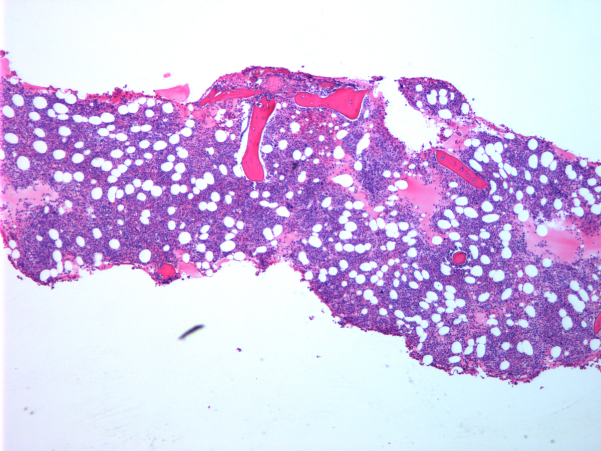 |
| Bone marrow biopsy (high power) | 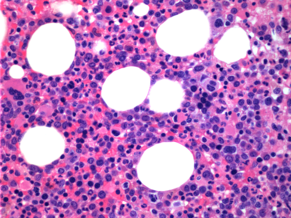 |
| CD34 (high power) | 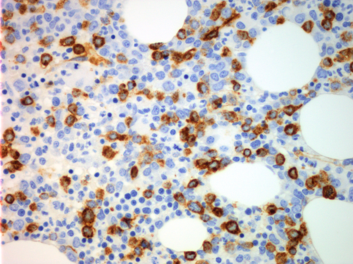 |
| CD117 (high power) | 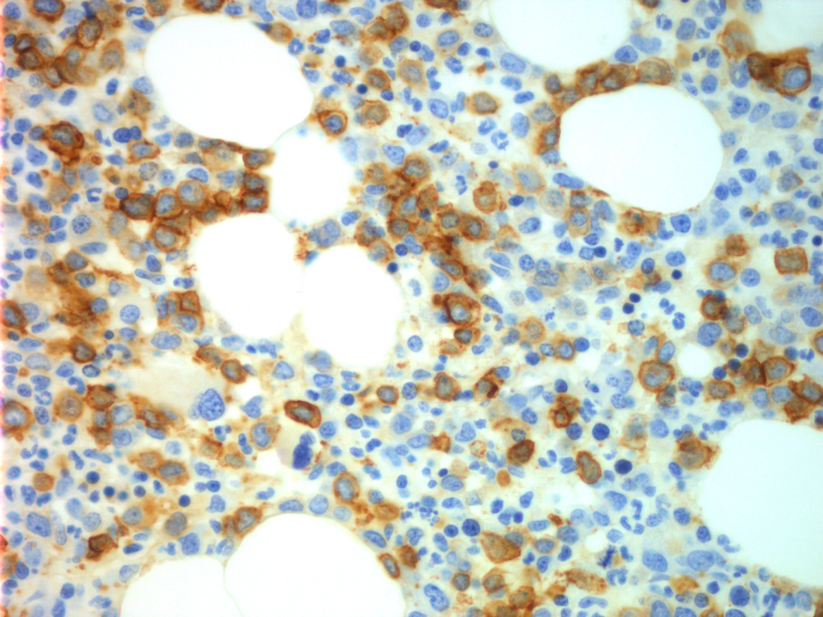 |
| CD42b (low power) | 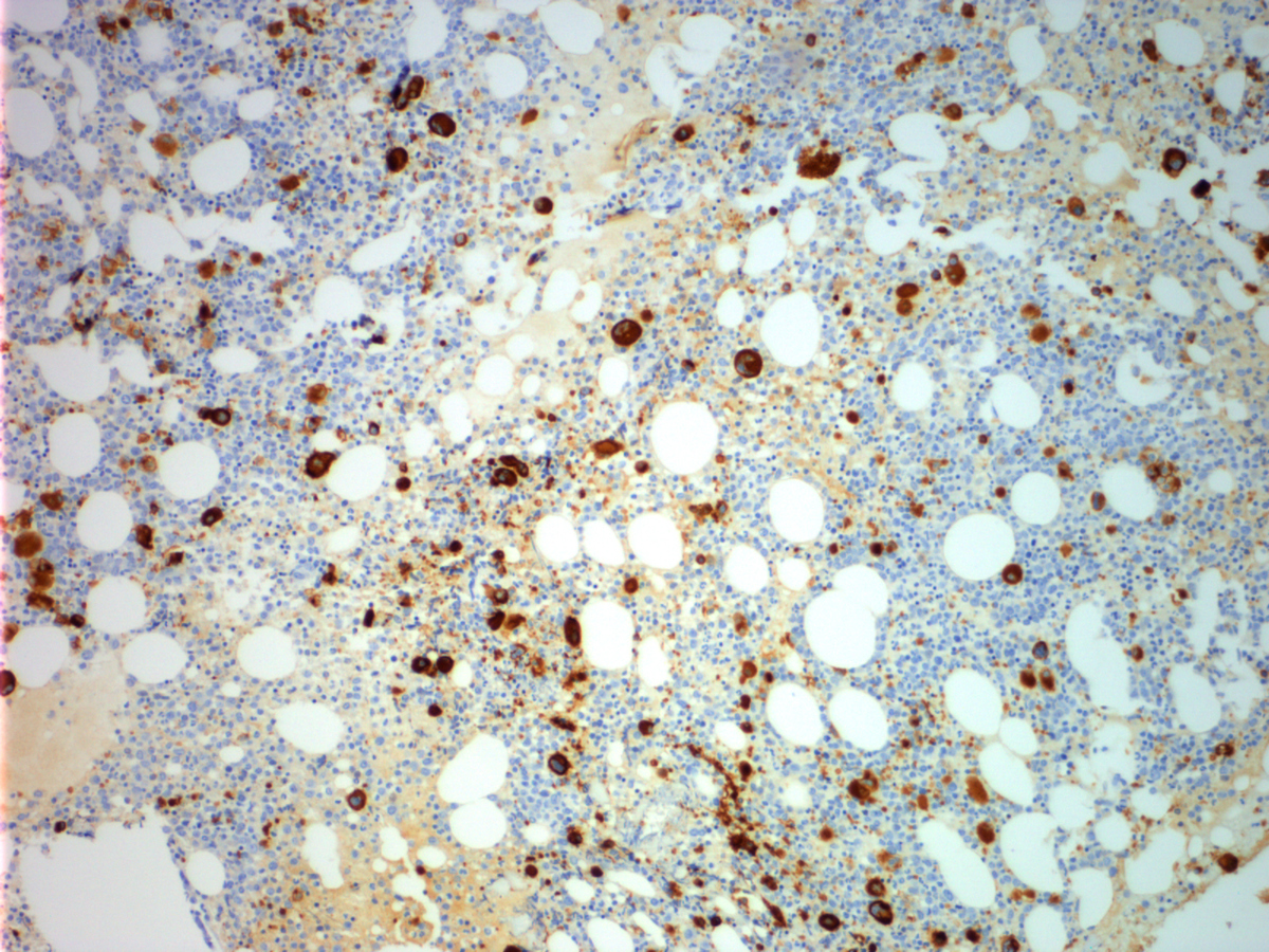 |
| CD42b (high power) |  |
| Bone marrow aspirate | 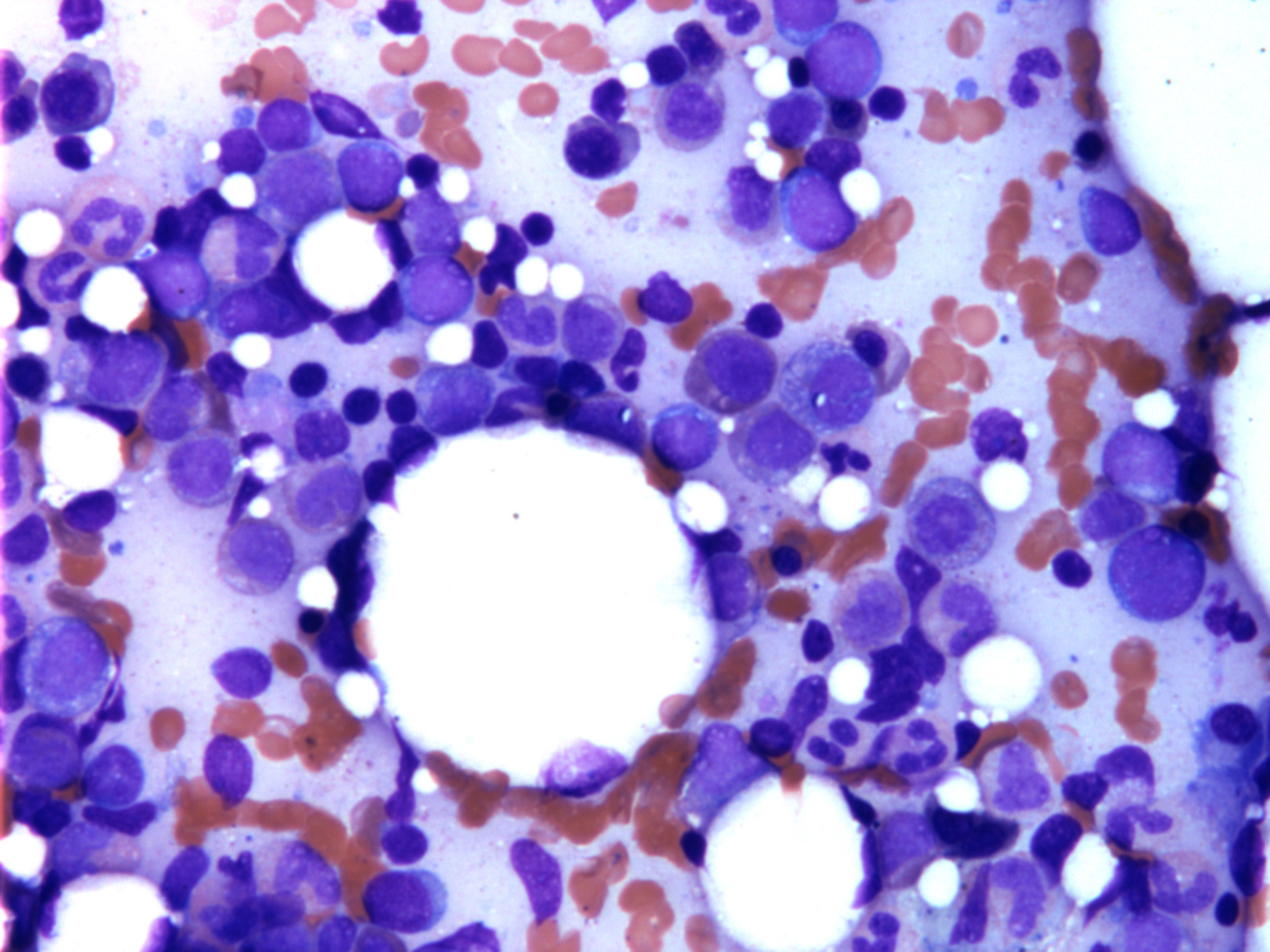 |
| Peripheral blood showing dysplastic neutrophils |  |
| Peripheral blood showing circulating blast | 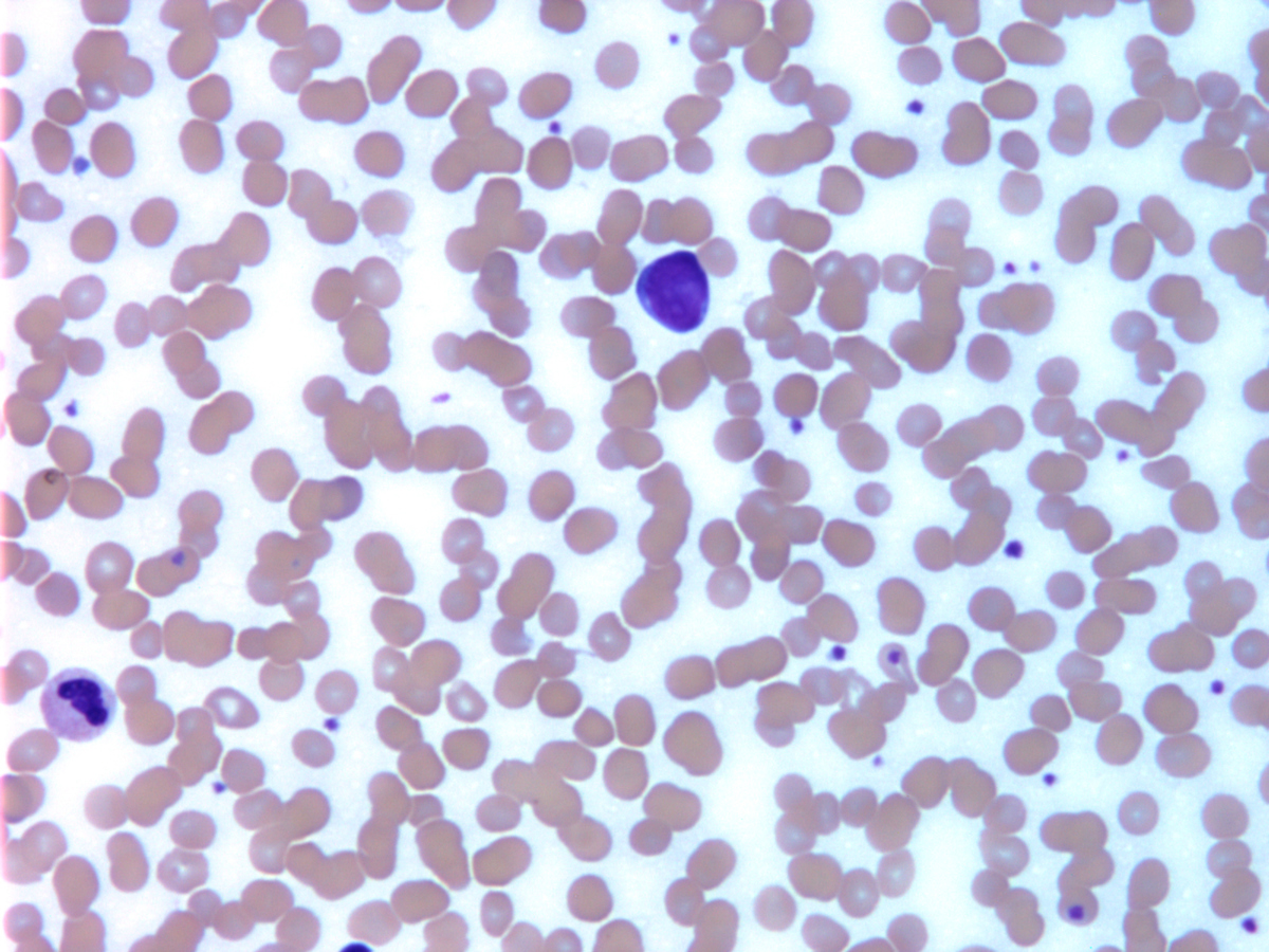 |
| Karyotype showing t(X;5)(q26;q31) | 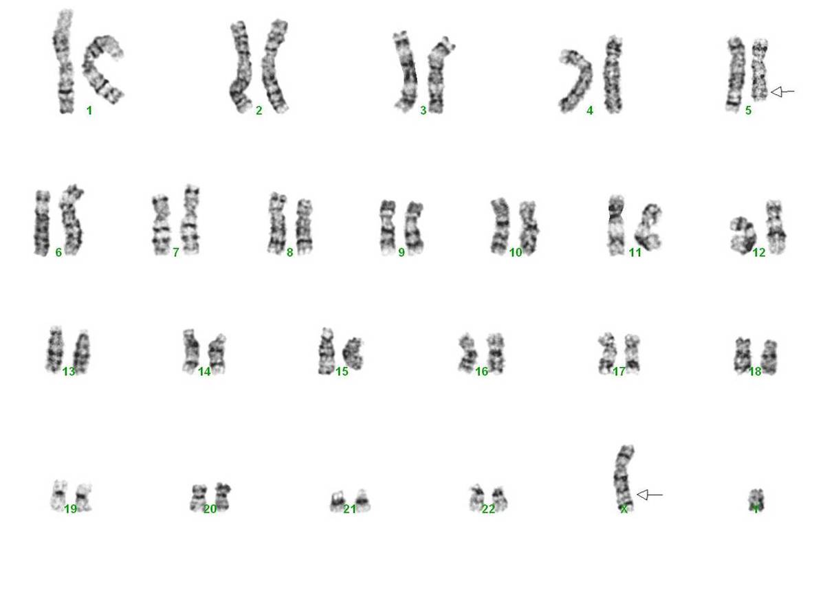 |
| FISH showing ERG probe on X chromosome | 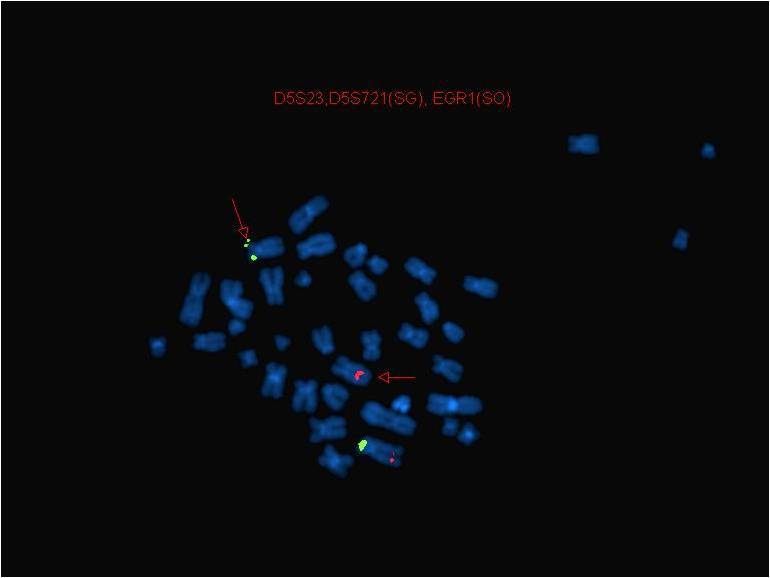 |