Institution: Massachusetts General Hospital
Additional authors:Robert Hasserjian, MD
Session: AML with recurrent genetic mutations Part II
HISTORY
79 year old man who presented with fatigue and weight loss. Peripheral blood analysis revealed a leukocytosis (WBC 11.7 x 10^9/L, 25% monocytes), anemia (Hgb 11.3 g/dL, MCV 101 fl), and normal platelets (platelets 163 x 10^9/L). A bone marrow biopsy showed chronic myelomonocytic leukemia-2 (CMML-2). The patient was treated supportively with darboepoeitin alfa and red blood cell transfusions. Three months later, he presented to the emergency department with extreme fatigue, dizziness, and anorexia. Peripheral blood analysis revealed a profound leukocytosis (WBC 165.5 x 10^9/L) with the following differential: 10% neutrophils, 11% lymphocytes, 44% monocytes, 1% eosinophils, 2% bands, 4% myelocytes, 2% promyelocytes, 8% promonocytes, and 18% blasts. The patient was anemic (Hgb 7.6 g/dL) and had thrombocytopenia (platelets 109 x 109/L). A bone marrow biopsy is performed.
DETAILS
Both specimens were bone marrow from the iliac crest. The biopsies were fixed in B plus fixative and submitted following decalcification. The aspirates were stained with Wright-Giemsa stain.
First bone marrow:
The biopsy is hypercellular for age (80% cellularity) with left shifted myeloids and erythroids. Megakaryocytes are slightly increased with overall normal morphology. A 200 cell count of the aspirate smear reveals 30% maturing myeloids, 40% erythroids, 2% lymphocytes, 10% monocytes, 1% eosinophils, 1% basophils, 13% blasts and promonocytes, and 3% plasma cells. There is moderate erythroid dysplasia
Second bone marrow:
The biopsy is >95% cellular with sheets of large blasts with irregular and lobated nuclei comprising about 90% of the cellularity.
A 200 cell count of the aspirate smear reveals 4% maturing myeloids 16% erythroids, 5% lymphocytes, 4% monocytes, 71% blasts and promonocytes, and 3% plasma cells. Enzyme cytochemistry revealed that many of the blasts are positive for both myeloperoxidase and non-specific esterase.
IMMUNOHISTOCHEMISTRY AND FLOW CYTOMETRY
First bone marrow: Flow cytometry showed 2% myeloid blasts. Immunohistochemistry for NPM1 showed aberrant cytoplasmic NPM1 staining indicating the presence of an NPM1 mutation.
Second bone marrow: Flow cytometry showed 25% myeloid blasts with myelomonocytic features (CD33+ CD13+ MPOdim+ CD117+ CD34+/- CD14- HLA-DR+ CD11c+ CD64+ CD4dim+ CD7dim+). Immunohistochemistry for NPM1 showed aberrant cytoplasmic NPM1 staining indicating the presence of an NPM1 mutation.
CYTOGENETIC FINDINGS
First bone marrow and second bone marrow: 46 XY [20]
MOLECULAR FINDINGS
Second bone marrow: Positive for FLT3 ITD and NPM1 mutations.
INTERESTING FEATURES
This is a case of acute myeloid leukemia (AML) arising from CMML and would be classified as AML with myelodysplasia-related changes (AML-MRC) according to the WHO 2008 classification. An NPM1 mutation was present in both the CMML and AML specimens. The patient rapidly progressed to AML within 3 months of his diagnosis of CMML. The NPM1 mutation is relatively uncommon in CMML (Bains A et al. AJCP 2011;135:62) but is one of the more common molecular abnormalities in AML. This, in addition to the rapid progression to AML, raises the possibility that the initially diagnosed CMML was actually an early de novo AML with mutated NPM1. AML-MRC has an inferior prognosis to NPM1-mutated AML, so this distinction is clinically relevant. Screening of newly diagnosed cases of CMML for the NPM1 mutation may be warranted, with close clinical follow-up of positive cases that could represent early presentation of NPM1-mutated AML.
PROPOSED DIAGNOSIS
AML with myelodysplasia related changes (versus AML with mutated NPM1)
CONSENSUS GROUP: ADDITIONAL INFORMATION/STUDIES
Additional immunostains performed by consensus group:
NPM1: Cytoplasmic positivity in blasts, proerythroblasts, megakaryocytes (multilineage involvement).
CONSENSUS DIAGNOSIS
Acute myeloid leukemia with NPM1 mutation and FLT3 ITD mutation
| Medium power view of first bone marrow biopsy showing a hypercellular marrow with left shifted myeloids and erythroids. |  |
| High power view of first bone marrow biopsy showing maturing trilineage hematopoiesis and increased monocytic cells. |  |
| High power view of first bone marrow aspirate showing erythroid dysplasia, monocytes, and occasional myeloid blasts. | 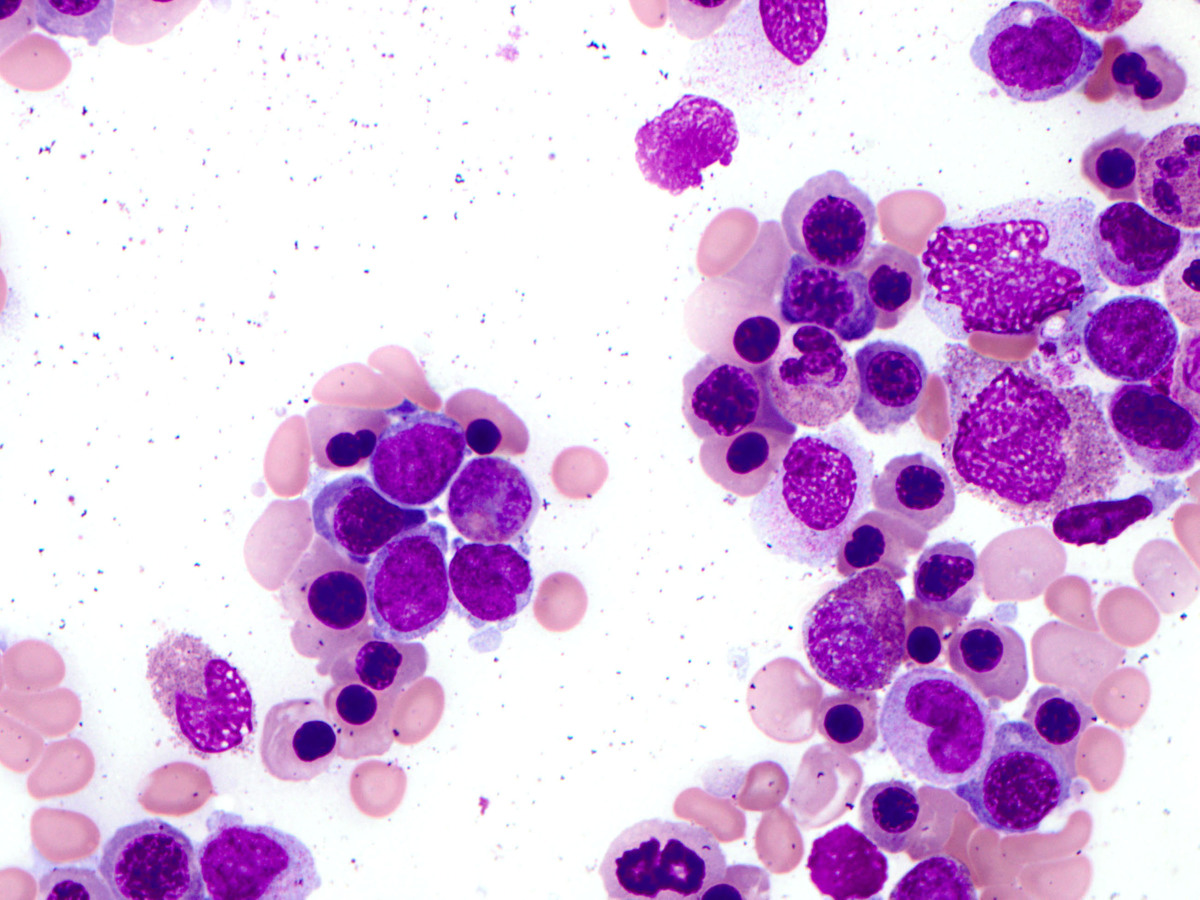 |
| NPM1 immunostain of first bone marrow biopsy showing aberrant cytoplasmic expression in hematopoietic elements. |  |
| Low power view of second bone marrow biopsy showing a hypercellular marrow composed mainly of blast forms. | 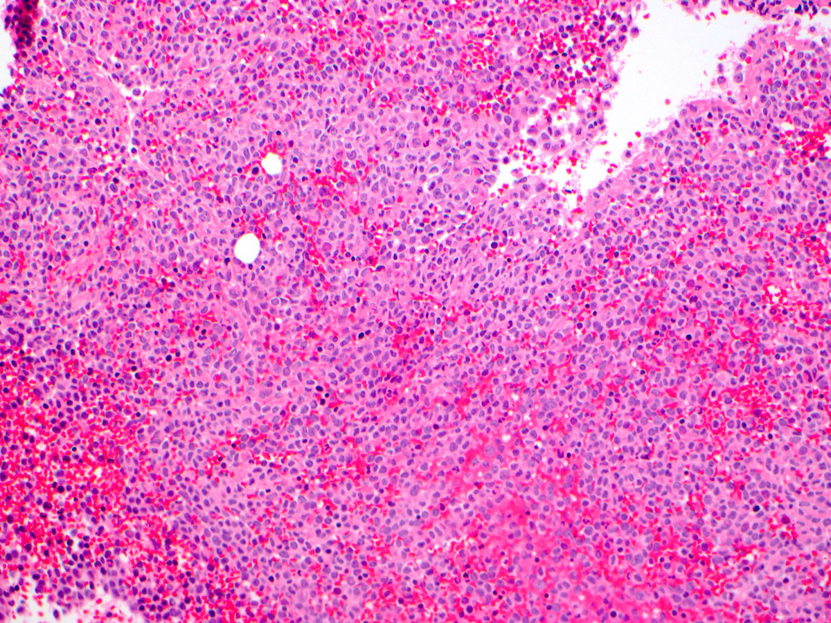 |
| High power view of second bone marrow biopsy showing sheets of immature monocytoid cells with irregular and lobated nuclei and prominent nucleoli, consistent with promonocytes and monoblasts. | 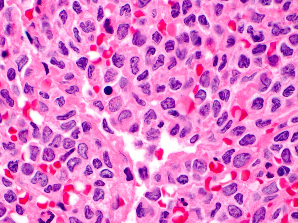 |
| High power view of second bone marrow aspirate showing predominantly blasts and promonocytes. |  |
| NPM1 immunostain of second bone marrow biopsy showing aberrant cytoplasmic expression in the blast population. |  |