Institution: Massachusetts General Hospital
Additional authors:Robert Hasserjian, MD
Session: AML secondary to myeloproliferative neoplasms and other types of disease progression in MPN
HISTORY
80 yo female who first presented with an elevated platelet count identified on routine medical check. Two years later she was noted to have a hemoglobin of 19 g/dL and a platelet count of 451 x 109/L and was diagnosed with polycythemia vera based on the hemoglobin level and the presence of a JAK2 V617F mutation She was treated with periodic phlebotomy with adequate control of her hematocrit and later hydroxyurea for symptomatic splenomegaly and splenic irradiation for splenic infarcts. Ruxolitinib (a JAK2 kinase inhibitor) was started 1 month following the last splenic radiation, with considerable reduction in her spleen size. Two months later, she was found to be anemic with a hemoglobin of 8.7 g/dL, and had worsening fatigue, abdominal pain, and intermittent fevers. She was found to have massive splenomegaly with a WBC of 14.1 x 10⁹/L (25% blasts), Hgb 10.5 g/dL and platelets 69 x 109/L. A bone marrow biopsy is obtained.
DETAILS
The specimen is bone marrow from the iliac crest. The biopsy was fixed in B plus fixative and submitted following decalcification. The aspirate was stained with Wright-Giemsa stain. Trephine biopsy: The marrow is hypercellular for age (approximately 90% cellular).The myeloid to erythroid ratio is increased. Megakaryocytes occur in focal small clusters and include frequent small to medium-sized forms with hyper-lobated and complex nuclei. Large clusters of blasts cells are present. Reticulin stain shows no increase in fibrosis. Aspirate smear: 400 cell count: 22% maturing myeloids; 31% erythroid precursors; 6% lymphocytes; 2% monocytes; 1% eosinophils; 1% basophils, 7% promyelocytes; and 30% blasts.
IMMUNOHISTOCHEMISTRY AND FLOW CYTOMETRY
Flow cytometry showed two distinct myeloid blast populations: CD117+ CD34dim+ CD33- CD13+ MPO- HLA-DR- (45% of total cells) and CD117+ CD34+ CD13+CD33dim+ HLA-DR+ MPOdim+ (14% of total cells).
CYTOGENETIC FINDINGS
A bone marrow karyotype showed 45,XX, -7[17]/46,XX[3]
MOLECULAR FINDINGS
NPM1 and FLT3 mutations were not detected. Snapshot sequencing revealed a KRAS mutation (Gly13Asp).
INTERESTING FEATURES
The case is an example of blastic transformation of a myeloproliferative neoplasm (polycythemia vera) 7 years following the initial diagnosis. Blastic transformation of BCR-ABL-negative myeloproliferative neoplasms such as polycythemia vera has a dismal prognosis (Tam CS et al. Blood 2008;112:1628). In the past, with the use of cytotoxic therapies such as alkylators or radioactive phosphorus, many cases of AML following myeloproliferative neoplasms were likely therapy-related (Finazzi G et al. Blood 2005;105:2664). In the modern era with infrequent use of such cytotoxic therapies, the incidence of AML is lower in these patients. The current patient had been treated with hydroxyurea intermittently and splenic irradiation. However, single agent hydroxyurea is not thought to increase the risk of AML in PV patients and the short latency period (3 months prior) of the radiation exposure makes this unlikely to have caused the AML. The role of recent ruxolitinib treatment in this patient’s blastic transformation is not known.
PROPOSED DIAGNOSIS
AML with myelodysplasia related changes (on the basis of a monosomy 7 cytogenetic abnormality).
CONSENSUS DIAGNOSIS
Blast phase of polycythemia vera: acute myeloid leukemia with monosomy 7 and NRAS G13D mutation
| Low power view of bone marrow biopsy showing hypercellular marrow and clustering of abnormal megakaryocytes. | 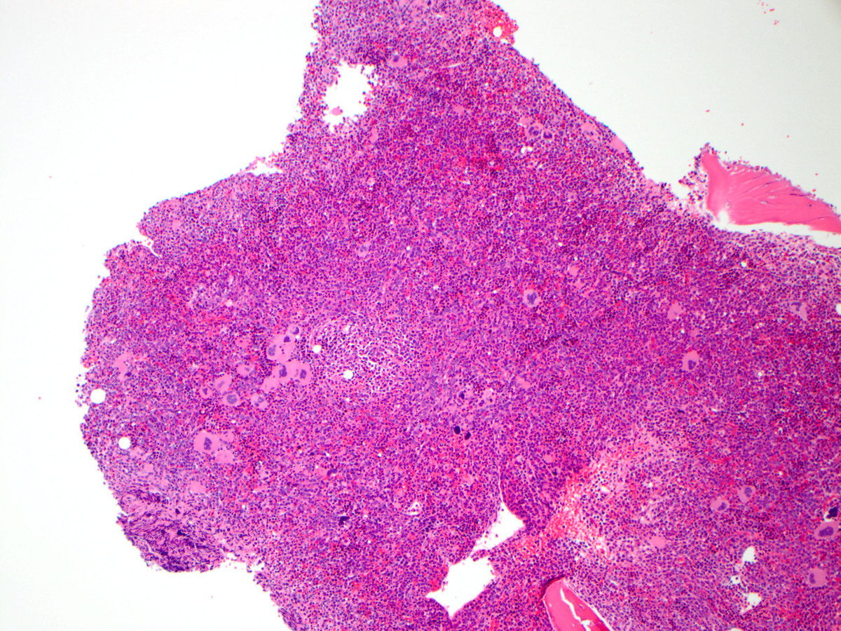 |
| High power view of bone marrow biopsy showing numerous blasts. | 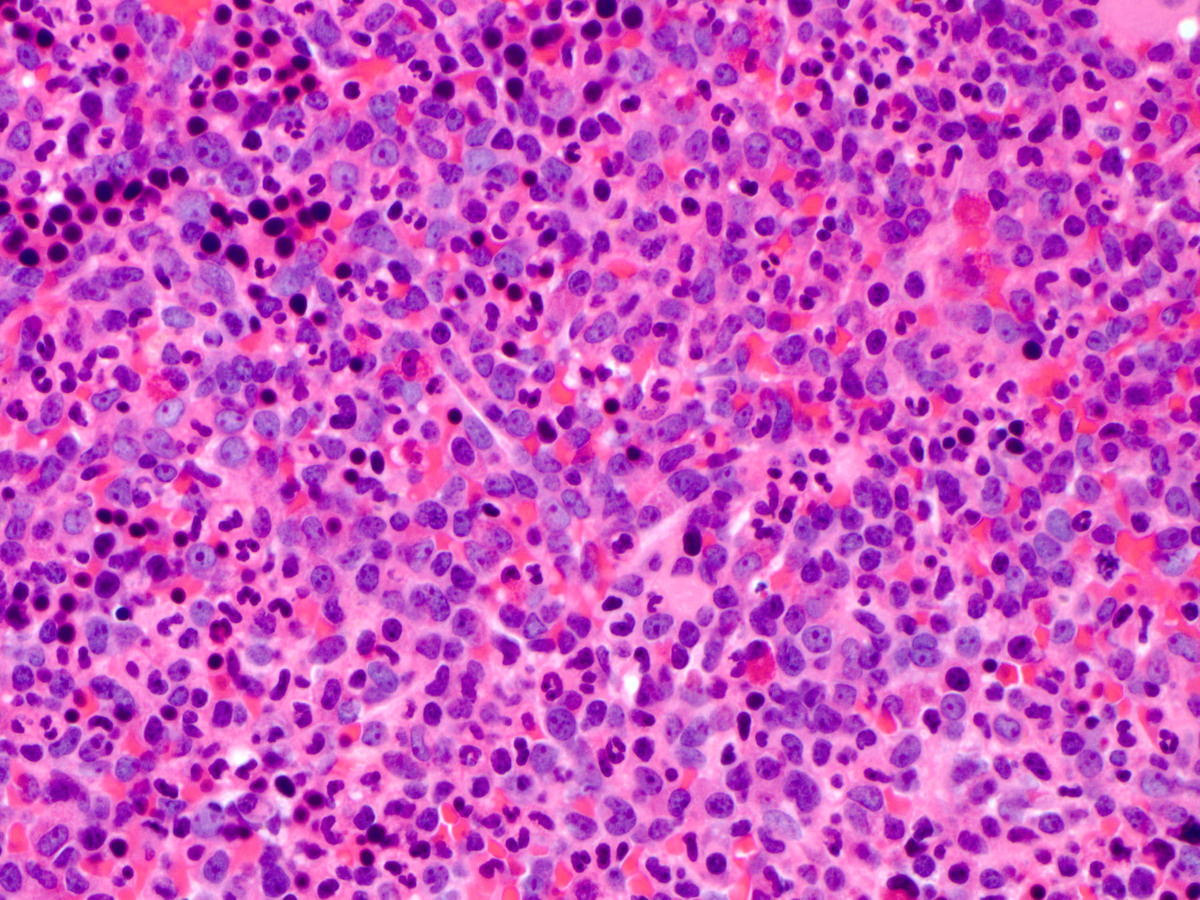 |
| High power view of bone marrow biopsy showing clustered megakaryocytes. | 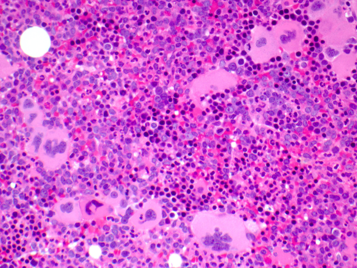 |
| High power view of aspirate smear showing numerous blast forms. | 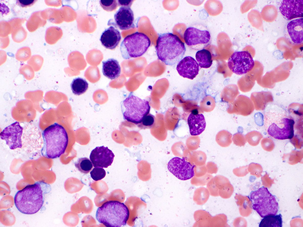 |
| Karyotype showing deletion of chromosome 7. | 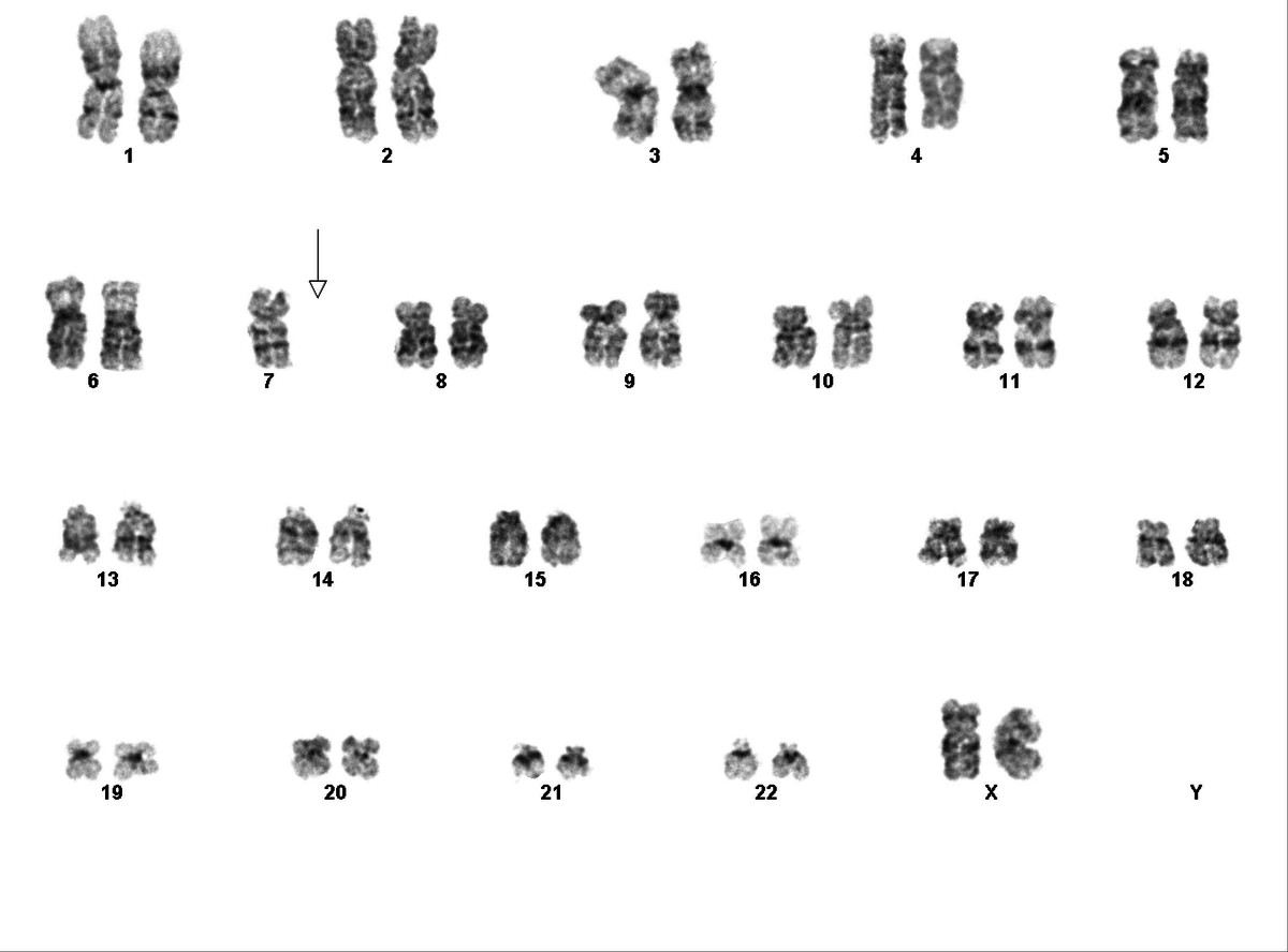 |