Institution: University of Michigan Health System
Additional authors:Amir Behdad, MD, Diane Roulston, PhD, David O. Ferguson, MD PhD, Megan Lim, MD PhD
Session: AML with recurrent genetic abnormalities Part I
HISTORY
A twenty-two year old previously healthy female sought medical attention at an outside facility for complaints of protracted sore throat, malaise and fatigue. Physical examination revealed no lymphadenopathy, organomegaly, petechiae or bruising. Laboratory studies demonstrated pancytopenia while outside bone marrow examination documented acute promyelocytic leukemia based upon cytomorphologic examination and supportive flow cytometric analysis (CD34 and HLA-DR negative; CD13, CD33, CD64 (dim), CD117 (dim) and bright myeloperoxidase positive; CD11b, CD11c, and CD14 negative). The patient was initiated on idarubicin and all-trans retinoic acid, but within 1-2 weeks of therapy developed infectious complications so was transferred to our facility for further management. At this time, fluorescent in-situ hybridization (FISH) studies returned and were negative for a PML-RARA fusion.
DETAILS
At our institution, a bone marrow core biopsy was received in acetic acid-zinc-formalin (H&E slides submitted for review) and was accompanied by marrow aspirate smears [digital images provided, Figure 1]. The marrow was hypocellular with a predominant population of atypical promyelocytes. Limited maturing but dysmorphic granulopoiesis was also noted.
IMMUNOHISTOCHEMISTRY AND FLOW CYTOMETRY
Flow cytometric analysis performed on the marrow aspirate revealed an abnormal population of progenitor cells with moderate CD45 positivity, moderate to high side scatter, absence of CD34 and HLA-DR, positivity for CD13, CD33 and myeloperoxidase as well as positivity for CD11b (bright), CD11c (moderate), CD14 (heterogeneous), and CD64 (moderate) [digital images provided, Figure 2].
CYTOGENETIC FINDINGS
Cytogenetic karyotypic and FISH studies from our institution documented an isochromosome 17q and absence of a t(15;17) rearrangement [digital images provided, Figures 3 and 4]. There was no evidence of a PML/RARA fusion with the Vysis Abbott dual fusion FISH probe set, however, an additional copy of RARA was demonstrated, consistent with the i(17q).
MOLECULAR FINDINGS
Real-time polymerase chain reaction (RT-PCR) analysis was positive for a PML/RARA fusion transcript while PCR was negative for FLT3 D835, FLT3 ITD, NPM1, CEBPA, KIT D816V, IDH1 and IDH2 mutations.
INTERESTING FEATURES
1. Acute promyelocytic leukemia with characteristic cytomorphologic features, but with cytogenetic karyotypic and FISH studies negative for the PML-RARA rearrangement. Diagnosis was confirmed only with molecular RT-PCR testing; hence, the importance of multimodality assessment.
2. ATRA alters the antigenic profile of leukemic promyelocytes with expression of markers characteristically associated with monocytic differentiation, a finding that has been previously documented in vitro, but that may confound clinical assessment and disease monitoring.PROPOSED DIAGNOSIS
Acute promyelocytic leukemia with a cryptic PML-RARA rearrangement as well as ATRA-induced maturation and phenotypic alterations
CONSENSUS DIAGNOSIS
Acute promyelocytic leukemia with cryptic t(15;17); PML-RARA and iso(17q)
| Fig 1A BMA atypical promyelocyte 1 |  |
| Fig 1B BMA left-shifted dysmorphic granulopoiesis |  |
| Fig 1C BMA atypical promyelocyte 2 | 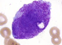 |
| Fig 1D BMA dysmorphic granulopoiesis | 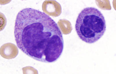 |
| Fig 1E BMA dysmorphic granulopoiesis |  |
| Fig 1F BMA atypical promyelocyte 3 | 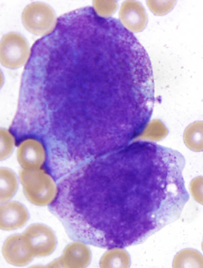 |
| Fig 2A CD45 vs ssc | 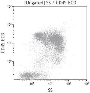 |
| Fig 2B cd33 vs cd34 |  |
| Fig 2C cd64 vs cd11b |  |
| Fig 2D cd117 vs cd7 |  |
| Fig 2E cd34 vs cd14 |  |
| Fig 2F cd13 vs cd19 | 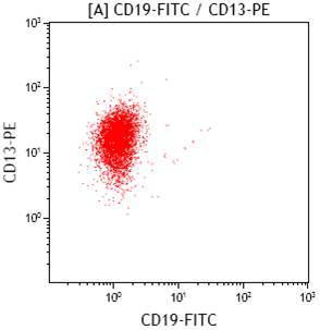 |
| Fig 2G cd11c vs cd14 |  |
| Fig 2H hla-dr vs cMPO |  |
| Fig 2I cd14 vs cMPO | 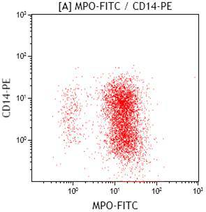 |
| Fig 3 cytogenetic karyotypic analysis |  |
| Fig 4 cytogenetic FISH analysis |  |