Institution: Fundacion Jimenez Diaz Universitary Hospital, Madrid, Spain
Additional authors:Gabriela Simona Ene, Maria Rodriguez Pinilla, Jose Luis Lopez Lorenzo, Elham Askari, Carlos Soto de Ozaeta, Elena Prieto Pareja, Teresa Villaescusa, Teresa Arquero, Cristina Serrano, Alicia Cazorla, Pilar Llamas Sillero
Session: B Lymphoblastic Leukemia/Lymphoma
HISTORY
A 52 years old women with a history of hypothyroidism and depression presented to our Emergency Department with malaise, high fever in the past 2 weeks, sore throat, dyspnea and non-productive cough. A month prior to her visit to the emergency room she consulted with her primary care physician for joint pain and malaise. Her blood work showed mild thrombocytopenia 118 x 10³ µl, normal WBC and Hgb, and high titer of Anti DNA antibodies>400 UI/ml (n.r.< 20 UI/ml), Anti SS-A antibodies (Ro) >240 U/ml ( nr:10 - 240), with the rest of the antibodies Anti SM (Smith ac.), Anti RNP 70 kD, Anti SS-B (La), Anti Scl-70 y Anti Jo-1 0.20 U/ml ) negative. Emergency room physical examination showed a necrotic plaque in the posterior pharynx and ulcers on both tonsils. No visceromegalies or adenopathies were observed. The rest of the physical exam was normal. Her blood work showed severe pancytopenia (WBC 0.20 x 10³ µl , Hgb 8.1 g/dl y Platelets 11 x 10³ µl) and peripheral blood smear did not show abnormal cells or parasites. Liver enzymes were abnormal ALT 190 UI/l, AST 153 UI/l, ALP 569 UI/l, GGT 291 UI/l, total bilirubin 4.4 mg/dl, direct bilirubin 3.4 mg/dl, total proteins 3.7 g/dl, Albumin 1.5 g/dl, LDH 3275UI/l, Prothrombin time 17.3 seconds, Quick 57.0 %, APTTT 27 seconds, Fibrinogen 492 mg/dl, CRP 43 mg/dl, Ferritin 3710 ng/mL, Tryglicerides 134 mg/dl. Chest x-ray was normal and abdominal USS showed mild hepatosplenomegaly. The patient was diagnosed with septic shock and immediately admitted to the ICU with antibiotic and anti-fungal treatment. Blood cultures and viral serology (hepatitis, HIV, EBV) were performed upon admission and were negative. In search of an infectious cause serologies were performed: Leishmania Ab (Leishmania donovani) negative, Parvovirus B19 IgG Ab positive, IgM Ab negative. The CMV serology was undetermined for IgG Ab and negative for IgM Ab. Malaria antigen detection test was also negative. Epstein-Barr virus IgG Ab were positive and IgM Ab were negative. Quantiferon-TB was negative. Based on her history of positive anti DNA antibodies, a screening for autoimmune diseases was performed, showing the same positivity for Anti DNA antibodies and Anti SS-A antibodies (Ro). Bone marrow biopsy was performed for persistent pancytopenia. The aspirate was dry (dry tap) and the bone marrow histology showed an intense hematopoietic hypoplasia of the bone morrow and haemophagocytosis of unclear/unknown cause. Given the initial findings of dry bone marrow aspirate that suggested medullary aplasia and the positivity for Anti DNA antibodies, a high dose steroid, Immunoglobulins and granulocyte colony stimulating factor (G-CSF) therapy were initiated When bone marrow results that suggested Haemophagocytic Lymphohistiocytosis were obtained, cyclosporine was added to the therapy. Two weeks later a second bone marrow biopsy was performed, showing hematopoietic hyperplasia with scattered images of hemophagocytosis and no evidence of microorganisms. Samples were sent for cultures that were negative. The patient had a favorable evolution and was discharged with the following CBC: WBC 5.40 x 10³ µl , Hgb 8.9 g/dL and Platelets 171x 10³ µl. The final diagnosis upon discharge: Septic shock. Severe pancytopenia caused by Haemophagocytic Lymphohistiocytosis syndrome.
DETAILS
First bone marrow biopsy (11/14/2011):
Right posterior iliac crest
Macroscopic description:
Bone marrow cylinder of 1,3/0,1 cm. (B5 Phormol-EDTA, x 1).
Mycroscopic description:
The bone marrow architecture was preserved with normal bone trabeculae. The adipose tissue was also preserved. The inter-trabecular space showed an extensive hemorrhage with hypocellularity of all three hematopoietic cell lines, especially of the myeloid one.
Histiocytic hyperplasia was present with images of erythrophagocytosis, as well as haemophagocytosis of other hematopoietic precursors and lymphocytes. No evidence of myeloid differentiation was observed.
The Wilder technique revealed a moderate increase in the reticuline frame/fibers.
The Ziehl-Neelsen stain was negative, while the PAS and Grocott stains did not show fungal infection/were negative.
In conclusion the bone marrow biopsy showed an intense hematopoietic hypoplasia of the bone morrow and haemophagocytosis of unclear/unknown cause.
Second bone marrow biopsy (11/25/2011):
Right posterior iliac crest
Macroscopic description:
Bone marrow cylinder of 1,8/0,1 cm. (B5 Phormol-EDTA, x 1).
Mycroscopic description:
The aspirate was hypocellular. Samples were sent for cultures that were negative.
The bone marrow had preserved its architecture. The relationship between the adipose and the hematopoietic tissues varied from area to area, between 85/15 and 10/90, but overall it is 50/50. The bone trabeculae were normal. Hematopoietic cell lines are clustered and maturation preserved. Regarding the previous biopsy, there was an
increased number of myeloid and megakaryocytic precursors. The reticulin frame was moderate and clustered.
Dispersed/Scaterred histiocytes with microvacuolae, and with scaterred images of hemophagocytosis. No granulomas were observed.
Tumoral infiltration was not present.
Ziehl Neelsen and PAS were negative.
Diagnosis:
Hematopoietic hyperplasia. Scattered images of hemophagocytosis. Negative for microorganisms.
Third bone marrow biopsy (6/22/2012):
Right posterior iliac crest
Macroscopic description:
Bone marrow cylinder of 1,2/0,1 cm. (B5 Phormol-EDTA, x 1).
iycroscopic description:
Hypercellular bone marrow with a slight increase in the reticulin frame where a monomorphic massive infiltrate with small lymphoblasts with scarce cytoplasm and irregular hyperchromatic nuclei without nucleolus could be observed. These cells expressed Pax-5 and TDT. Most of them expressed CD99 and some were positive for CD20. All blasts cells were myeloperoxidase positive, but negative for CD117 and CD56. There was no residual hematopoiesis. CD 68 positive hystiocytes were present, but no images of hemophagocytosis could be observed. There was a diffuse and intense positivity for CD 10, weakly and clustered for CD 34, and the cells were negative for CD 45 and EBER. Retrospective analysis was performed on the anterior bone marrow biopsies to check positivity for TdT but the previous samples were negative.
IMMUNOHISTOCHEMISTRY AND FLOW CYTOMETRY
First bone marrow biopsy (11/14/2011): Immunohistochemistry (IHC): - Glycophorin : ++ in 40-50% of the preserved erythroid line. - Myeloperoxidase: positive in approximately 5% …… - CD61: positive in 10-15%. - FVIII: positive in 5-10% of the megakaryocytic line - CD79a and CD20: scattered B lymphocytes and some plasma cells (CD 79)). - CD3: scattered T lymphocytes. - CD34: negative - EBER: scattered positive nuclei - CMV and Parvovirus: negative. - CD68: positive in the majority of the histiocytes - Kappa y Lambda: policlonal. Second bone marrow biopsy (11/25/2011): Immunohistochemistry (IHC): - Glycophorin : ++ in 30% - Myeloperoxidase: positive in approximately 60% - FVIII: positive in 5% of the megakaryocytic line - Kappa y Lambda: policlonal. - CD68: positive in the majority of the histiocytes, less than the previous biopsy - CD79a and CD20: positive in few B lymphocytes and some plasma cells. - CD3: scattered T lymphocytes grouped in small clusters between trabecules. - MIB1: 95% proliferation - EBER: negative Flow cytometry of the bone marrow revealed a decrease in the erythroid line and an increase of the myeloid one, with a significantly higher number of mature myeloid forms, and a small percentage of stem cell precursors (0.06% of all cellularity). Amongst lymphoid cells, T as well as B lymphocytes expressed normal maturation antigens and were polyclonal. These features/characteristics discard the presence of a lymphoproliferative syndrome. Third bone marrow biopsy (6/22/2012): Hypercellular bone marrow with a slight increase in the reticulin frame where a monomorphic massive infiltrate with small lymphoblasts with scarce cytoplasm and irregular hyperchromatic nuclei without nucleolus could be observed. These cells expressed Pax-5 and TDT. Most of them expressed CD99 and some were positive for CD20. All blasts cells were myeloperoxidase positive, but negative for CD117 and CD56. There was no residual hematopoiesis. CD 68 positive hystiocytes were present, but no images of hemophagocytosis could be observed. There was a diffuse and intense positivity for CD 10, weakly and clustered for CD 34, and the cells were negative for CD 45 and EBER. Retrospective analysis was performed on the anterior bone marrow biopsies to check positivity for TdT but the previous samples were negative. The inmunophenotype of the bone marrow showed 83% of blasts with the following profile: CD19+, CD45+weakly, HLA-DR++, CD79a+, CD22s+, CD34+heterogenous, CD10+, CD20+ (20%), TdT+, mic-, light chains -. CD15-, CD11b, CD33-, CD13-, CD64-. CD117-, CD16-, CD56-, CD36-, CD14-, CD61-, GlyA-, CD8-, CD5-, CD7-, CD2-, CD3s.The inmunophenotype was compatible with a LAL BII of the FAB classification.
CYTOGENETIC FINDINGS
Third bone marrow biopsy (6/22/2012): No metaphases were obtained for the karyotype. FISH for 11q23 (gen MLL) re-arrangement and for BCR/ABL were negative.
MOLECULAR FINDINGS
INTERESTING FEATURES
Most published cases of HLH and T- or B-cell lymphoblastic leukemia in pediatric patients occurred after the diagnosis of acute lymphoblastic leukemia, during induction therapy and can persist after consolidation therapy even with remission from leukemia. Usually, in the case of malignancy, HLH is a complication of the primary disease, not a preceding diagnosis. Several case reports of ALL presenting with HLH as the first manifestation of leukemia have been published. Our patient had five diagnostic criteria for HLH: fever, splenomegaly, cytopenias affecting ≥ 2 lineages, haemophagocytosis in the bone marrow, with no evidence of malignancy, and ferritin ≥ 500 μg/L. After immunosuppressive and antibiotic therapy was initiated, the cytopenias and fever resolved. When levofloxacin was discontinued, the fever and respiratory symptoms reappeared. At the same time, M. tuberculosis was recovered from blood cultures performed five weeks before, and tuberculostatic therapy was indicated for resolution of the symptoms. The newly diagnosis of a Precursor B-cell acute lymphoblastic leukemia seven months after the haemophagocytic syndrome episode may imply that our patient suffered from preclinical leukemia or that this syndrome appeared in the context of a real predisposition for suffering an ulterior lymphoid malignancy. We were unable to identify lymphoid blast cells in the first bone marrow biopsy. Nevertheless, the HLH diagnosis involves the need for subsequent monitoring of the patient for malignances, even if the HLH resolves.
PROPOSED DIAGNOSIS
Acute lymphoblastic leukemia ALL BII (FAB classification) in a patient with history of Haemophagocytic Lymphohistiocytosis syndrome secondary to a disseminated infection with M. tuberculosis.
CONSENSUS DIAGNOSIS
B-acute lymphoblastic leukemia in a patient with history of haemophagocytic lymphohistiocytosis syndrome secondary to disseminated infection with M. tuberculosis
| first bone marrow |  |
| first bone marrow |  |
| first bone marrow | .jpg) |
| first bone marrow EBER | 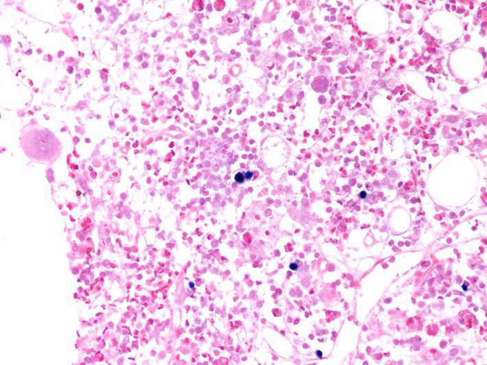 |
| first bone marrow TdT | 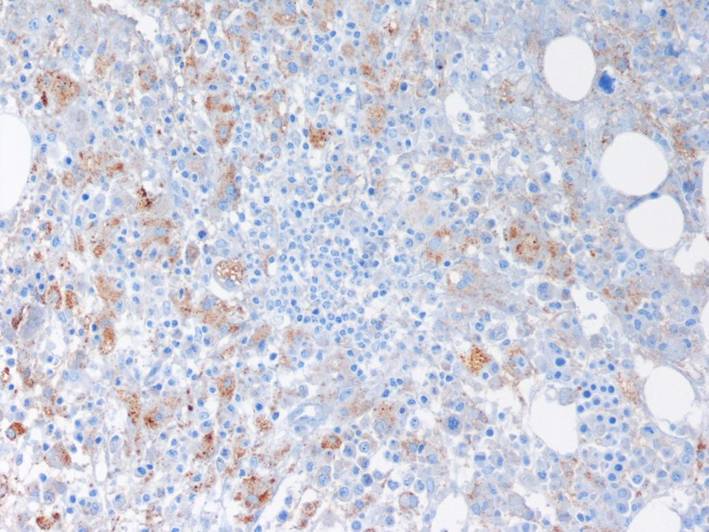 |
| second bone marrow | 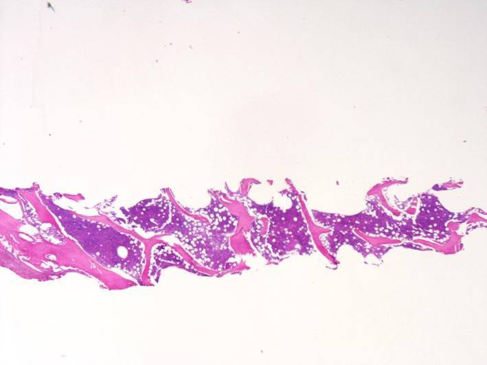 |
| second bone marrow | 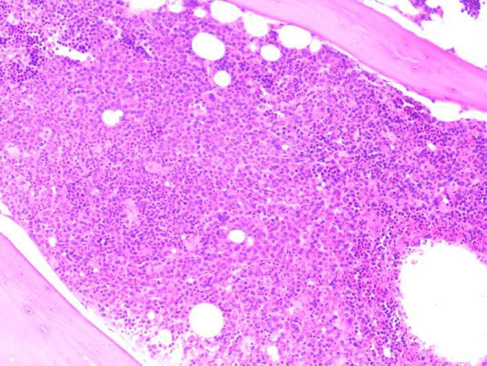 |
| second bone marrow TdT | 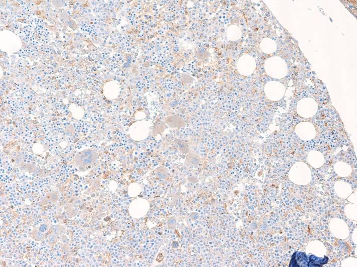 |
| third bone marrow biopsy | 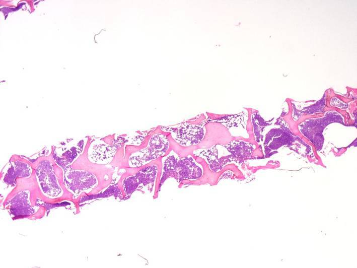 |
| third bone marrow biopsy | 3.jpg) |
| third bone marrow biopsy CD10 | 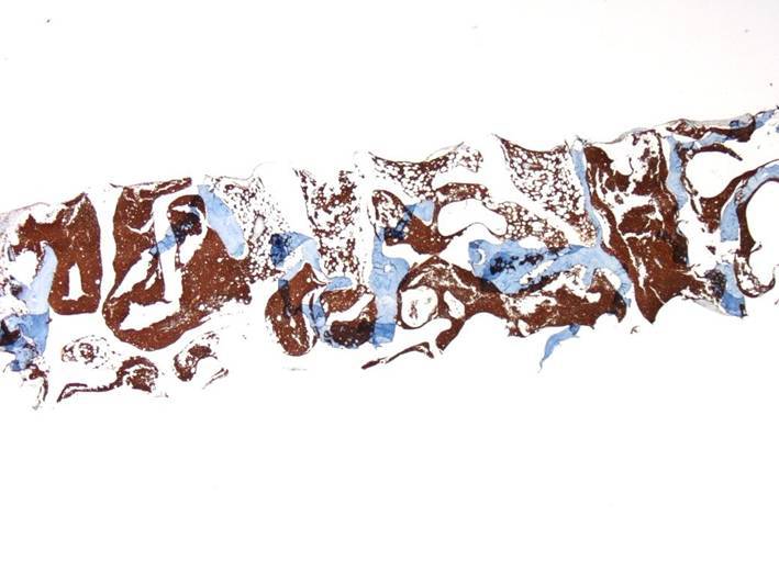 |
| third bone marrow biopsy PAX 5 | 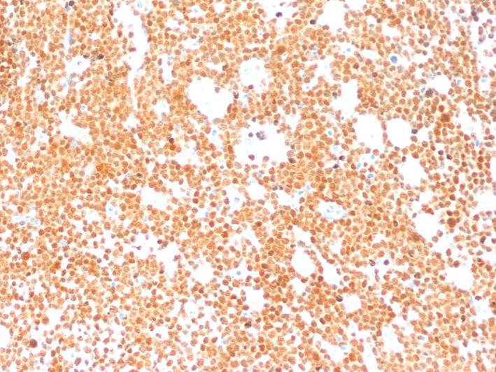 |
| third bone marrow biopsy TdT | 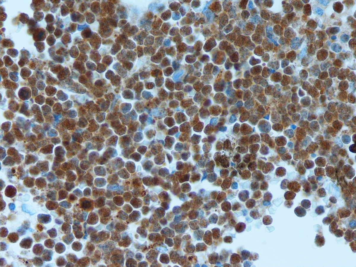 |
| Peripheral blood extension ALL | 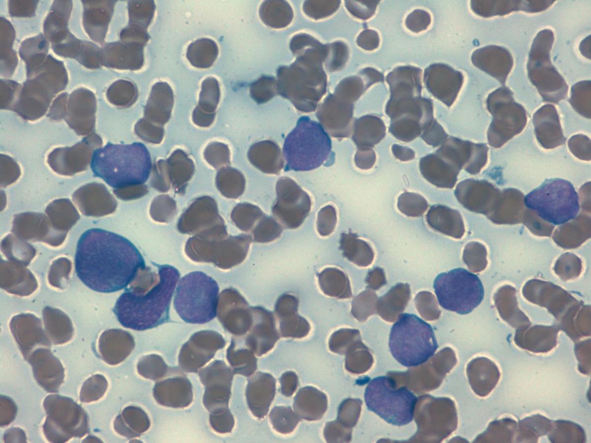 |