Institution: Columbia University Medical Center
Additional authors:Daniela Hoehn, M.D., Ph.D., Bachir Alobeid, M.D., Govind Bhagat M.D.
Session: B Lymphoblastic Leukemia/Lymphoma
HISTORY
82-year-old Caucasian female with a history of Breast Cancer status post right mastectomy, endometrial cancer status post hysterectomy and bilateral salpingo-oophorectomy 6 years previously presented to an outside hospital with recent onset of generalized weakness (x2 weeks). On examination, she was found to be pancytopenic (CBC: WBC 0.7 x10⁹/L, Hct 17.3%, Plts 74x109/L and 91% lymphocytes on differential count). Bone marrow biopsy (2/8/2012) showed an extensive infiltrate of lymphocytes with blastoid features. The patient received one round of Cytoxan, Vincristine and Adriamycin with no recovery in counts. Due to non response to therapy, the patient was transferred to our institute for further management. The patient received multiple rounds of chemotherapy with a follow-up bone marrow biopsy showing no evidence of disease. She subsequently relapsed (10/4/2012) with peripheral blood and CSF involvement by her lymphoid neoplasm. No further therapy was given and the patient was transferred to a hospice where she died one month later.
DETAILS
Biopsy fixation details: Bouins
Details of microscopic pathology Diagnostic biopsy: The bone marrow core biopsy was markedly hypercellular for age (80% cellularity). An extensive infiltrate of small to medium sized lymphocytes was present, accounting for 90% of all marrow nucleated elements. The lymphocytes had round to ovoid nuclei, fine chromatin, variably prominent single or multiple nucleoli, and scant cytoplasm. Occasional large lymphocytes were seen admixed. Scattered mitotic figures and apoptotic cells were noted. Rare scattered colonies of maturing erythroid and myeloid elements were present and focal, small clusters of normal appearing megakaryocytes were also noted.The bone marrow biopsy sample at disease relapse (10/4/2012) exhibited an extensive infiltrate of immature appearing lymphoid cells with similar features as observed in the diagnostic biopsy.IMMUNOHISTOCHEMISTRY AND FLOW CYTOMETRY
Diagnostic biopsy and aspirate clot preparation: The neoplastic cells had the following phenotype: CD34-, CD19+, CD20-, CD10+, BCL6+, BOB1+, LMO2+, MUM1+, TdT(subset ~30%)+, CD5-, CD23-, cyclinD1-, CD43+, BCL2+. In-situ hybridization for Epstein-Barr virus (EBER) was negative. The proliferation index, as determined with a stain for Ki-67, was markedly elevated (>90%).
The immunophenotype of neooplastic cells at relapse differed slightly from the diagnostic sample. The phenotype was: CD34-, CD19+, CD10+, MUM1-, with rare cells expressing TdT and a minor component expressed CD20 (5%). An immunohistochemical stain for cMYC showed ~40% nuclear cMYC expression.By flow cytometric analysis (at relapse), the neoplastic cells had the following phenotype: CD34-, TdT-, CD19+, cCD79a+, CD20-, FMC7-, CD10+/-, CD5-, CD23-, CD38+, HLA-DR+, CD43+/-, CD11c-, CD52+/-, CD30-, cIgM+, sIgM(small subset)+, and sIgD-. A small subset showed cytoplasmic and surface kappa light chain restriction.CYTOGENETIC FINDINGS
Cytogenetics: No karyotype was available for the diagnostic biopsy sample. Karyotype of the relapse sample was:
46,XX,del6(?q13q21)[4]/46,idem,del(16)(q12-13)[8]/46,XX[8]. Four metaphases show an interstitial deletion on the long arm of chromosome 6 (deletion 6q), while eight metaphases display deletion 6q and deletion 16q, suggestive of clonal evolution.FISH analysis using MYC, BCL2, BCL6, IGH, MLL, BCR/ABL and TP53/CEP17 probes showed no evidence of rearrangement, numerical abnormality or amplification.MOLECULAR FINDINGS
Polymerase chain reaction (PCR) for the Immunoglobulin heavy chain gene showed clonal IgH gene rearrangement.
INTERESTING FEATURES
This case likely represents a case of mature lymphoblastic leukemia with unusual cytogenetic abnormalities that was refractory to therapy. The phenotype of the neoplastic cells raised the possibility of an unclassifiable aggressive B-cell non-Hodgkin lymphoma with blastoid features presenting in leukemic phase.
PROPOSED DIAGNOSIS
Lymphoid neoplasm with blastoid features, suggestive of mature lymphoblastic leukemia.
CONSENSUS DIAGNOSIS
B-acute lymphoblastic leukemia, with partial immunophenotypic shift at relapse
| Extensive bone marrow infiltrate by neoplastic cells (hematoxylin and eosin, 400x and 1000x(insert)) |  |
| Bone marrow aspirate smear morphology (Wright-Giemsa, 1000x | 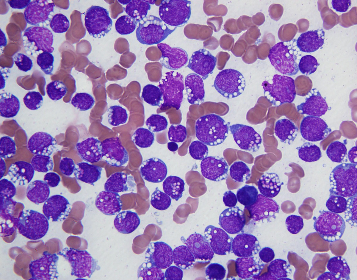 |
| Neoplastic cells demonstrating CD19 positivity (400x) |  |
| Neoplastic cells demonstrating CD10 positivity (400x) |  |
| . Neoplastic cells demonstrating partial TdT positivity (400x, insert showing loss of TdT expression on neoplastic cells in relapse specimen) | 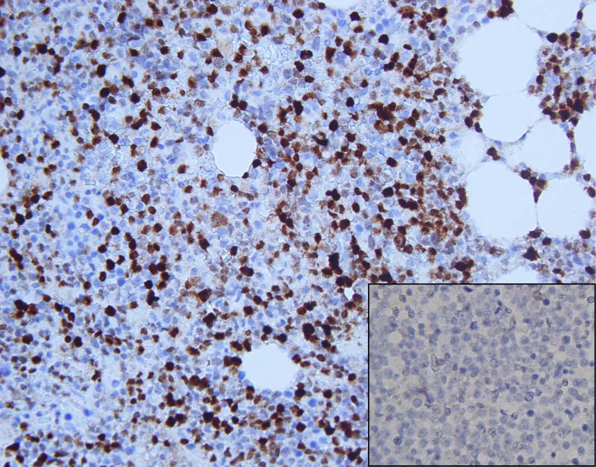 |
| Neoplastic cells demonstrating partial MUM1 positivity (400x, insert showing loss of MUM1 expression on neoplastic cells in relapse specimen) | 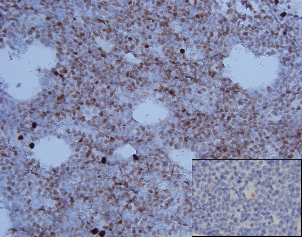 |
| Neoplastic cells demonstrating high proliferation index Ki-67 (400x) |  |
| Small subset of neoplastic cells demonstrating CD20 positivity in relapsed specimen (400x) | 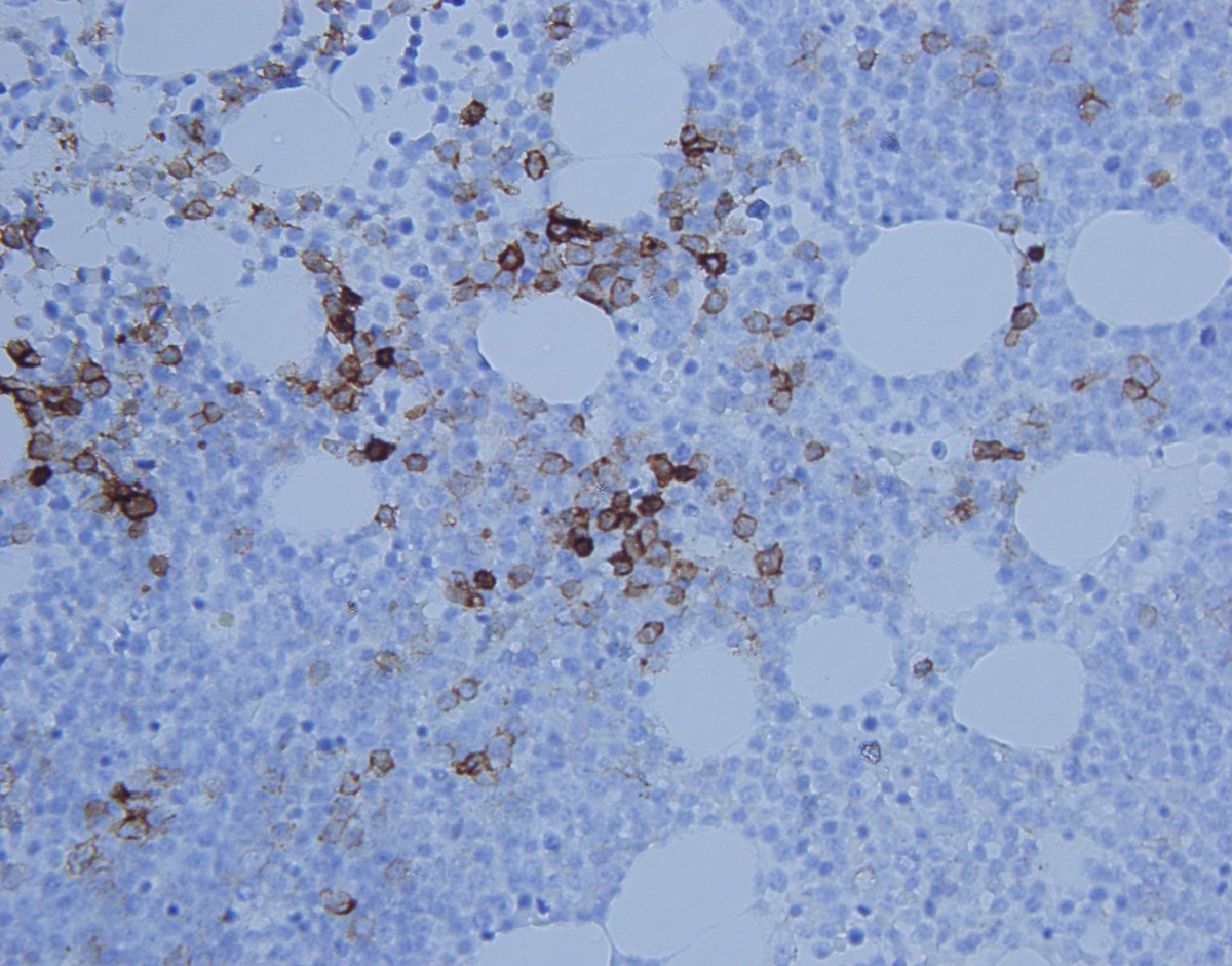 |
| CD34 immunostain highlights vessels with CD34 negative neoplastic cells in background (400x) | 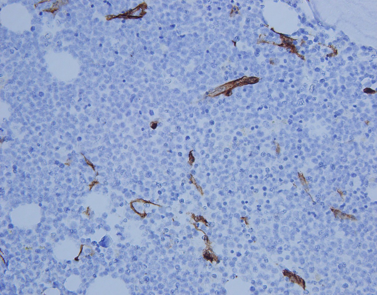 |
| cMYC immunostain demonstrates strong nuclear staining in ~40% of neoplastic cells (400x) | 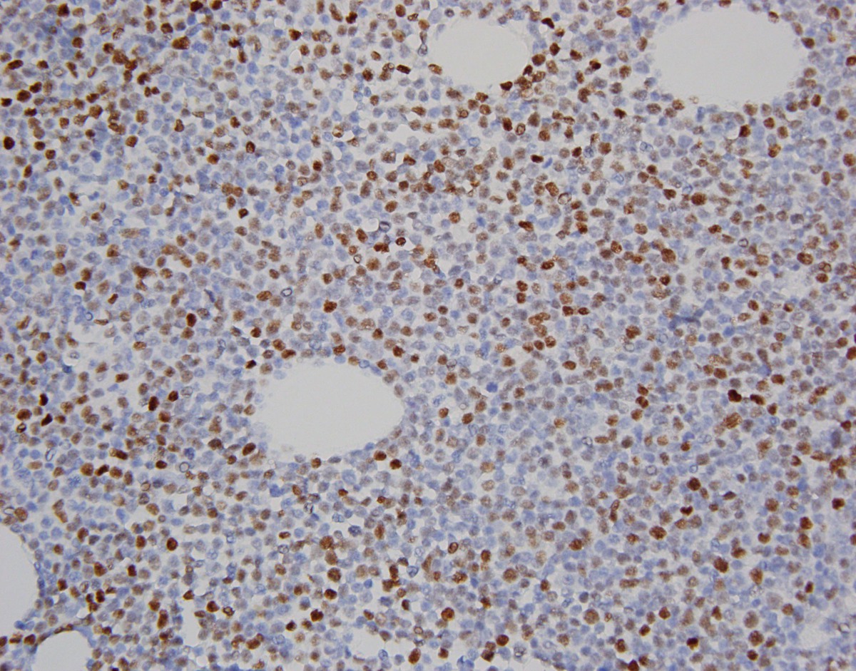 |