Institution: Hospital of the University of Pennsylvania
Additional authors:David L. Porter, M.D., Bruce L. Levine, Ph.D., Michael Kalos, Ph.D., Carl H. June, M.D., and Adam Bagg, M.D.
Session: B Lymphoblastic Leukemia/Lymphoma
HISTORY
A 66-year-old man was originally diagnosed with chronic lymphocytic leukemia (CLL) in 1996. Following a six-year period of asymptomatic surveillance, he experienced progressive leukocytosis and lymphadenopathy requiring therapy. Between 2002 and 2010 various agents, including rituximab, fludarabine, bendamustine, and alemtuzumab, were used with approximately 2-year progression-free intervals between treatments. However, his disease continued to evolve with persistent widespread lymphadenopathy. In July 2010, the patient was enrolled in a phase I clinical trial evaluating chimeric antigen receptor-modified T cells directed against CD19 (CART-19). In September 2010, he received pentostatin/cytoxan induction and a total of 1.42×10^7 autologous CART-19 cells (infused over a three day period). The patient initially experienced fevers, chills, anorexia, nausea and diarrhea, which was followed by delayed tumor lysis syndrome on day 22. Bone marrow studies performed on day 23 showed no evidence of CLL by histology, immunophenotype, and cytogenetics/FISH. By day 28 the patient no longer had palpable adenopathy. Subsequent serial CT scans confirm sustained clinical remission, and bone marrow studies demonstrate no residual disease. The patient is now 2 years and 4 months post autologous CART-19 therapy without any evidence of residual disease, using sensitive (10^-6) molecular techniques. He does have sustained hypogammaglobulinemia, which is treated with periodic IVIG therapy.
DETAILS
Bone marrow biopsies were fixed in B5 and decalcified prior to processing and H&E staining. The first marrow submitted to the workshop (labeled HS-10-10547) was obtained five months prior to CART-19 therapy, which showed a hypercellular marrow (60%) extensively involved (40%) by CLL. Post-therapy bone marrow studies up to two years following infusion, including the second bone marrow submitted for review (labeled HS-11-6512) obtained at six months post treatment, have all remained histologically negative*. (*Note: Since this patient is part of an ongoing clinical trial, with a need to perform multiple additional studies on the limited bone marrow biopsies, we are unfortunately unable to submit unstained slides to the workshop.)
IMMUNOHISTOCHEMISTRY AND FLOW CYTOMETRY
Day 0 immunohistochemical stains on the bone marrow core biopsy show numerous CD20+ lymphoid aggregates, while flow cytometry performed on the bone marrow aspirate shows an expansion (65%) of variably-sized bright surface kappa restricted CD5+ CD10- CD19+ CD20+ CD23+ CD38- IgM+ B cells. Bone marrow samplings obtained at day +23 and +180 reveal virtually no B cells by immunohistochemistry and flow cytometry. Subsequent bone marrow biopsies up to one-year post therapy continue to demonstrate a virtual absence of B cells and certainly no immunophenotypic evidence of CLL by flow cytometry.
CYTOGENETIC FINDINGS
One day prior to receiving CART-19 infusion, cytogenetics studies were performed on a bone marrow aspirate, and showed 46,XY,del(17)(p12)[5]/46,XY,der(17)t(17;21)(q10;q10)[5]/46,XY[14]. FISH studies confirmed the deletion of TP53. Follow-up cytogenetics studies of the bone marrow up to two years post therapy demonstrate a normal male karyotype, 46,XY, and FISH remains negative for deletion of TP53.
MOLECULAR FINDINGS
Real-time PCR was performed on peripheral blood to detect DNA encoding the anti-CD19 chimeric antigen receptor starting on day 1 of CART-19 infusion. Using this approach, by day 21 a 3-log expansion of the modified CART-19 T cells was seen in vivo. At this peak transgene time-point, CART-19 cells accounted for over 20% of circulating lymphocytes, which also coincided with clinical tumor lysis syndrome. Copies of the transgene in peripheral blood remained high six months post infusion. The circulating half-life of these T cells is estimated to be 35 days. Quantitative PCR also detected transgene DNA in the bone marrow up to two years post therapy. Next generation sequencing of the IGH@ gene (with a sensitivity of ~10^-6) shows no minimal residual disease in the bone marrow two years post CART-19 infusion.
INTERESTING FEATURES
This is the first ever case to demonstrate the utility of a novel and highly effective form of therapy, which can also be applied to treat other CD19+ neoplasms, including B lymphoblastic leukemia. Unlike first-generation CART cells, the modified receptor against CD19 used in this case is linked to the 4-1BB domain of CD137 and the cytoplasmic zeta domain of CD3, which are T cell-specific co-stimulatory and signaling domains. These CART cells are observed to have unique qualities such as a capacity for in vivo replication, acquisition of a memory T cell phenotype, and long-term in vivo persistence resulting in sustained tumor control. This patient is now two years and four months post therapy with durable clinical, pathologic, and exquisitely sensitive molecular remission.
PROPOSED DIAGNOSIS
Refractory chronic lymphocytic leukemia apparently cured by autologous chimeric antigen receptor-modified T cell therapy against CD19 (CART-19).
CONSENSUS DIAGNOSIS
Refractory chronic lymphocytic leukemia apparently cured by autologous chimeric antigen receptor-modified T cell therapy against CD19 (CART-19)
| Bone marrow core biopsy (Case HS-10-10547), 5 months prior to CART-19 therapy, H&E, 10x: The bone marrow biopsy obtained at 5 months prior to CART-19 therapy (submitted for review) demonstrates a hypercellular marrow (~60%) involved by a nodular interstitial expansion of small mature lymphocytes (~40% of total cellularity). |  |
| Bone marrow aspirate, Day 0 prior to CART-19 infusion, flow cytometry: Flow cytometry performed on the bone marrow aspirate obtained immediately prior to CART-19 therapy reveals an expansion (65% of total cellularity) of variably-sized bright surface kappa restricted CD5+ CD10- CD19+ CD20+ CD23+ CD38- IgM+ B cells. |  |
| Bone marrow aspirate, Day 0 prior to CART-19 infusion, cytogenetics: Cytogenetics performed at this time shows an abnormal karyotype 46,XY,del(17)(p12)[5]/46,XY,der(17)t(17;21)(q10;q10)[5]/46,XY[14]. Shown here is the deletion of the short arm of chromosome 17 at p12, which includes the TP53 locus. | ![Bone marrow aspirate, Day 0 prior to CART-19 infusion, cytogenetics: Cytogenetics performed at this time shows an abnormal karyotype 46,XY,del(17)(p12)[5]/46,XY,der(17)t(17;21)(q10;q10)[5]/46,XY[14]. Shown here is the deletion of the short arm of chromosome 17 at p12, which includes the TP53 locus.](cases/264/images/Day0_cytogenetics1.jpg) |
| Bone marrow aspirate, Day 0 prior to CART-19 infusion, cytogenetics: Additionally, a derivative chromosome 17 is identified, which is composed of the long arm of chromosome 21 replacing the short arm of chromosome 17. This rearrangement leads to monosomy 17 from 17p10 through 17pter and trisomy 21, and loss of the TP53 locus. |  |
| The day -270 bone marrow core demonstrates a markedly hypercellular bone marrow (90%) composed of a dense lymphocytic infiltrate. The day 0 specimen is also hypercellular (60%), and shows a mostly interstitial expansion of lymphocytes. The day +23 bone marrow biopsy continues to be hypercellular, now with occasional interstitial aggregates of lymphocytes accounting for 10% of cellularity. The day +180 bone marrow is normocellular and contains scattered groups of small lymphocytes (<5% of cellularity). |  |
| CD20 staining shows an overwhelming B cell infiltrate in the bone marrow specimen obtained 9 months prior to CART-19 therapy. CD20 also highlights the interstitial aggregates of B cells in the day 0 specimen. The day +23 bone marrow contains scattered lymphoid aggregates that are partially composed of CD20+ B cells. Finally, the day +180 bone marrow shows rare B cells. |  |
| Background CD3+ T cells can be seen admixed within the dense B cell infiltrate in the bone marrow core biopsy obtained 9 months and day 0 prior to CART-19 infusion. CD3 staining confirms that the lymphoid aggregates seen on the day +23 bone marrow are composed of both B and T cells. Finally, the day +180 specimen shows scattered CD3+ T cells, likely accounting for the majority of the lymphocyte population at this time. |  |
| Detection of modified antigen receptor transgene over time: Genomic DNA isolated from serial samplings of the patient’s whole blood (A), and bone marrow aspirate (B) was subjected to quantitative real-time polymerase-chain-reaction (PCR) analysis to detect transgenic DNA. Whole blood specimens were also subjected to flow cytometric analysis to detect the presence of CART-19 T cells (A). Both assays show that the modified T cells expanded to levels greater than 1000x as high as initial engraftment levels in the peripheral blood and bone marrow. Further, the number of copies of transgene stayed elevated even at day +180 post infusion. | 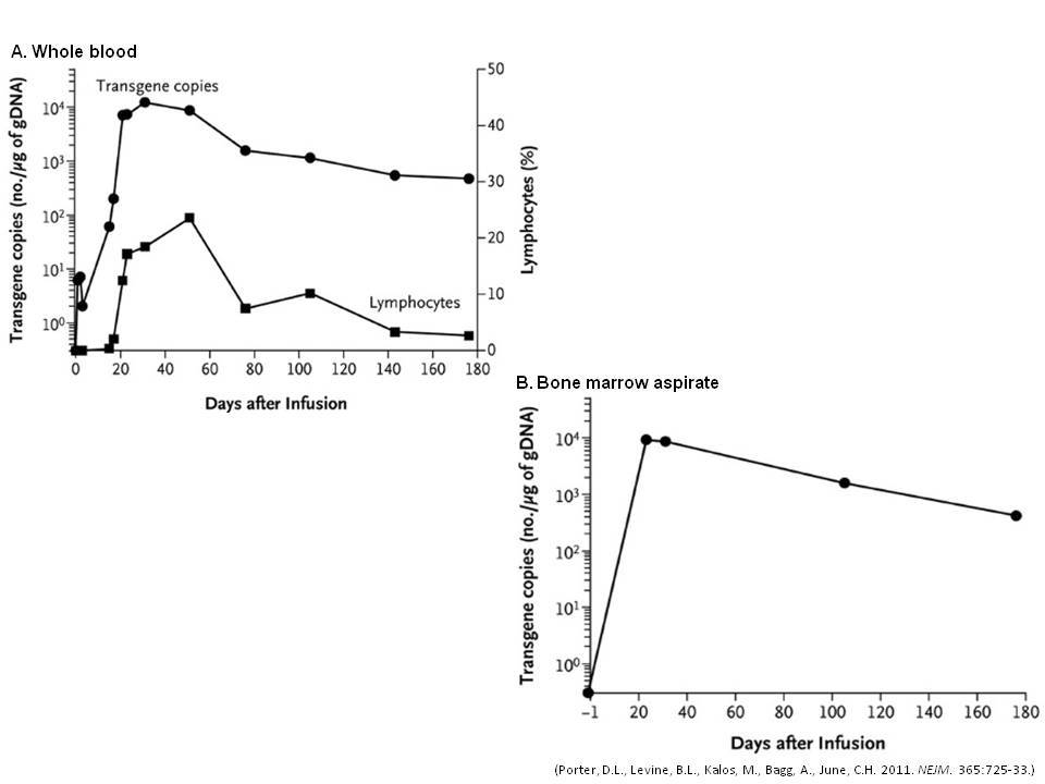 |
| Bone marrow core biopsy (Case HS-11-6512), 6 months (day +180) post CART-19 therapy, H&E, 20x: Bone marrow biopsy obtained at six months post CART-19 therapy (submitted for review) shows normocellular marrow with trilineage hematopoiesis, and no morphologic evidence of CLL. |  |
| Bone marrow aspirate, 1 year post CART-19 therapy, flow cytometry: Flow cytometry performed on the bone marrow aspirate obtained 1 year post CART-19 therapy shows CD5+ T cells and a virtual absence of mature B cells. |  |
| Bone marrow aspirate, 2 years post CART-19 therapy, cytogenetics: Cytogenetics performed on the bone marrow aspirate obtained 2 years post CART-19 therapy shows a normal male karyotype 46,XY[25]. FISH confirmed the absence of TP53. | ![Bone marrow aspirate, 2 years post CART-19 therapy, cytogenetics: Cytogenetics performed on the bone marrow aspirate obtained 2 years post CART-19 therapy shows a normal male karyotype 46,XY[25]. FISH confirmed the absence of TP53.](cases/264/images/2years_cytogenetics.jpg) |