Institution: University of New Mexico Health Sciences
Additional authors:David R. Czuchlewski, M.D.
Session: T Lymphoblastic Leukemia/Lymphoma
HISTORY
The patient is a 11-year-old male with a history of constipation of 'chronic' duration who presented to the emergency department following several febrile episodes accompanied by vomiting and increasing abdominal pain. Radiographic evaluation demonstrated 'grape-like' masses throughout the duodenum and an anterior mediastinal mass with some mild compression of the superior vena cava. CBC reveals: WBC=5.2; RBC=4.34; Hgb=12.2; Hct=35%; MCV=81 and Plt=320. Manual differential cell count includes: 58% neutrophils, 17% lymphocytes, 15% blasts, 9% monocytes and 1% eosinophils.
DETAILS
Duodenal mucosal biopsies demonstrated marked expansion of the lamina propria by an infiltrative lymphoid process. Atypical lymphoid cells were characterized by increased nuclear-cytoplasmic ratio, immature chromatin, prominent nucleoli and frequent mitotic figures with an overall blastic appearance.
Wright-stained bone marrow aspirate demonstrated increased blasts of intermediate size (47% of total cells) with an appearance similar to that described in the previous duodenal biopsies. The otherwise normocellular bone marrow biopsy demonstrated intact trilineage hematopoeisis.IMMUNOHISTOCHEMISTRY AND FLOW CYTOMETRY
Tissue based immunohistochemistry (IHC) on paraffin embedded duodenal mucosal biopsies demonstrates an atypical lymphoid infiltrate that is immunoreactive with TdT, CD3, CD5 (weak), CD43 and CD7.
Subsequent peripheral blood flow cytometry demonstrated 13% of total cells expressing CD45 (dim), cytoplasmic CD3, CD5 (dim), CD7 and CD117. A subset of lymphoblasts expressed B-cell antigens including: CD19 and cCD79a. Otherwise, lymphoblasts did not express: CD34, MPO, HLA-DR, CD13, CD33, CD1a, CD2, surface CD3, CD8, CD56, CD57, T-cell receptors (TCR alpha/beta and TCR gamma/delta), CD20, CD22 or CD10.CYTOGENETIC FINDINGS
45~47,XY, add(1)(p13), add(4)(q35), -5,-7,-8, add(9)(p22), +19, +1mar, +1~3r[cp7]/
45-47, XY, add(4)(q35), -5,-7,-8, add(9)(p22), +10, +1~2mar, +1~2[cp6]/46, XY [7]Fluorescence in-situ hybridization (FISH) is negative for BCR/ABL1 fusion associated with t(9;22)(q34;q11.2) and rearrangement of the MLL gene region at 11q23.MOLECULAR FINDINGS
DNA PCR analysis for T-cell gamma receptor rearrangement was negative. DNA PCR analysis for FLT3 internal tandem duplication (FLT3-ITD), FLT3 D835 variant and NPM-1 exon 12 mutation were all negative. DNA PCR analysis for codons 816 or 820-825 in exon 17 of the KIT gene was negative.
INTERESTING FEATURES
This case demonstrates immunophenotypic findings consistent with T-lymphoblastic leukemia, early T-cell precursor ALL (ETP-ALL). ETP-ALL represents a newly recognized subset of T-ALL whose immunophenotype suggests origin from very early thymocytes. ETP-ALL reflects a multipotent stem cell repertoire and may express T and myeloid antigens. The diagnosis of ETP-ALL requires: absence of CD1a and CD8 expression, weak CD5 expression and gain of one or more of the myeloid/stem cell markers including CD117, CD34, HLA-DR, CD13, CD33, CD11b or CD65. The pattern of antigen expression in ETP-ALL may raise the possibility of acute leukemia of ambiguous lineage; however, the WHO 2008 criteria for such a diagnosis are not fulfilled. As in the current case, ETP-ALL may lack T-cell gene rearrangements and may demonstrate complex cytogenetic findings. ETP-ALL is clinically characterized by a decreased clearance of leukemic cells following induction chemotherapy, as was the case in our patient.
PROPOSED DIAGNOSIS
T-lymphoblastic leukemia, early T-cell precursor ALL (ETP-ALL)
CONSENSUS DIAGNOSIS
T-acute lymphoblastic leukemia, early T-cell precursor immunophenotype
| Bone Marrow Aspirate, Core Biopsy and Immunohistochemical Stain | 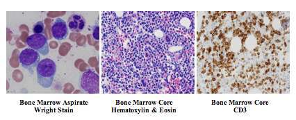 |
| Flow Cytometry of Peripheral Blood | 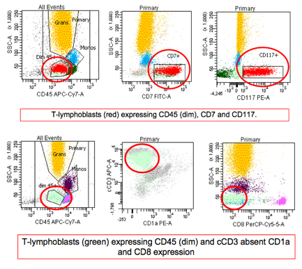 |
| Additional slide 1 |  |
| Additional slide 2 |  |