Institution: University of Michigan Health System
Additional authors:Daniel Leino, MD, Lina Shao, MD PhD, Jason X. Cheng, MD PhD
Session: AML with recurrent genetic abnormalities Part I
HISTORY
A 68 year old male with untreated low grade follicular lymphoma and prostatic adenocarcinoma status post prostatectomy presented with a one month history of fatigue and a 10 pound weight loss and was found to be leukopenic and anemic with associated thrombocytosis. Bone marrow examination and ancillary studies established a diagnosis. The patient was treated with a small molecular inhibitor (AT-406), but without response. The disease remained refractory to more aggressive therapies and, nine months after diagnosis, lead to the patient’s demise.
DETAILS
A bone marrow biopsy (fixed in acetic acid-zinc formalin), aspicular hemodilute aspirate smears and peripheral blood smears were reviewed at the time of diagnosis [digital images provided, Figure 1]. The marrow was hypercellular with left-shifted granulopoiesis and increased blasts, megaloblastoid erythroid maturation with dyserythropoiesis, and megakaryocytic hyperplasia with dysmegakaryopoiesis, the latter characterized by numerous small hypolobated megakaryocytes. The peripheral blood smear confirmed thrombocytosis with numerous giant hypogranular platelets, circulating normoblasts and dysmorphic myelomonocytic elements. Follow-up examination after multiple aggressive therapies revealed persistent pronounced megakaryocytic hyperplasia and dysplasia with megakaryocytes clustered within sinusoids. Peripheral blood smear now revealed leukoerythoblastic features with numerous blasts, circulating micromegakaryocytes and hypogranular platelets. [H&E sections of the marrow core and digital images of the marrow and peripheral blood provided, Figure 2]
IMMUNOHISTOCHEMISTRY AND FLOW CYTOMETRY
Flow cytometric analysis of marrow demonstrates an abnormal population of myeloid blasts that exhibit dim expression of CD45 and low side scatter, are positive for CD38 (dim), CD34 (moderate), CD117 (moderate-dim), CD13 (dim-moderate) and CD33 (dim), and aberrantly coexpress CD7 (dim) and CD56 (partial dim) [digital images provided, Figure 3].
CYTOGENETIC FINDINGS
Cytogenetic karyotypic analysis revealed the following: 45,XY,t(3;3)(q21;q26.2),-7[20] [digital images provided, Figure 4].
MOLECULAR FINDINGS
Molecular polymerase chain reaction analysis was negative for FLT3 D835, FLT3 ITD, NPM1, CEBPA, IDH1, IDH2 and KIT D816V mutations.
INTERESTING FEATURES
This case represents a classic presentation of the rare entity, AML with t(3;3)(q21;q26.2) and highlights characteristic cytomorphologic features manifest at diagnosis and with disease progression.
PROPOSED DIAGNOSIS
Acute myeloid leukemia with t(3;3)(q21;q26.2)
CONSENSUS DIAGNOSIS
Acute myeloid leukemia with t(3;3)(q21;q26.2); RPN1-EVI1, and -7
| Fig 1A BMX 40x | 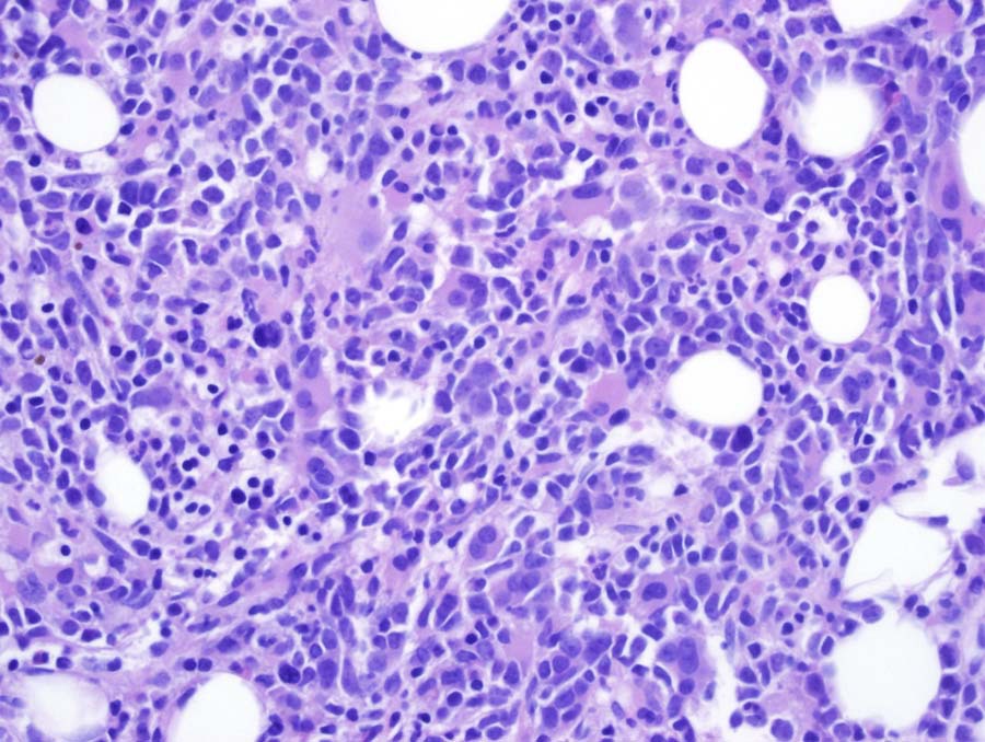 |
| Fig 1B BMX 100x immature mononuclear cells | 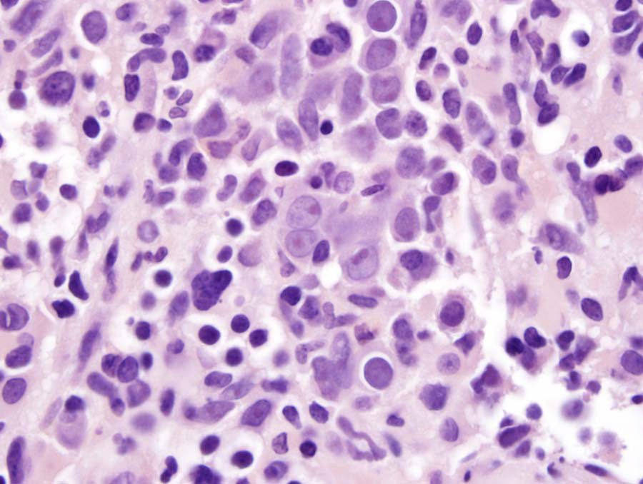 |
| Fig 1C BMX 100x dysplastic megakaryocytes | 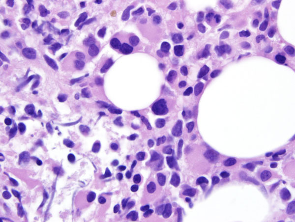 |
| Fig 1D BMA 100x dysplastic erythropoiesis | 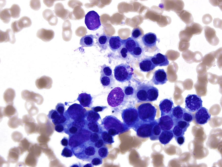 |
| Fig 1E BMA 100x blasts and dysmyelopoiesis | 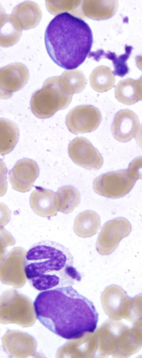 |
| Fig 1F BMA 100x blasts and dysmonopoiesis | 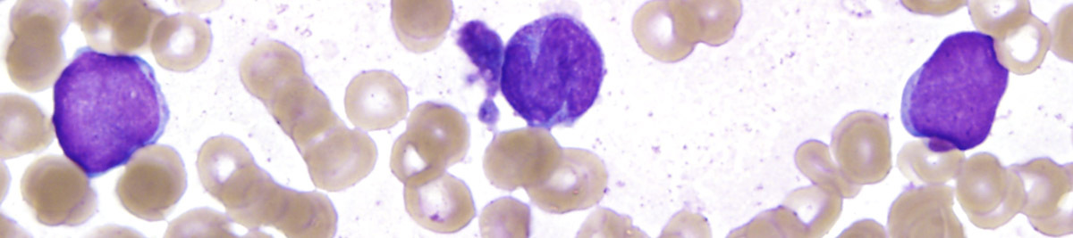 |
| Fig 1G PB blast |  |
| Fig 1H PB blast |  |
| Fig 1I PB giant hypogranular platelet | 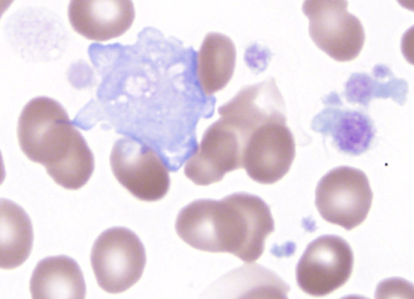 |
| Fig 1J PB dysplastic monocyte | 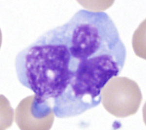 |
| Fig 1K PB circulating normoblast | 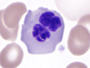 |
| Fig 2A Refract dis BMX intrasinusoidal megakaryocytes 10x | 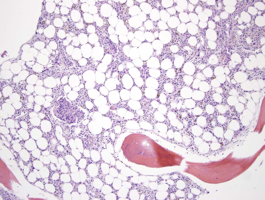 |
| Fig 2B Refract dis BMX intrasinusoidal megakaryocytes 40x | 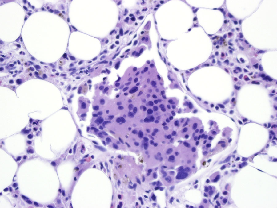 |
| Fig 2C Refract dis BMX intrasinusoidal megakaryocytes CD61 | 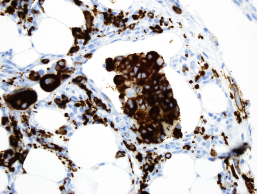 |
| Fig 2D PB blasts |  |
| Fig 2E PB circulating micromegakaryocyte | 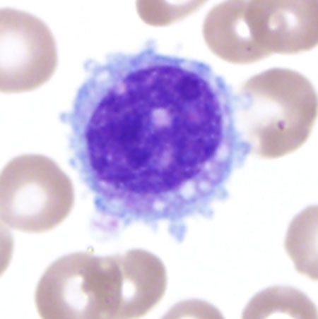 |
| Fig 2F PB giant hypogranular platelets |  |
| Fig 3A CD45 vs SSC | 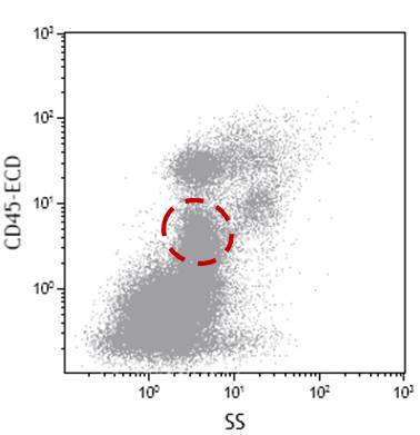 |
| Fig 3B CD33 vs CD34 | 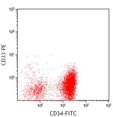 |
| Fig 3C CD19 vs CD38 |  |
| Fig 3D CD13 vs CD19 | 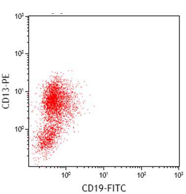 |
| Fig 3E CD117 vs CD7 | 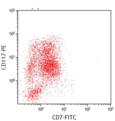 |
| Fig 3F CD2 vs CD56 |  |
| Fig 4 Cytogenetic karyotypic analysis | 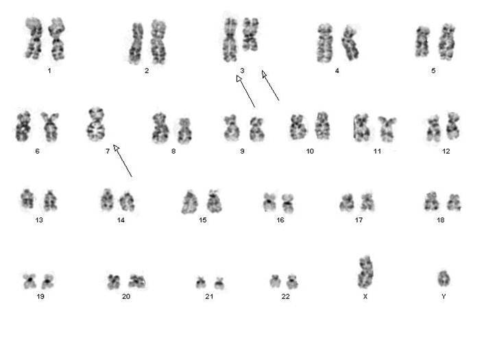 |