Institution: Weill Medical College of Cornell University
Additional authors:Julia Geyer, Atillio Orazi, Jiong Yan, Susan Mathew, Wayne Tam, Mikhail Roshal
Session: Acute leukemias of ambiguous lineage
HISTORY
A 52 year-old male, in his usual state of health, had a routine physical because his wife requested it. At an outside institution, he was found to have abnormal labs, sent to the ER and had a bone marrow which showed acute leukemia. He was transferred immediately to our institution and had a second marrow biopsy; because it was a dry tap, a peripheral blood specimen was sent for flow cytometry and cytogenetic analysis. The diagnosis rendered was acute leukemia of ambiguous lineage.
His PMH was significant for 125 pack/year h/o cigarettes, asthma and food allergies. He had a family history of a father and one brother dead of pancreatic carcinoma, a mother and two sisters dead of breast carcinoma and two additional sisters alive and well, both having had chemotherapy for breast carcinoma. He also has three brothers alive and cancer-free.
LABS:
WBC: 0.4 / Hb: 9.0 / Hct: 26.8 / Plt: 17
PT: 12.4.
The clinical team considered this to be a “newly diagnosed biclonal AML and ALL with the ALL being biphenotypic B and T.” A complete remission was achieved with 7+3 induction therapy; biopsies on day 14 and day 28 were negative, including negative for MRD by 10 color flow cytometry. The patient is now 35 days s/p diagnosis. His brothers are being worked up as possible donors for allo-transplantation.
DETAILS
The specimen consisted of a dry-tap marrow, with a large/intact core biopsy showing 3+ reticulin fibrosis. (Therefore, flow cytometry and cytogenetic analyses were performed on peripheral blood specimens.) The cellularity was 90%, the normal cellular elements were replaced by sheets of blasts, composed of two, dimorphic populations. There were approximately equal numbers of large blasts with a moderate amount of cytoplasm and small blasts with scant cytoplasm. Some areas were composed mainly of large blasts, while others were composed mainly of small blasts; in most areas, however, the two populations were intermingled. Two post-therapy biopsies (day 14 and day 28) were negative, including negative for MRD by 10 color flow cytometry.
IMMUNOHISTOCHEMISTRY AND FLOW CYTOMETRY
Immunoperoxidase staining showed distinct, non-overlapping antigen expression in the large and small blast populations (Figures 1 and 2). The large blasts expressed CD117 and MPO, while the smaller blasts expressed PAX5, CD79a, CD22, and CD20. By immunohistochemistry, expression of TdT and cCD3 were negative or equivocal in both populations. Ten-color flow cytometric analysis showed two distinct, non-overlapping leukemic populations (Figure 3). The populations were flow-sorted, in order to produce pure aliquots of each, for molecular analyses and FISH. The 1st population (42% of analyzed cells) had relatively higher side scatter with strong expression of CD7, CD13, CD33, CD34; they had moderate-intensity expression of CD38; weak or partial expression of CD4, CD11b, CD36, CD64, CD117, HLA-DR and myeloperoxidase; they lacked expression of all other antigens, including all B-cell antigens. The 2nd population (19% of analyzed cells) shows extremely low side scatter, with strong expression of CD10, CD19, CD20, CD38, and HLA-DR. They showed weak/partial expression of CD2, CD5, CD7, CD33, cytoplasmic CD3 and TdT; they lacked expression of all other antigens, including sIg, and cIg.
CYTOGENETIC FINDINGS
The karyotype was normal and FISH was negative for a BCR-ABL translocation on flow-sorted samples of both components.
MOLECULAR FINDINGS
Analyses are underway for BCR-ABL (p210 and p190), Tg, Jh, and Jk. Each are being performed on flow-sorted populations. This analysis may determine whether the two populations have and/or share clonal abnormalities.
INTERESTING FEATURES
The patient was well, without signs or symptoms at the time of diagnosis. He was prompted to get a physical examination by his wife, only because he had not had one in years. The CBC was normal except for normocytic anemia (Hb 9.5g/dL), which prompted the marrow examination. It was a great surprise that the marrow was 90% cellular, consisting of sheets of blasts. From a lineage categorization perspective, it is a very rare case because it appears to be composed not of a single clone with divergent subpopulations, but rather, of two separate leukemias. The fist, if viewed in isolation, is a fairly typical AML, NOS. The second, if viewed in isolation, would best be described as B/T lymphoblastic leukemia/lymphoma.
PROPOSED DIAGNOSIS
We believe these features fit best into the WHO category, acute leukemia of ambiguous lineage. An alternative would be to diagnose separate neoplasms; the first would be AML, NOS and the second could be descriptive, “B/T acute lymphoblastic leukemia/lymphoma.” We may revise our interpretation after the molecular analyses are performed. Depending on those findings, mixed phenotype acute leukemia with t(9;22) (q34;q11.2); BCR-ABL1 may be a possibility.
CONSENSUS DIAGNOSIS
Mixed phenotype acute leukemia (bilineal, B/myeloid and ?T)
| Some areas were composed mainly of large blasts with abundant cytoplasm. They expressed CD117 and MPO, but not lymphoid antigens. | 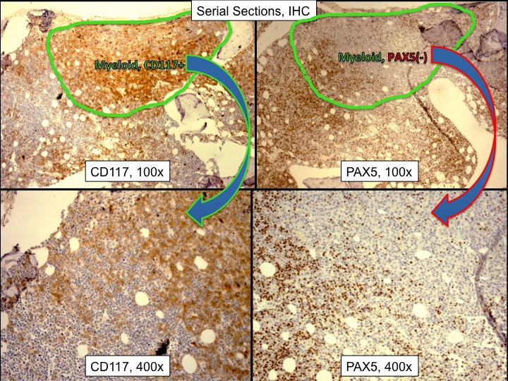 |
| Other areas were composed mainly of small blasts with scant cytoplasm. They expressed B cell antigens, including PAX5, CD79a and CD20. They did not express CD117 or MPO. | 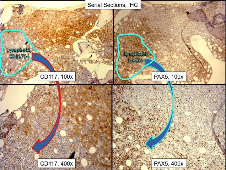 |
| 10 color flow cytometric analysis showed separate, distinct populations. One population had a myeloid phenotype with CD34 and CD13, but not CD10 or CD19. The second population expressed CD10 and CD19, but not CD34 or CD13. | 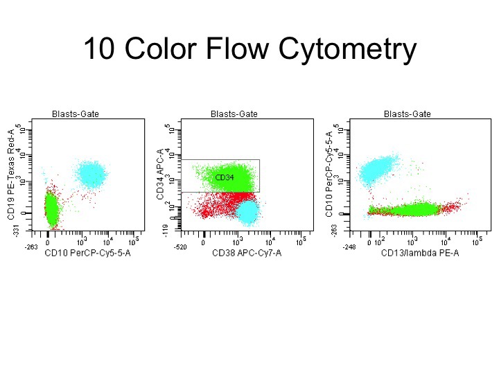 |