Institution: Tennessee Valley Healthcare System VA Hospitals and Vanderbilt University Medical Center
Additional authors:Ferrin C. Wheeler, PhD
Session: Extramedullary manifestations of myeloid neoplasms
HISTORY
This 57 year old white male presents to the Emergency Department complaining of increasing dyspnea on exertion, new onset rash, “neck lumps” and extreme fatigue. The patient has a long history of coronary artery disease and recent cardioablation for symptomatic premature ventricular contractions. He has also had a history of thrombotic thrombocytopenic purpura responsive to plasmapheresis in the distant past as well as a 3 year history of “cyclic neutropenia” for which he had had a bone marrow biopsy 9 months ago. That bone marrow biopsy showed a normocellular bone marrow with trilineage hematopoiesis with a full range of maturation. There was no overt dysplasia and no increase in blasts although the patient was neutropenic at the time of that biopsy. Karyotype and FISH showed a normal male karyotype with no abnormalities of chromosome regions 5p, 5q, 7c, 7q, 8c, and 20q. At the time of the current presentation, the patient is anemic, thrombocytopenic and has a leukocytosis. (WBC 12,500/uL; HGB 9.5 g/dL; HCT 28.2%; PLT 32,000/uL; MCV 95.9 fL). Of note, this represents a drop in hematocrit from 42% in the last 4 weeks since his last cardiology visit. During work-up it was discovered that in fact this patient has been pancytopenic, and not just neutropenic, for at least 3 years prior to the current presentation (see CBC charts Figure 1).
Physical examination showed multiple lymph nodes including axillary, cervical and inguinal areas. Skin exam showed an 8cm ill-defined blanchable erythematous nodule on the abdomen with many smaller 4mm crusted papules elsewhere on the abdomen and chest. There is a 3cm firm, violaceous nodule on the right shoulder. Also notable on exam was palpable hepatosplenomegaly and evident scleral icterus.DETAILS
Punch biopsies (0.5cm diameter) are taken of the right shoulder nodule and an excisional biopsy is taken of the right axillary lymph node. A bone marrow biopsy with aspirate and peripheral blood smear review are performed as well. Skin and lymph node are fixed in 10% buffered formalin as well as B+ fixative. The bone marrow biopsy was fixed in B+ after decalcification.
Review of the blood smear (Figures 2 – 5) shows a marked reduction in granulocytes which are dysplastic with Pelgeroid forms, uneven distribution of granules in the cytoplasm and left-shift. There is a relative increase in monocytes. Most notable is a population of blastoid cells comprising approximately half of the nucleated cells. These blastoid cells are moderate in size, with moderate grey-blue cytoplasm which is agranular and occasionally vacuolated. The cells have round to oval nuclei with smooth chromatin. Occasional cells show irregular, folded or even bi-lobed nuclei.H&E and PAS stained bone marrow biopsy sections (Figures 6, 7) show near total effacement by a monomorphous infiltrate of medium sized cells with irregular nuclei similar to that seen in the blood. There are only rare elements of trilineage hematopoiesis in the background of these cells. Wright’s stained bone marrow aspirate smears and stained touch preparations show a large number of medium sized immature cells with scant to moderate, agranular, gray-blue cytoplasm, irregular nuclei with fine chromatin, and discernable nucleoli (Figure 8). The lymph node measures 5 x 4 x 2.7cm and is red-pink on cross-sectioning. The architecture of this lymph node is completely effaced and replaced with a monomorphous infiltrate of medium sized cells with round to oval nuclei, smooth chromatin and surrounded by scant cytoplasm (Figures 9 - 11). The skin punch biopsies showed a reddish central nodule in the dermis on cross sectioning. These skin biopsies all demonstrate florid involvement with a monomorphous infiltrate of medium sized cells with irregular nuclei and smooth chromatin. There is a high rate of mitotic activity. These cells are morphologically identical to the recent bone marrow biopsy and excised lymph node (Figure 12-14).IMMUNOHISTOCHEMISTRY AND FLOW CYTOMETRY
Immunohistochemical and flow cytometric results across these specimens are concordant and are summarized as follows:
Flow cytometry shows a population of cells in the typical myeloid blast gate that express CD56, dim CD4, CD2, HLA-DR, dim CD71, and bright CD38 but lacks expression of surface and cytoplasmic CD3, surface and cytoplasmic CD5, surface and cytoplasmic CD19, surface and cytoplasmic CD22, cytoplasmic CD79a, glycophorin A, CD61, CD41a, TdT, Myeloperoxidase, CD7, CD57, CD25, CD52, CD30, CD33, CD13, CD16, CD11b, CD15, CD64, CD14, CD34 or CD117.Immunohistochemistry confirms that these cells express CD4 and CD56 (Figures 15, 16) but are negative for CD3, CD138, CD68, PAX5, AE1/AE3, CD8, S100, CD1a and CD79a. Moreover, the tumor cells express TCL1 and CD123 (Figures 17, 18).CYTOGENETIC FINDINGS
Cytogenetic analysis of this patient's lymph node and bone marrow specimens showed a normal male karyotype. Blasts were sorted from the patient’s lymph node using CD45/SSC gating to separate the blasts from the rare lymphocytes left in the lymph node. Whole genome SNP microarray analysis using the Affymetrix CytoScan HD array on these sorted cells showed multiple copy number changes including a large interstitial 13q deletion (35 Mb) and multiple other smaller deletions on 7p, 9p (CDKN2A), 12p (ETV6) and 14q (detailed analysis available for presentation if necessary).
MOLECULAR FINDINGS
The cells are negative for FLT3-ITD and JAK2 V617F mutations.
INTERESTING FEATURES
Given the patient’s extended history of pancytopenia, the question arises whether this blastic plasmacytoid dendritic cell neoplasm arose from an underlying myelodysplasia. Although a bone marrow biopsy taken 9 months prior to presentation showed a normal bone marrow, a bone marrow biopsy taken 4 weeks after induction chemotherapy due to delayed count recovery showed a hypocellular bone marrow with marked megakaryocytic dysplasia and 14% myeloblasts with no evidence of a blastic plasmacytoid dendritic cell neoplasm (Figures 19 – 23). Because this was felt to be a hypoproliferative myeloid neoplasm by the treating hematologist, all chemotherapy including growth factor support was held for an additional two weeks. At that time, the bone marrow biopsy showed a hypercellular bone marrow with persistent megakaryocytic dysplasia but the blast count had fallen to 4%. Of note, this may reflect dilution of the blasts by increased numbers of maturing cells in the later biopsy. The patient is consistently anemic and thrombocytopenic with delayed count recovery during consolidation chemotherapy.
PROPOSED DIAGNOSIS
Blastic Plasmacytoid Dendritic Cell Neoplasm
CONSENSUS DIAGNOSIS
Blastic plasmacytoid dendritic cell neoplasm
| Figure 1: CBCs charted |  |
| Figure 2 Blood 40x |  |
| Figure 3 Blood 100x1 | 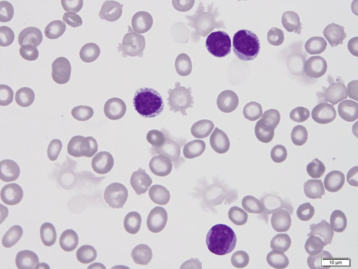 |
| Figure 4 Blood 100x2 |  |
| Figure 5 Blood 100x3 |  |
| Figure 6 BM 2x |  |
| Figure 7 BM 40x | 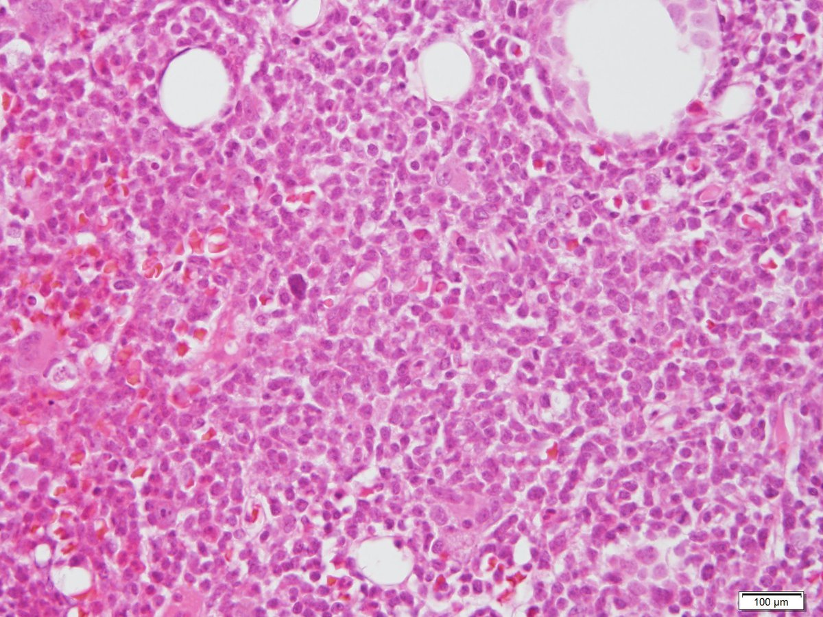 |
| Figure 9 LN 2x | 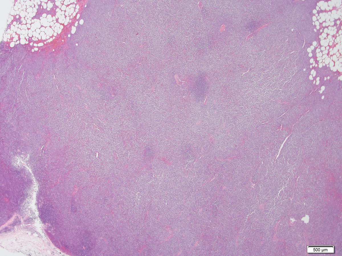 |
| Figure 10 LN 20x | 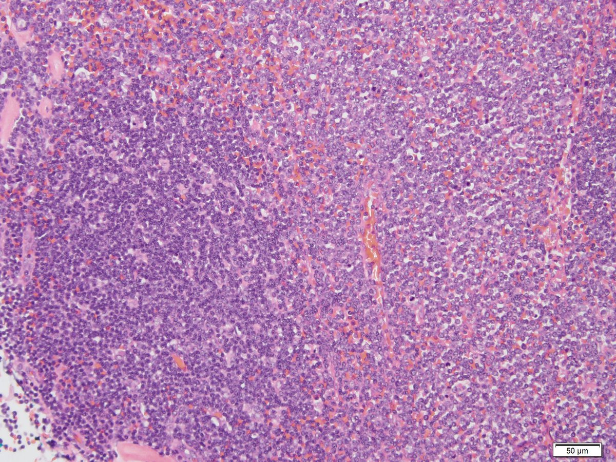 |
| Figure 11 LN 40x |  |
| Figure 12 skin 2x |  |
| Figure 13 skin 20x |  |
| Figure 14 skin 100x |  |
| Figure 15 CD4 |  |
| Figure 16 CD56 | 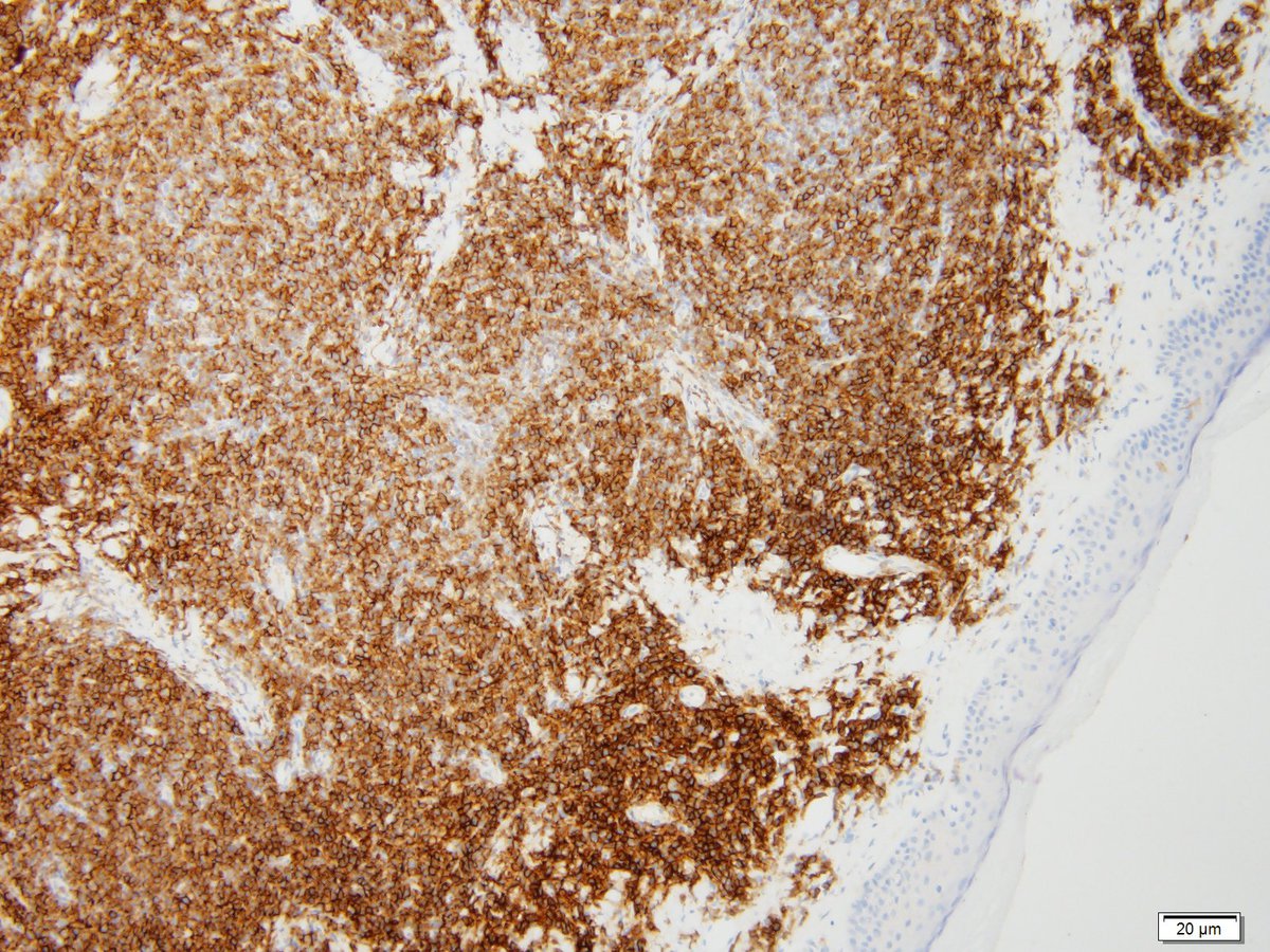 |
| Figure 17 TCL1 | 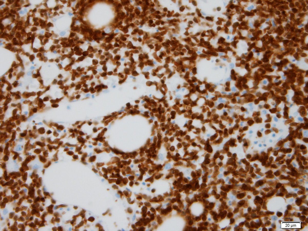 |
| Figure 18 CD123 | 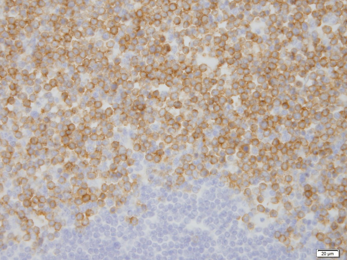 |
| Figure 19 Recovery BMBx 10x |  |
| Figure 20 Recovery BMBx 40x | 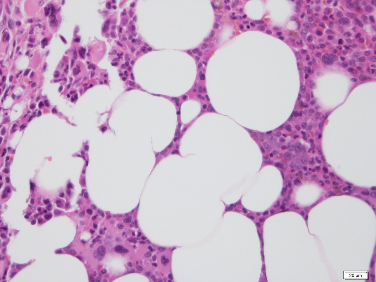 |
| Figure 21 Recovery BMBx 40x 3 | 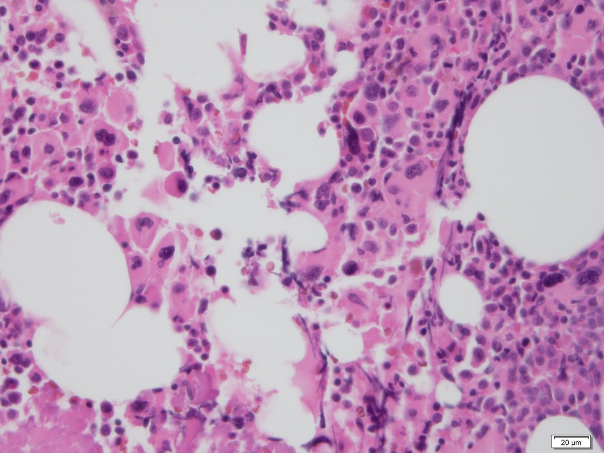 |
| Figure 22 Recovery PAS Clot 40x | 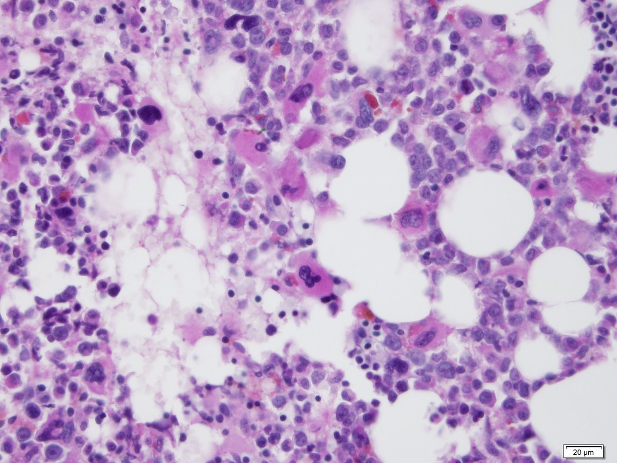 |
| Figure 23 Recovery Aspirate | 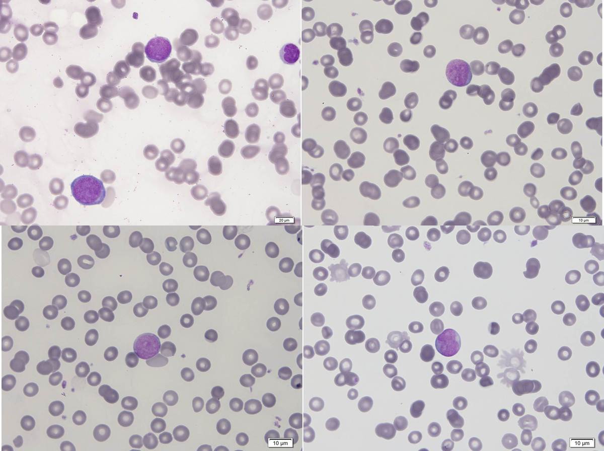 |
| Figure 8 Diagnostic Aspirate 100x | 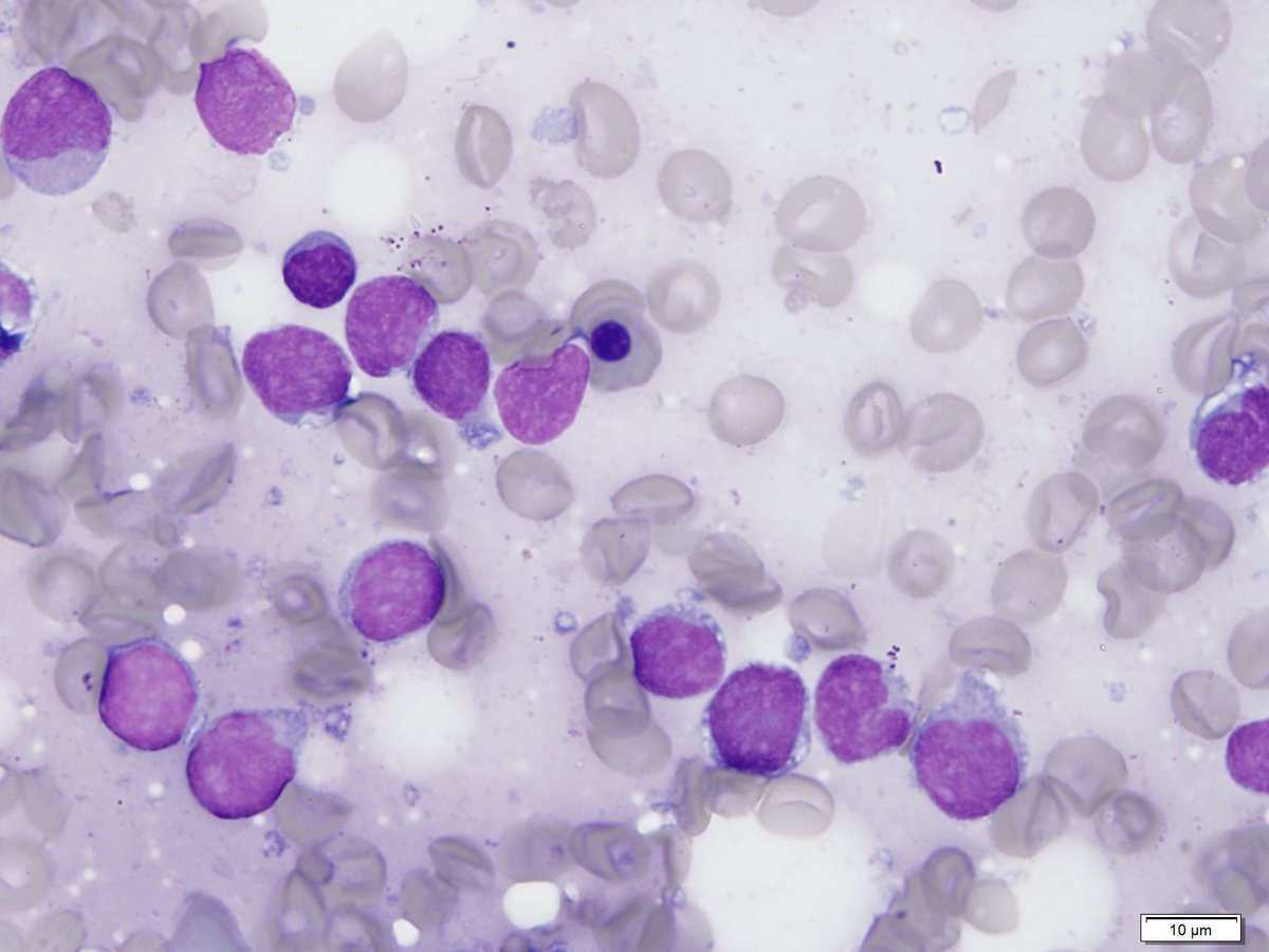 |