Institution: University of Wisconsin School of Medicine and Public Health
Session: Erythroleukemia and megakaryoblastic AML and mimics
HISTORY
This 57 year old woman underwent liver transplantation for cirrhosis due to hepatitis C with associated hepatocellular carcinoma. Ten months after transplant hepatitis C recurred, and she was treated for 8 months with interferon and ribavirin, with erythropoietin support. 18 months after transplant, as she completed interferon and ribavirin therapy, she developed a nasal mass. Biopsy showed a post-transplant lymphoproliferative disorder, monomorphic large B-cell type, EBER negative. A bone marrow was performed as part of the staging evaluation. CBC at this time showed WBC 2.2 K/uL with absolute neutrophil count of 1.58 K/uL, hemoglobin 7.8 g/dl, MCV 105 fL, reticulocyte count 106K/uL, platelet 104 K/uL. Folate and B12 levels were normal. She was treated with R-CHOP for 6 cycles, and was disease free on follow up scans. At follow up, 6 months after completing ribavirin and interferon treatment, CBC showed WBC 6.3 K/uL with absolute neutrophil count of 5.21 K/uL, hemoglobin 12.3 g/dl, MCV 101 fL, reticulocyte count 135K/uL, platelet 103 K/uL.
DETAILS
Peripheral blood smear, bone marrow aspirate and biopsy from posterior iliac crest obtained at staging were reviewed. Wright Giemsa stained smear and aspirate slides, and H&E stained biopsy slides were reviewed. Peripheral blood shows pancytopenia with moderate red cell anisopoikilocytosis and tear drop cells. Neutrophils and platelet counts are low but without dysplastic changes. Bone marrow aspirate is hypercellular with erythroid hyperplasia (Granulocyte: Erythroid ratio of 1.2). Erythroid cells show megaloblastoid changes, bizarre multinucleated forms, nuclear budding and karyorrhexis in > 20% of cells. Ringed sideroblasts are not identified. Granulocytes show some giant bands and megakaryocytes show hyperlobation. The biopsy is hypercellular for age (70%) and reflects the aspirate. Repeat examination of peripheral blood and bone marrow 6 months after completion of ribavirin and interferon therapy still shows a hypercellular marrow with erythroid hyperplasia (G: E 1.3) but without significant dysplastic changes in any lineage.
IMMUNOHISTOCHEMISTRY AND FLOW CYTOMETRY
Not performed
CYTOGENETIC FINDINGS
46, XX, normal female karyotype
MOLECULAR FINDINGS
Not performed
INTERESTING FEATURES
This marrow is hypercellular marrow with significant dysplastic changes in red cell precursors. With peripheral cytopenias and in the absence of B12 and folate deficiency, these changes would be very concerning for myelodysplasia. In this case, history of ribavirin and interferon therapy suggested the changes seen could be secondary to drug treatment. After cessation of ribavirin and interferon, the patient’s anemia and neutropenia resolved. Follow up marrow showed resolution of the dyserythropoiesis. Peripheral blood cytopenias with marrow morphology that meets criteria for myelodysplasia can be seen associated with drug therapy. Evaluation of clinical history, particularly in complex situations, with reevaluation after drug cessation can help to prevent over diagnosis of myelodysplasia.
PROPOSED DIAGNOSIS
Drug induced dyserythropoiesis mimicking myelodysplasia
CONSENSUS DIAGNOSIS
Drug-induced dyserythropoiesis
| Peripheral blood | 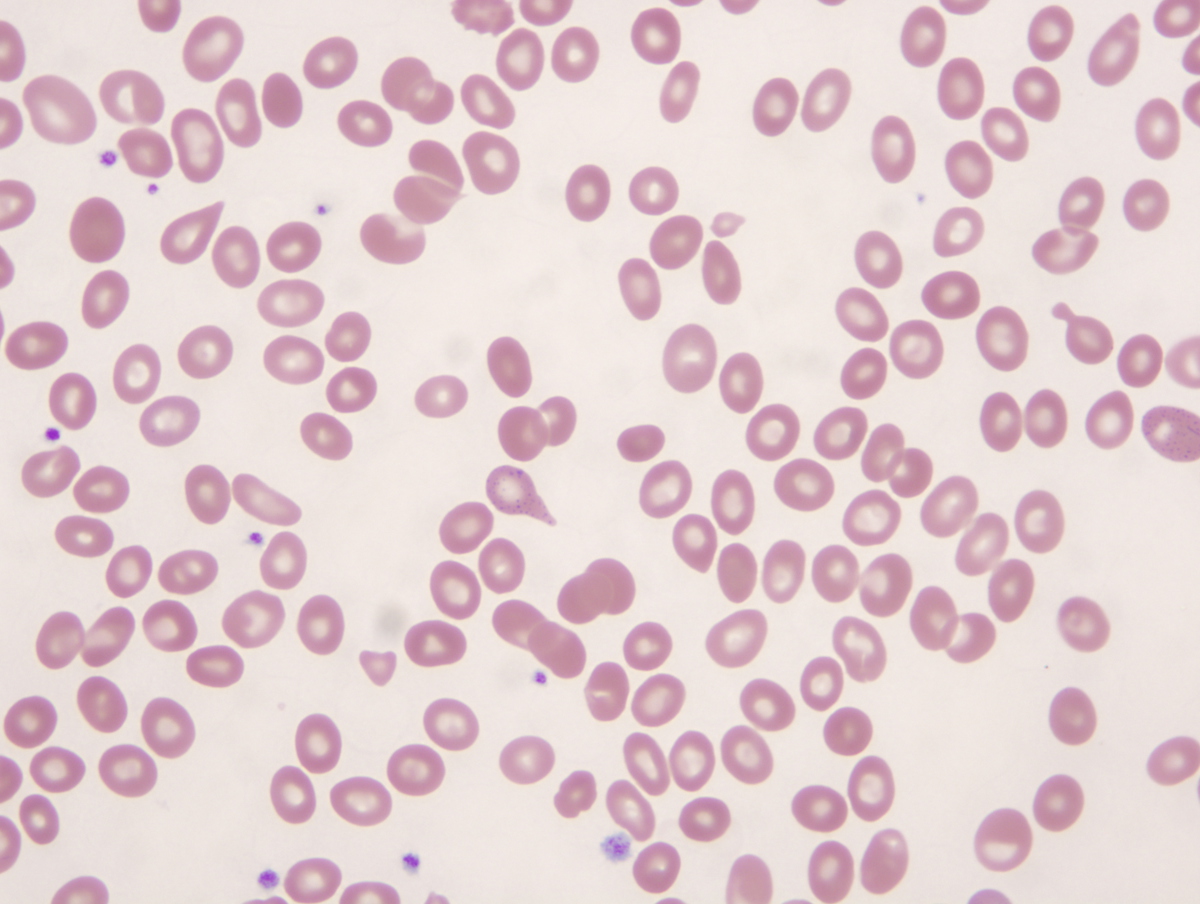 |
| Peripheral blood | 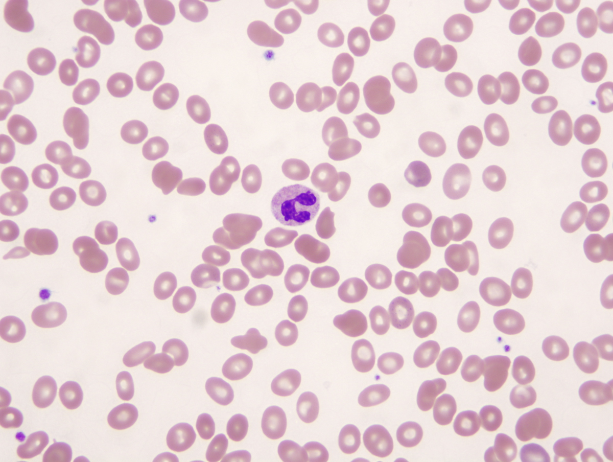 |
| Bone marrow aspirate low | 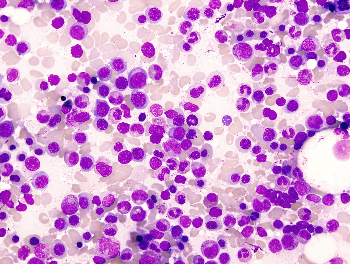 |
| Bone marrow aspirate | 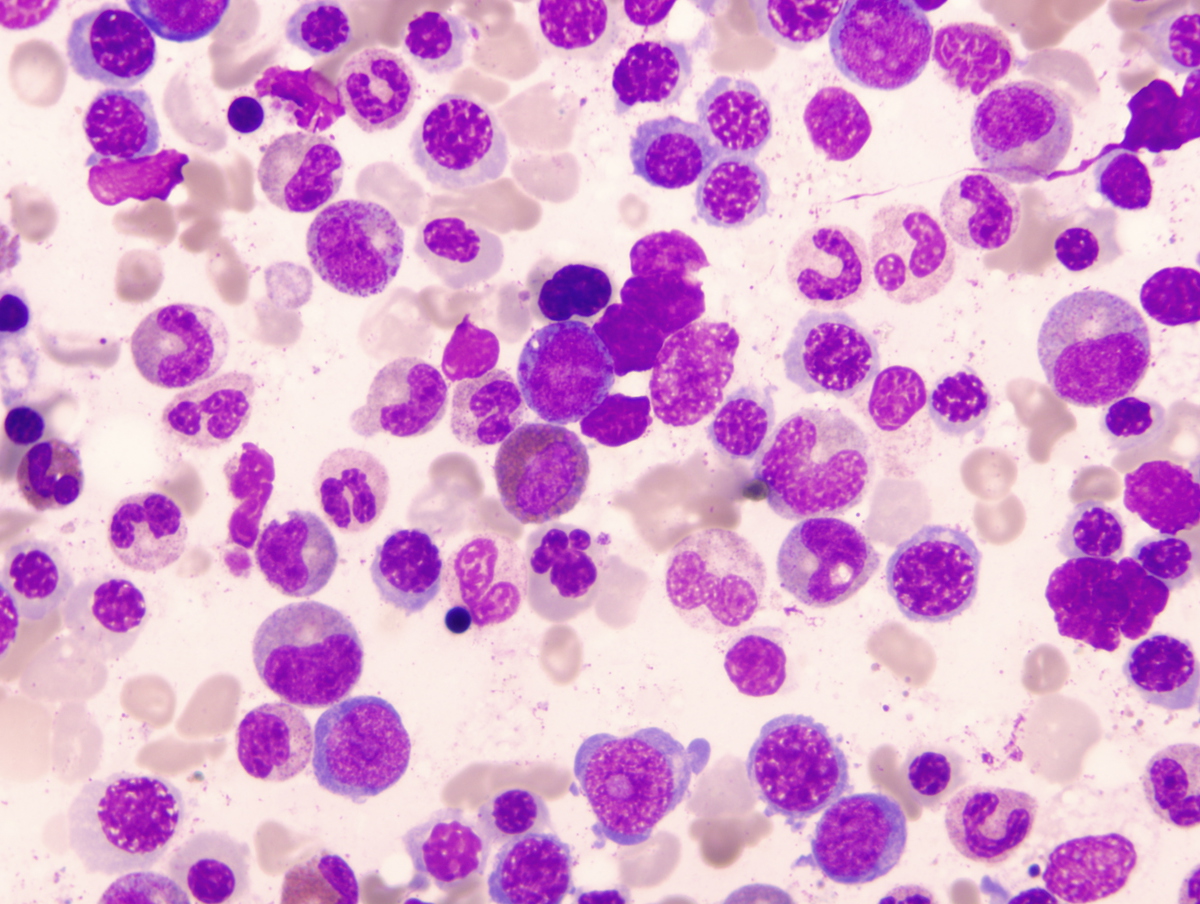 |
| Bone marrow aspirate | 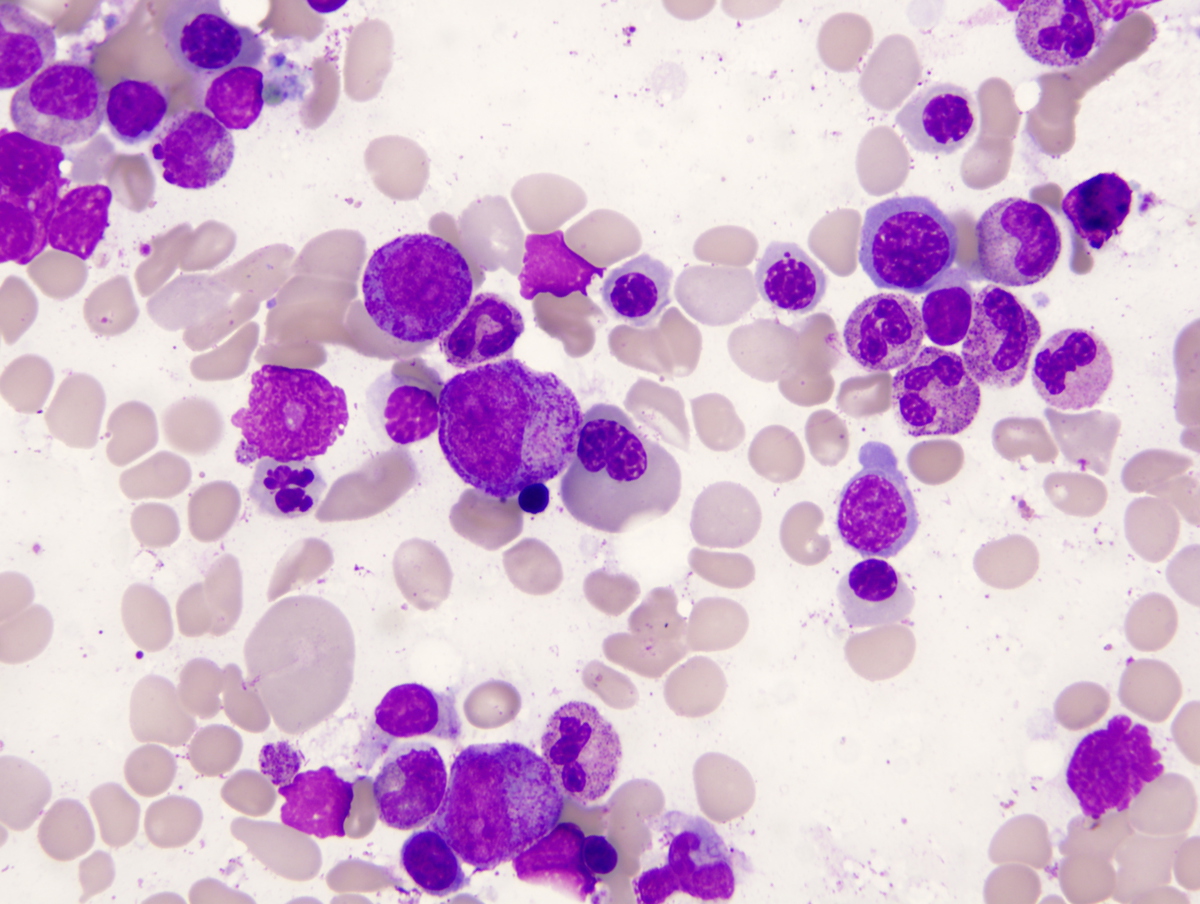 |
| Bone marrow aspirate | 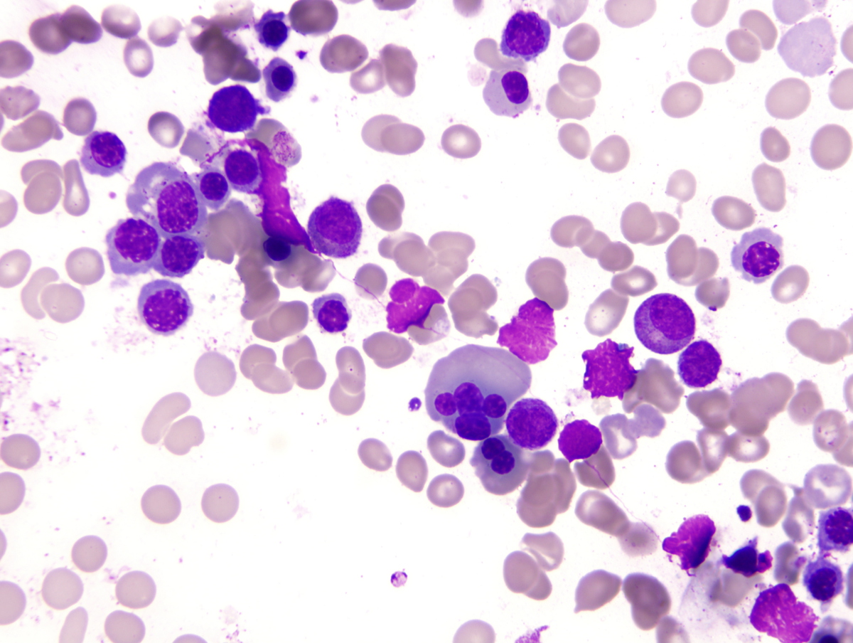 |
| Bone marrow after cessation of medication |  |