Institution: The Children's Hospital of Philadelphia
Additional authors:Gerald Wertheim, Michele E. Paessler
Session: T Lymphoblastic Leukemia/Lymphoma
HISTORY
This patient is a 13 year old boy who presented in April 2009 with a four month history of worsening cough, intermittent fevers, pallor, and a 14 pound weight loss. CBC revealed a WBC of 300,800/uL, Hgb 4.2 g/dL, platelets 214,000/uL, and peripheral blasts of 82%. An initial chest CT showed a 13 x 9 cm anterior mediastinal mass with associated axillary, hilar, subcarinal, and celiac axis lymphadenopathy and splenomegaly. Bone marrow evaluation revealed a diagnosis of T lymphoblastic leukemia (details below) with 92% blasts and 8% nRBCs. He received induction chemotherapy with prednisone, vincristine, daunorubicin, pegylated asparaginase, and intrathecal cytarabine and methotrexate. Minimal residual disease flow cytometry was negative at Day 29. In addition to vincristine and pegylated asparaginase, he also received cyclophosphamide, cytarabine, doxorubicin, dexamethasone and mercaptopurine during his therapeutic course.
In October 2009, day 30 of delayed intensification, he presented for chemotherapy and was found to have blasts on his peripheral smear. He had also been complaining of recent bone and joint pain. WBC was 5,900 with 15% blasts. He was also in DIC with a PT/INR of 26s/2.6, PTT 50.8s, and a fibrinogen of <45 mg/dL. Bone marrow evaluation revealed acute myeloid leukemia, with no evidence of T lymphoblastic leukemia (details below). Bone marrow differential showed 93% blasts, 1% myelocytes, 1% neutrophils, 3% monocytes, and 2% lymphocytes.DETAILS
Bone marrow, aspirates and biopsies (decalcified and fixed in AZF).
April 2009: Sheets of small to medium sized blasts with round nuclei, moderately condensed chromatin and scant cytoplasm. (Image 1)October 2009: Sheets of large blasts with irregular, occasionally bilobed nuclei, fine chromatin, prominent nucleoli and moderate amounts of cytoplasm with occasional granules. (Image 5)IMMUNOHISTOCHEMISTRY AND FLOW CYTOMETRY
April 2009:
Flow cytometry: 90% blasts (low side scatter) with the following immunophenotype: positive for cCD3, CD4, CD5(dim), CD7, CD38, cCD79a, TdT while negative for CD2, sCD3, CD8, CD19, CD56, CD34, HLA-DR, MPO. (Image 3)Immunohistochemistry: Blasts positive for CD3, negative for MPO. (Image 2)October 2009:Flow cytometry: 70% blasts (high side scatter) with the following immunophenotype: positive for CD4, CD56, CD33, CD38, CD11b(variable), MPO(bright) while negative for CD34, cCD3, CD5, CD7, TdT, HLA-DR. (Image 7)Immunohistochemistry: Blasts positive for MPO, negative for CD3. (Image 6)CYTOGENETIC FINDINGS
April 2009: 46,XY,t(5;14)(q35;q32),del(9)(p21.3),del(13)(q14.11q21.1)[7].ish
t(5;14)(IGH+;IGH-),del(9)(p16-)/46,idem,del(7)(p1?3p?21)[6]/45,idem,dic(7;8)(p1?3;p11.2),del(10)(q22)[6].ish dic(7;8)(7ptel-,7cen+,D7S486+,8cen+,MYC+)nuc ish 12p13(TELx2),21q22(AML1x2)[200/200]nuc ish 11q23(MLLx2)[200/200]nuc ish 9q34(ABLx2),22q11.2(BCRx2)[200/200]nuc ish 4cen(CEP4x2)[200/200]nuc ish 10cen(CEP10x2)[200/200]nuc ish 17cen(CEP17x2)[200/200]nuc ish 9p21(p16x1),9cen(CEP9x2)[139/200]nuc ish 9p21(p16x1),9cen(CEP9x1)[47/200]The leukemic clone showed evidence of extensive clonal evolution. The key findings in this leukemic clone were noted to be to be the deletion of 9p, and the t(5;14)(q35;q32). (Image 4)October 2009: 67,XY,+Y,der(4)del(4)(q13.1)inv dup(4)(p12pter),del(7)(p11.2p14.1),+8,-9,del(9)(p21.3),-10,-11,-14,-15,der(15)t(11;15)(q13.1;q26),+19,+22[9]ish der(15)t(11;15)(MLL+,PML+)nuc ish 11q23(MLLx3)[89/100]nuc ish 15q22(PMLx2),17q21.1(RARAx3)[94/100]nuc ish 9p21(p16x1),9cen(CEP9x2)[90/100]FISH studies were performed on this relapse specimen using the IGH/MYC/CEP8 probe. Metaphases showed that IGH was detected on one of the number 5 chromosomes, thus confirming the same translocation between chromosomes 5 and 14 as was seen in the originaldiagnostic study. Although there are many differences between the original leukemic clone and this clone, the most significant abnormality at the time of diagnosis was the deletion of 9p, and loss of p16, which is also present in this leukemia. (Image 8)Molecular analysis: High resolution SNP array analysis confirmed similarities, as well as several differences, between the two leukemic clones. (Image 9)INTERESTING FEATURES
This is an interesting case of childhood T lymphoblastic leukemia (T ALL) that underwent lineage switch to acute myeloid leukemia (AML) following 6 months of chemotherapy. Although mixed phenotype acute leukemias (MPAL), especially of the T/myeloid subtype are uncommon (<1% of acute leukemias), they are well documented in the literature. However, MPAL comprises cases in which the blast population either expresses markers of multiple lineages, or consists of two, immunophenotypically distinct blast populations identified concurrently. In this case, the initial leukemic clone showed a T lymphoblastic immunophenotype with no evidence of a concurrent myeloblast population. Similarly, the relapsed clone showed a pure myeloblast population, without cCD3 expression, and showed no evidence of a residual T lymphoblast population by flow cytometry.
Confirming lineage switch in acute leukemia can be challenging, and relies on identification of genetic markers present at both diagnosis and relapse. In spite of the relatively short time from therapy to diagnosis of the patient’s myeloid leukemia (6 months), this was initially assumed to be a therapy related AML. However, karyotype, FISH, and SNP array studies demonstrated the presence of several genetic aberrations common to both clones. In spite of obvious clonal evolution from the original T ALL to the subsequent AML, the persistence of a t(5;14)(q35;q32) as well as del 9p were most compatible with a common progenitor for these two leukemias. Although the t(5;14)(q35;q32) which involves the TLX3/BCL11B genes has been described in approximately 20% of T ALL, it has only been reported once, to our knowledge, in a myeloid or mixed lineage leukemia (1). Interestingly, this reported case was a 9 year old boy with a bilineage leukemia showing both myeloid and T lymphoid blast populations, both of which were found to harbor the translocation (1). Of note, the patient in this case report shared some, albeit common, clinical features with our case including presenting with a mediastinal mass and lymphadenopathy. The morphology of the myeloid blasts in this report was not described in detail; however, they were noted to have magenta colored cytoplasmic granules, similar to those seen in our case. The authors of this report raise the possibility that the TLX3 gene, which is known to alter myeloid differentiation when transduced into bone marrow progenitors, may be playing a role in maturation arrest in the myeloblast population (1). Our case raises the intriguing possibility that the association between this translocation (which is typically seen in T ALL) and the myeloid differentiation seen in these leukemias is non-random. Because of the rarity of phenotypic switch in leukemia, data on optimal therapy and prognosis is limited. However, anecdotal data suggests that these patients do poorly, even when therapy is altered to reflect the phenotypic switch. Our patient was treated initially with T ALL therapy and then switched to AML therapy. He went on to develop leukemia cutis and did not achieve a second remission in spite of therapy. He died approximately 2 months following the diagnosis of AML. Reference:1. Oliveira JL, Kumar R, Khan SP, Law ME, Erickson-Johnson M, Oliveira AM, Ketterling RP, Dogan A. Pediatr Blood Cancer. 2011 Mar;56(3):467-9PROPOSED DIAGNOSIS
T lymphoblastic leukemia with t(5;14)(q35;q32) with lineage switch into acute myeloid leukemia.
Comment: This case could likely be discussed under several different categories of the workshop.CONSENSUS DIAGNOSIS
T-acute lymphoblastic leukemia with t(5;14)(q35;q32) with lineage switch into acute myeloid leukemia
| Image 1 | 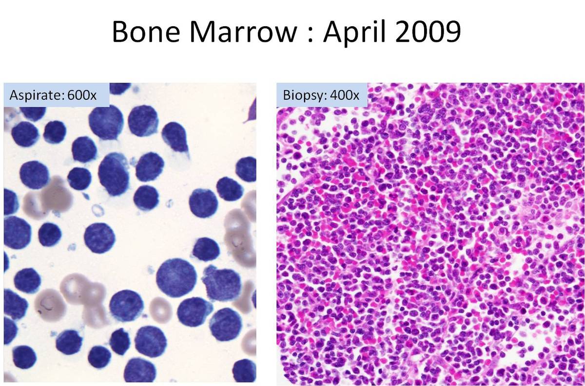 |
| Image 2 |  |
| Image 3 | 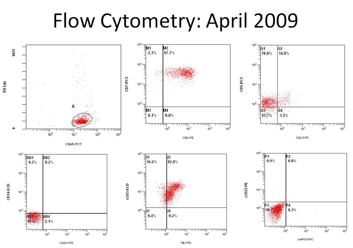 |
| Image 4 | 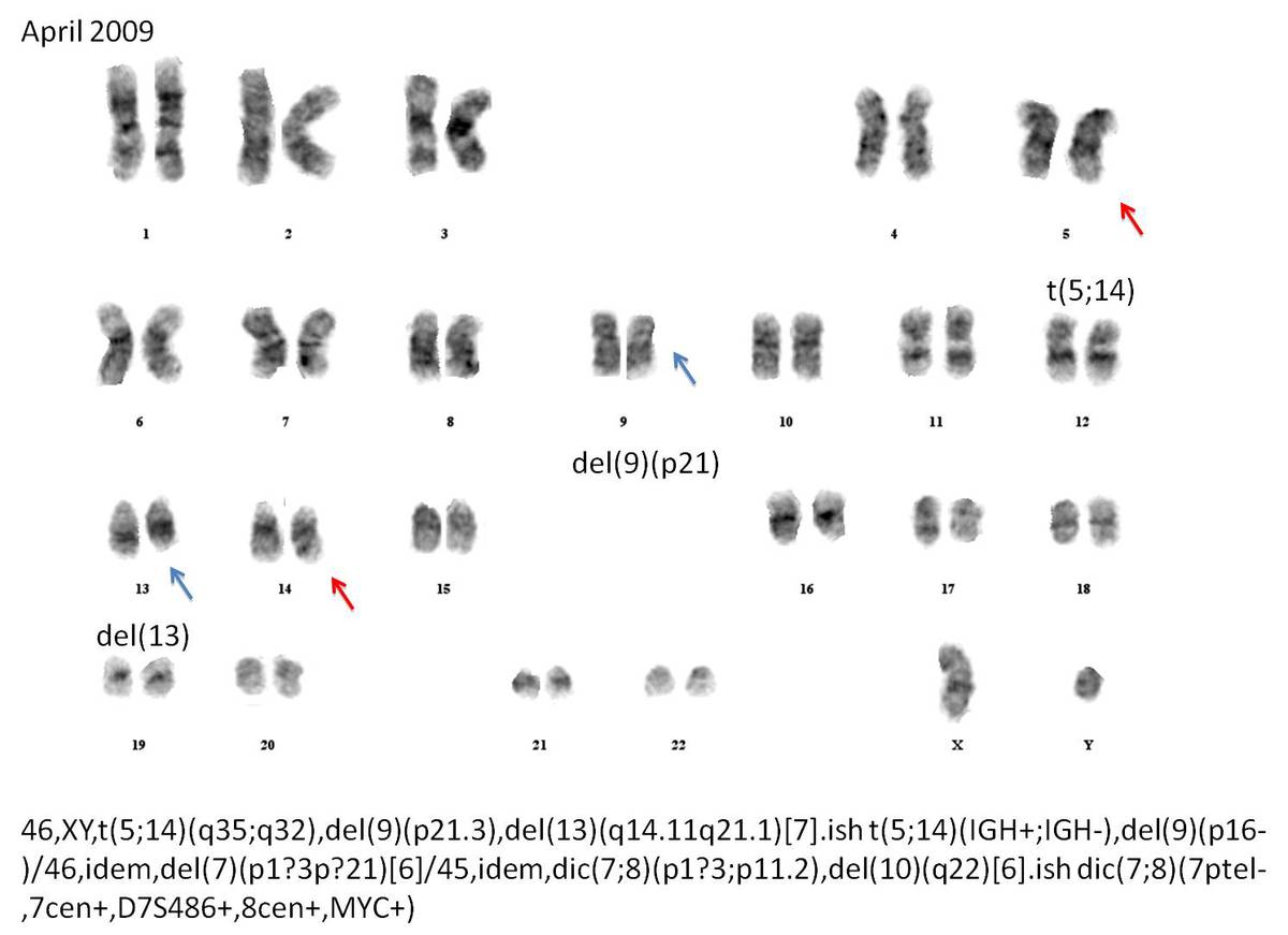 |
| Image 5 | 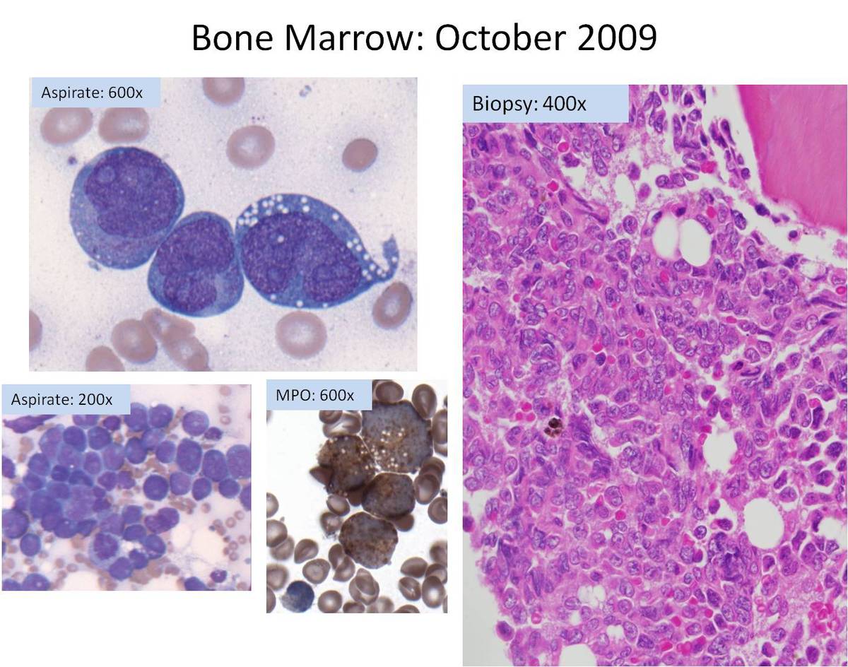 |
| Image 6 | 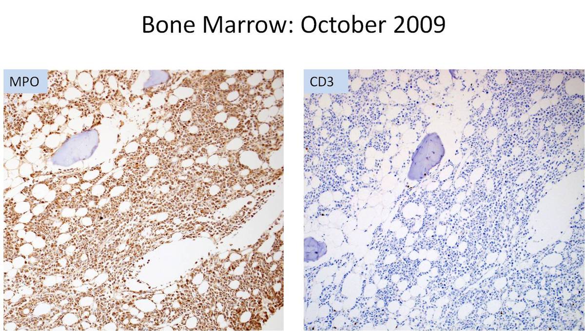 |
| Image 7 |  |
| Image 8 | 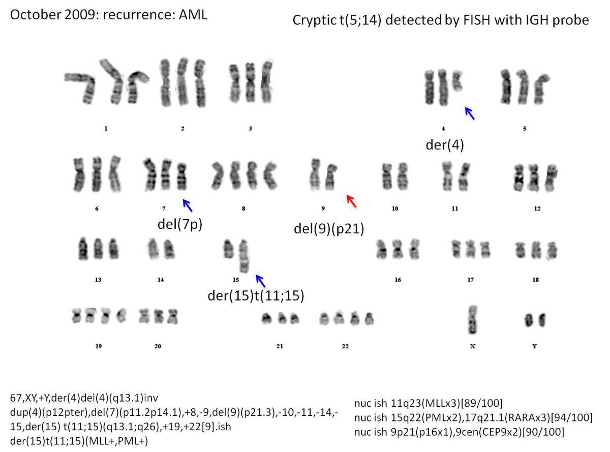 |
| Image 9 | 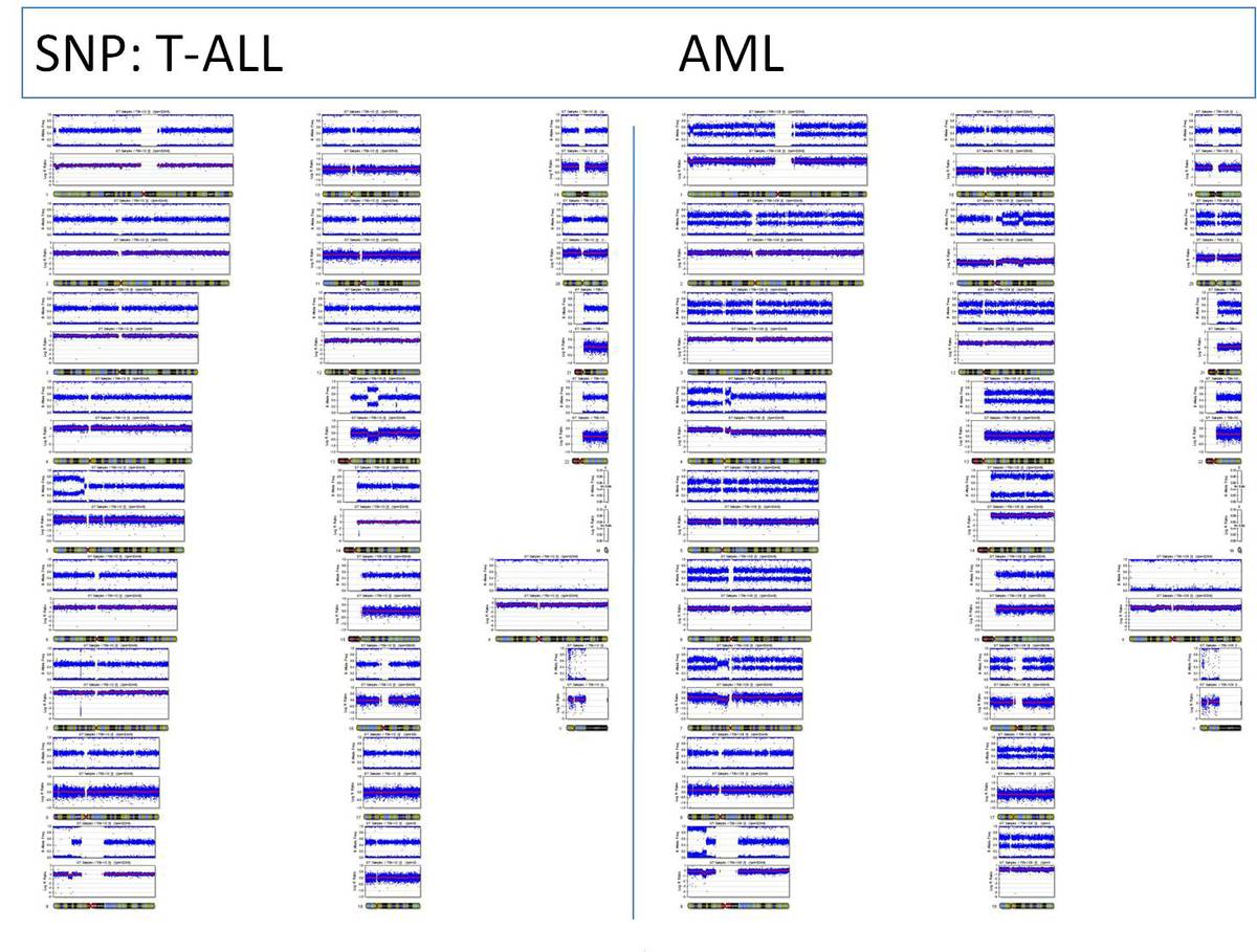 |
| Image 10 - TLX3 BCL11b fusion | 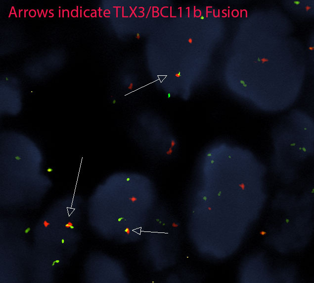 |