Institution: University of Michigan
Additional authors:Charles W Ross Diane Roulston Elena Ivan
Session: AML with recurrent genetic abnormalities Part I
HISTORY
The patient is a 20 year-old male who presented to his local hospital after experiencing progressive fatigue and syncopal episodes. Physical exam was significant for pallor, tachycardia, and scattered ecchymoses over the lower extremities. Initial laboratory workup was significant for marked anemia and marked thrombocytopenia (hemoglobin 4.2 g/dL, platelets 6000/mm3). The white blood cell count was 11000/mm3. He was subsequently transfused with two units of red blood cells and a single five-pack unit of pooled platelets.
DETAILS
The Wright-Giemsa stained peripheral blood smear showed numerous blasts (44% of the white blood cells) with rare Auer rods present (Fig.1) and granulocytic dysplasia including pelgeroid nuclear morphology (Fig. 2). Marked normocytic anemia and thrombocytopenia were also evident. A bone marrow trephine biopsy and aspirate were obtained from the right posterior iliac crest. The bone marrow core biopsy was fixed in acetic-acid-zinc-formalin (AZF). The haematoxilin and eosin (H&E) stained sections of the marrow biopsy demonstrated increased cellularity (approximately 95%) with increased number of myeloid precursors and markedly decreased number of the erythroid and megakaryocytic elements (Fig. 3). A manual differential count of the Wright-Giemsa stained bone marrow aspirate revealed 43.2% blasts. These blasts were intermediate in size with irregular nuclear contours and sparsely granulated basophilic cytoplasm. Rare long Auer rods and large pink granules were present within these blasts (Fig. 4). Maturing granulocytic precursors at the myelocyte stage and beyond displayed homogeneous salmon-colored cytoplasm surrounded by a distinctive rim of blue cytoplasm (Fig. 5, 6). These maturing granulocytic precursors comprised 38.2% of cells. Eosinophils and their precursors were slightly increased, comprising 4.8% of cells on differential count.
IMMUNOHISTOCHEMISTRY AND FLOW CYTOMETRY
Flow cytometry immunophenotyping performed on the bone marrow aspirate revealed an increased percentage of myeloid blasts with expression of CD34 (moderate), CD33 (dim, subset), and CD117 (dim-moderate) and aberrant co-expression of CD19 (dim) and CD56 (moderate) in a subset of blasts (Fig. 7-12).
CYTOGENETIC FINDINGS
Chromosome analysis at the 350 band level of resolution using the GTG banding method revealed a three way translocation involving chromosomes 3, 8 and 21. The karyotype was as follows: 46,XY,t(3;21;8)(q29;q22;q22) (Fig.15).
MOLECULAR FINDINGS
Fluorescent in situ hybridization (FISH) studies were performed on the bone marrow aspirate using Vysis LSI RUNX1/RUNX1T1 Dual Color Dual Fusion Probe (Abbott Molecular), specific for RUNX1 labeled with SpectrumGreen and RUNX1T1 labeled with SpetrumOrange respectively. Interphase FISH revealed a RUNX1/RUNX1T1 fusion-positive variant signal pattern (2R,2G,1F) in 77.5% (155/200) of the cells examined (Fig. 13). FISH performed on metaphase chromosomes demonstrated RUNX1T1 signals on the abnormal chromosome 3 and the normal chromosome 8, RUNX1 signals on the normal and abnormal chromosomes 21, and a single RUNX1-RUNX1T fusion on the abnormal chromosome 8 (Fig. 14). The PCR studies performed on the bone marrow aspirate were negative for KIT D816V mutation, and also negative for CEBPA, NPM1, IDH1, IDH2 and FLT3 mutations and for BCR/ABL1 and PML/RARA.
INTERESTING FEATURES
This case displays morphologic features that are often associated with the t(8;21): blasts with basophilic cytoplasm, some with large azurophilic granules and occasional forms with slender Auer rods, myelocytes and metamyelocytes with homogenous salmon-colored granules and a rim of basophilia in the cytoplasm, increased number of eosinophils and dysplastic neutrophils. The immunophenotypic features present in this case are also characteristic for acute myeloid leukemia with t(8;21): subpopulation of myeloblasts with relatively high intensity of CD34, weak expression of CD33 and aberrant expression of CD19. A subset of the blasts also expressed CD56, that can be seen in AML with t(8;21). A unique feature seen in this case is the three-way translocation that involves the chromosomes 3, 8 and 21. Although variant translocations are present in approximately 3-4% of cases of AML with t(8;21), this three-way translocation involving chromosome 3 has not been described before. Since variant rearrangements involving the RUNX1 and RUNX1T1 genes can be subtle and sometimes overlooked by traditional cytogenetic analysis, FISH studies using probes that specifically detect the RUNX1-RUNX1T1 fusion gene and RT-PCR can be helpful in the detection or confirmation of variant t(8;21). These can be especially helpful when morphologic and immunophenotypic features characteristic of AML with t(8;21) are present, yet the question of whether the RUNX1-RUNX1T1 fusion gene is present cannot be definitively answered by conventional cytogenetic studies.
PROPOSED DIAGNOSIS
Acute myeloid leukemia with variant t(8;21)(q22;q22); RUNX1-RUNX1T1
CONSENSUS DIAGNOSIS
Acute myeloid leukemia with variant t(8;21)(q22;q22); RUNX1-RUNX1T1
| Fig.1 | 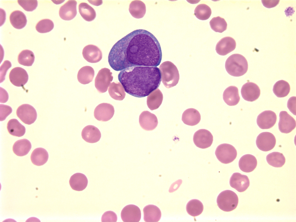 |
| Fig 2 | 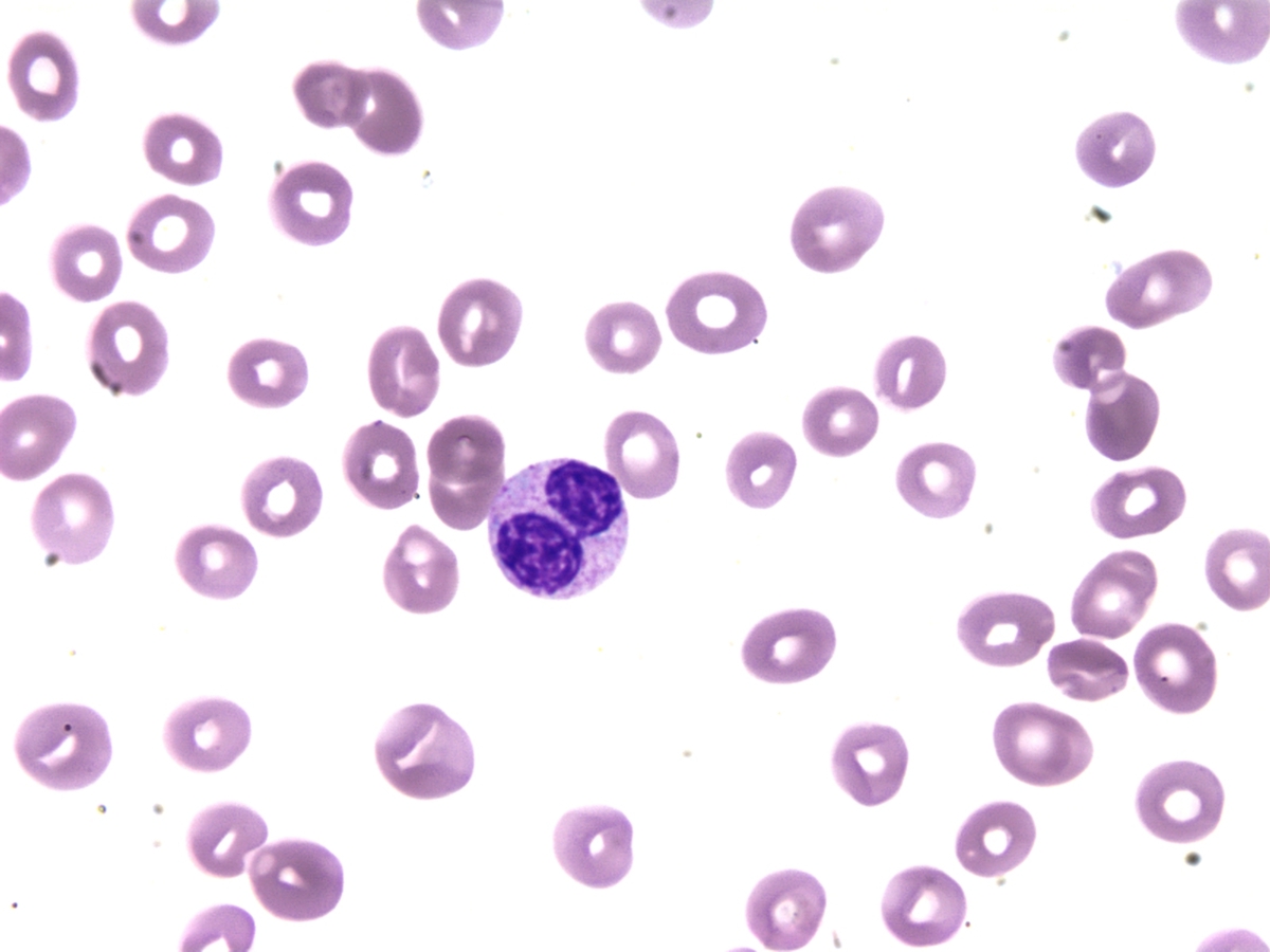 |
| Fig3 | 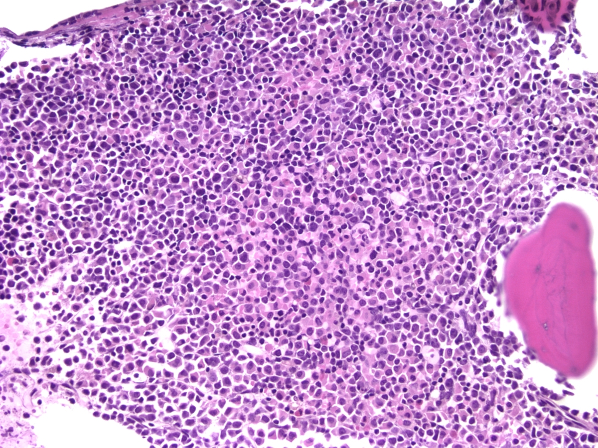 |
| Fig 4 | 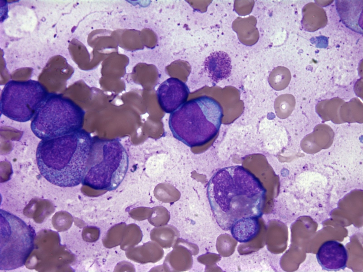 |
| Fig 5 | 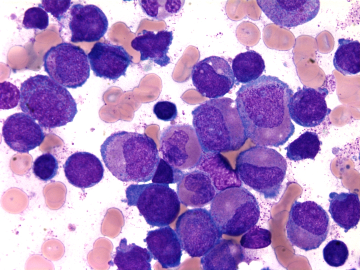 |
| Fig 6 | 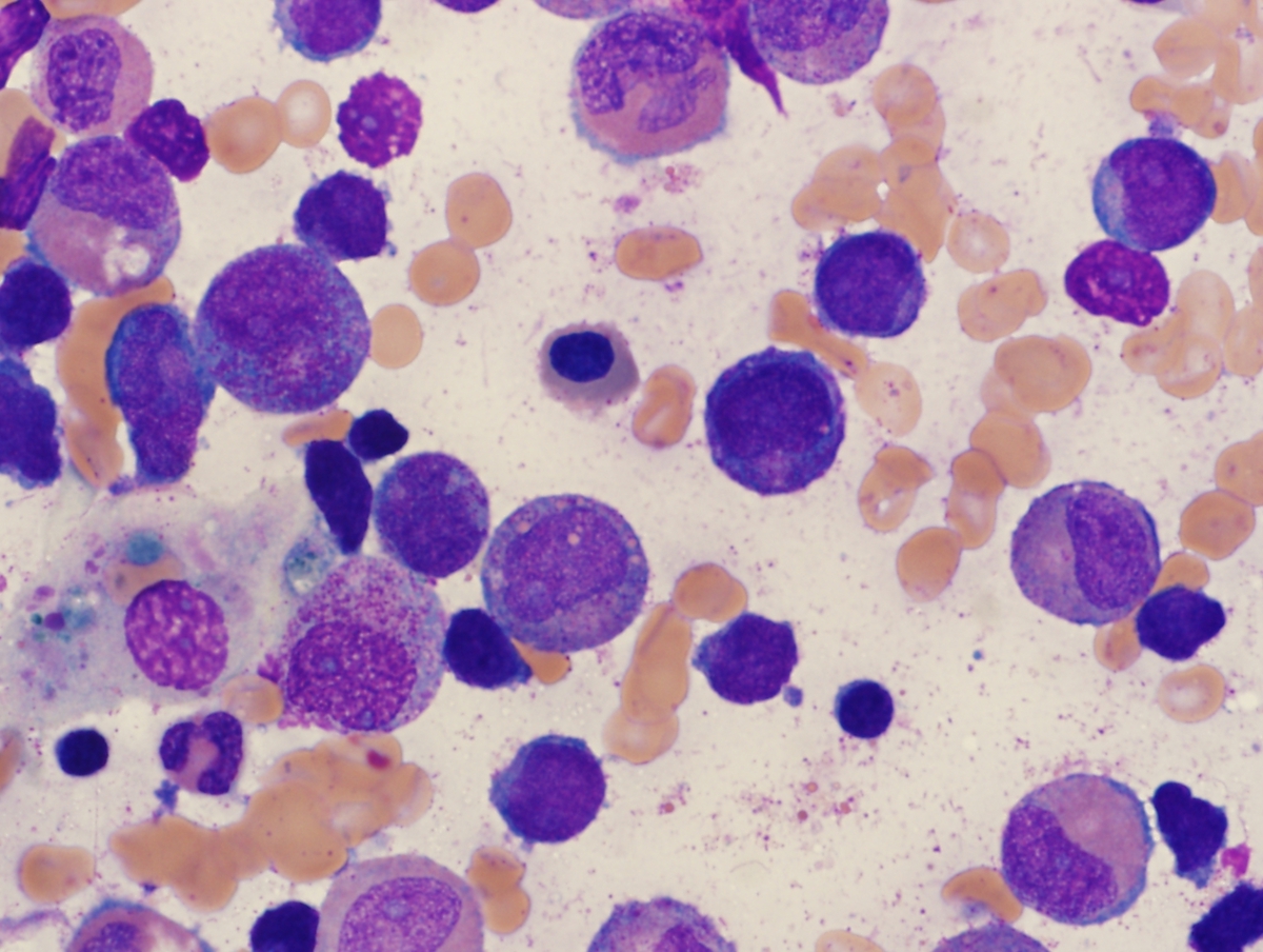 |
| Fig 7 | 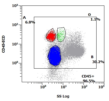 |
| Fig 8 | 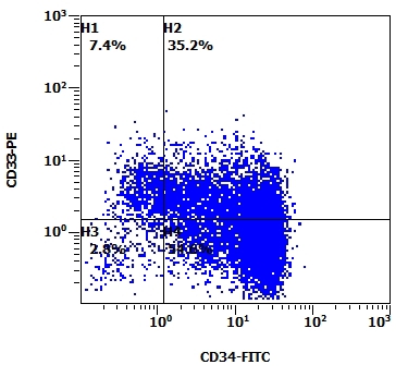 |
| Fig 9 | 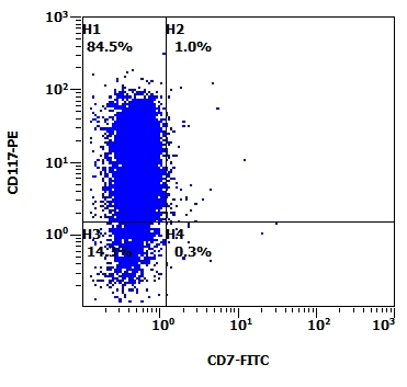 |
| Fig 10 | 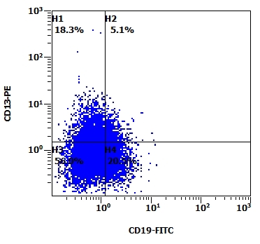 |
| Fig 11 | 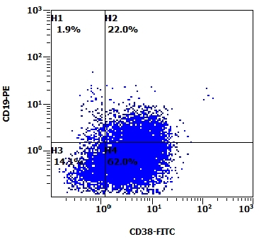 |
| Fig 12 | 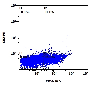 |
| Fig13 | 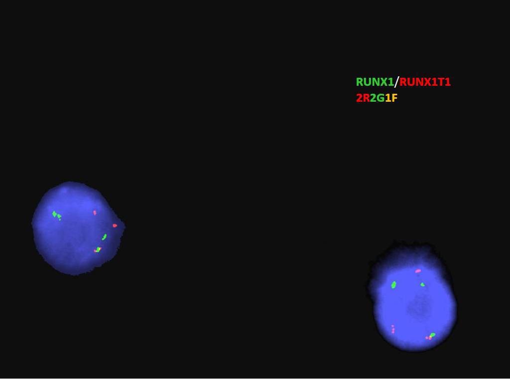 |
| Fig 14 | 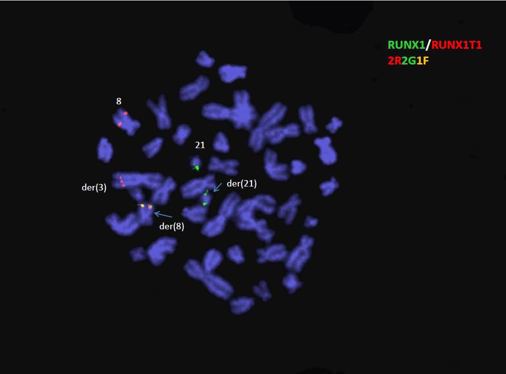 |
| Fig 15 | 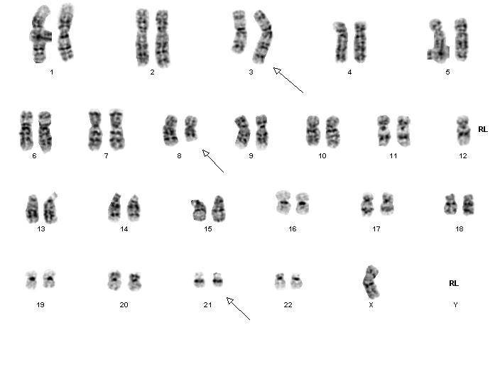 |