Institution: University of Texas Southwestern Medical Center
Additional authors:Charles Zaremba, UT Southwestern Medical Center Prasad Koduru, UT Southwestern Medical Center
Session: Acute leukemias of ambiguous lineage
HISTORY
The patient is a 40 year-old African American man without significant past medical history who presented to his primary care physician with a one month history of worsening fatigue and shortness of breath. In the week prior to presentation, he was able to walk only 50 feet before having to rest. On the day of presentation, he complained of headache and blurry vision. Physical examination revealed no organomegaly. CBC values were WBC 145,000 x 10(3)/uL with 56% blasts, hemoglobin 5.0 g/dl, and platelets 40,000/uL. He received packed red blood cell transfusion and therapeutic leukopheresis. A bone marrow biopsy was performed the morning after admission to the hospital.
DETAILS
A left iliac crest bone marrow aspirate and core biopsy (0.6 cm long) was obtained. The aspirate clot was fixed in 10% neutral buffered formalin, and the trephine core biopsy was fixed in B-plus fixative for 2 hours followed by formalin with 30 minutes of decalcification.
CBC values at the time of the marrow biopsy were WBC 79.5 x10(3)/uL, hemoglobin 7.0 g/dL, and platelets 24,000/uL. The peripheral blood white cell differential count revealed 45% blasts, 26% segmented neutrophils and bands, 12% myelocytes, 4% lymphocytes, 2% monocytes, 2.5% eosinophils, and 8.5% basophils. Review of the peripheral blood showed blasts were variably sized with round to irregular nuclei, prominent nuclei, and occasional cytoplasmic granules. Auer rods were not seen. The left-shifted granulocytes showed dysplastic features such as abnormal nuclear segmentation and some forms with hypogranular cytoplasm. Absolute basophilia was confirmed. The red blood cells showed mild anisocytosis, and platelets were morphologically unremarkable. The bone marrow aspirate differential count included 58% blasts with features similar to the blood, 21% maturing granulocytes with dysplastic features, 7% eosinophils, 8% basophils, 1% erythroid precursors, and 5% lymphocytes. Only a few megakaryocytes were identified, and they were morphologically unremarkable. The core biopsy was insufficient for evaluation, with only scant marrow elements. The aspirate clot section had several variably-sized particles with left-shifted granulocytic maturation, increased blasts, and increased eosinophils. A myeloperoxidase (MPO) cytochemical stain performed on the aspirate smear demonstrated rare blasts with MPO-positive granules.IMMUNOHISTOCHEMISTRY AND FLOW CYTOMETRY
Immunohistochemical stains were not performed. Immunophenotypic analysis of the bone marrow aspirate by flow cytometry revealed a 40% population of variably-sized cells with the following immunophenotype: CD2(-), surface CD3(-), cytoplasmic CD3(-), CD4(predominantly -), CD5(-), CD7(+), CD8(-), CD10(-), CD11b(partial +), CD11c (partial +), CD13(+), CD14(-), CD15(partial +), CD16(-), CD19(partial +), CD20(-), CD22(partial +), cytoplasmic CD22(partial +), CD33(+), CD34(+), CD36(-), CD38(variably +), CD45(+), CD56(-), CD64(-), CD79a(partial +), CD117(+), HLA-DR(+), MPO(small subset +; 6-8% of the total population), TdT (partial +). In addition, flow cytometry identified 33% maturing granulocytic elements with decreased orthogonal light scatter properties consistent with hypogranularity and 7.0% monocytes that lack expression of CD13 and show decreased expression of CD36.
CYTOGENETIC FINDINGS
The classical cytogenetic study revealed an abnormal karyotype with 46,XY,inv(3)(q21q26.2),inv(9) in each of the 20 cells examined. An AML FISH panel analysis showed no evidence of (8;21) translocation, MLL gene rearrangement, PML/RARA gene rearrangement or inversion/translocation of chromosome 16. The BCR/ABL1 FISH analysis showed no evidence of a BCR/ABL1 gene rearrangement.
MOLECULAR FINDINGS
There was no evidence of a CCAAT/enhancer binding protein alpha (CEBPA) mutation.
INTERESTING FEATURES
Inversion of chromosome 3, identified in this patient’s sample, is a recurrent cytogenetic abnormality associated with overexpression of EVI1 in acute myeloid leukemia and myelodysplasia. The dysplastic granulocytic maturation, absolute eosinophilia, absolute basophilia, and expression of myeloid markers by flow cytometry suggest evidence for myeloid differentiation. The degree of B-lymphoid differentiation present on the neoplastic blasts is quite unusual for this entity and meets the 2008 World Health Organization criteria for a mixed phenotype acute leukemia, B/myeloid. To our knowledge, this is the first report of acute leukemia with inv(3)(q21q26.2) presenting with a mixed phenotype. In addition, inv(3)(q21q26.2) typically presents with abnormal megakaryocytes, which were not a prominent feature in this case.
Based on morphologic features alone, the marked leukocytosis with neutrophilia and basophilia may at first suggest transformation of a chronic myeloproliferative neoplasm. The dysplastic neutrophilic maturation may suggest AML with maturation and possibly another recurrent cytogenetic abnormality, t(8;21). The chromosome 9 inversion that was also identified is a common constitutional variant in the general population and is of no known clinical significance.PROPOSED DIAGNOSIS
Mixed phenotype acute leukemia, B/myeloid, with inv(3)(q21q26.2)
CONSENSUS GROUP: ADDITIONAL INFORMATION/STUDIES
Additional immunostains performed by conference consensus group:
MPO: Positive in blasts
Lysozyme: Positive in blasts
CD19: Weakly positive in subset of blasts
CD79a: Negative
PAX5: Negative
CONSENSUS DIAGNOSIS
Mixed phenotype acute leukemia, B/myeloid (with inv(3)(q21q26.2))
| 1. Peripheral blood | 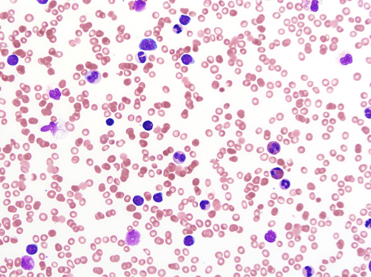 |
| 2. Aspirate smear 50x oil | 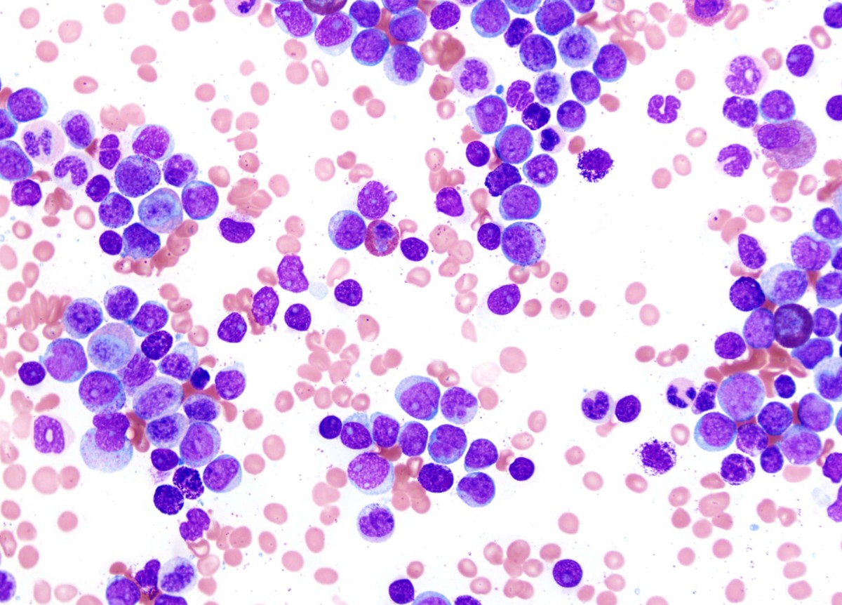 |
| 3. Aspirate smear 100x oil | 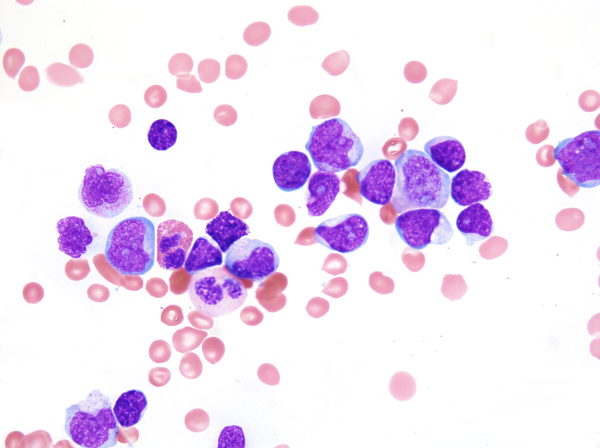 |
| 4. Aspirate smear 100x oil | 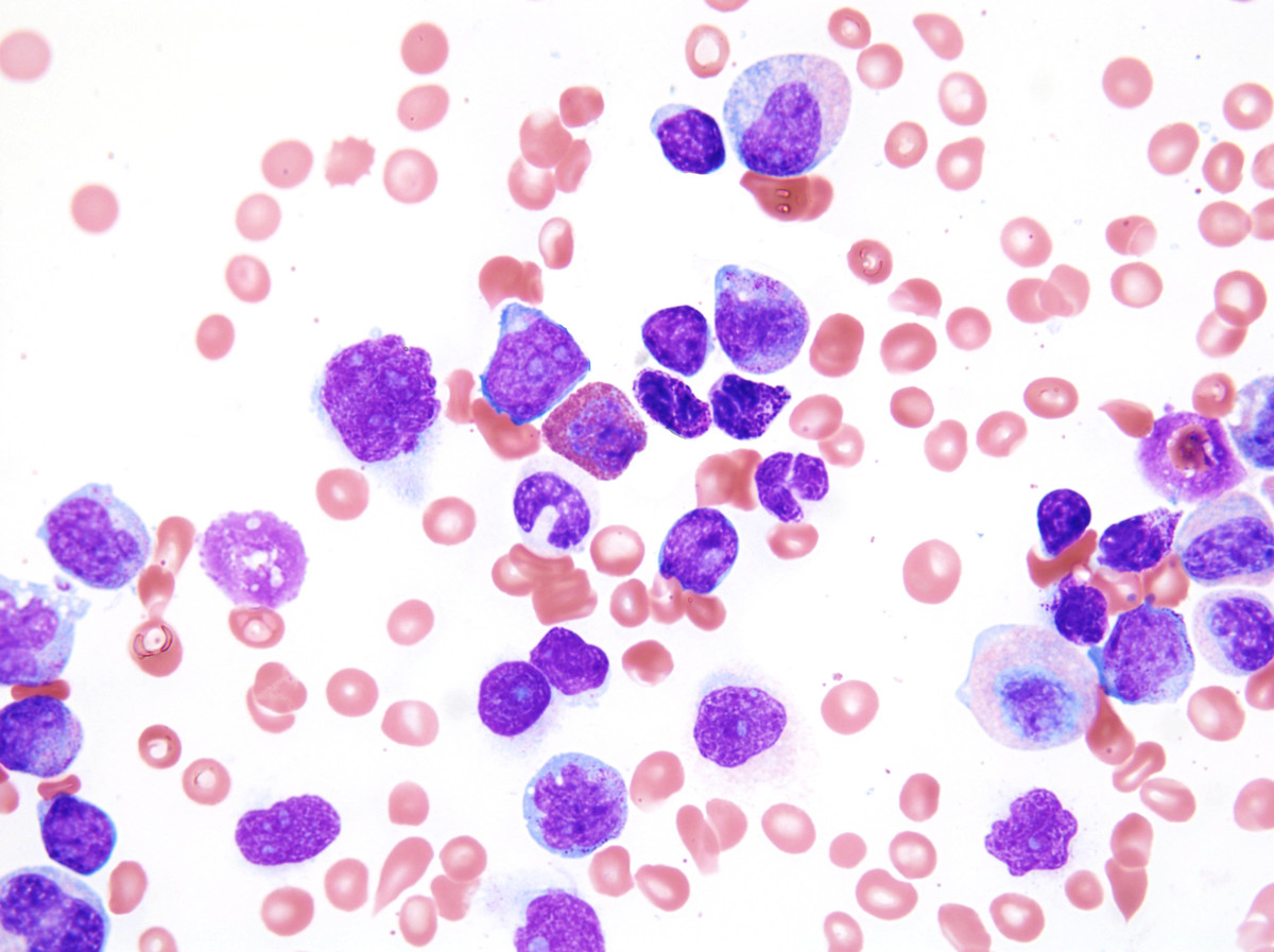 |
| 5. Clot section | 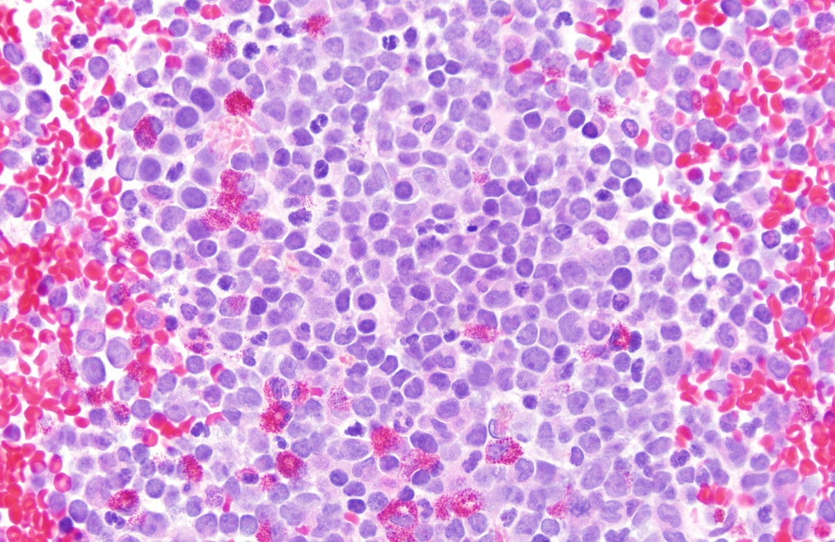 |
| 6. Representative flow cytometry histograms | 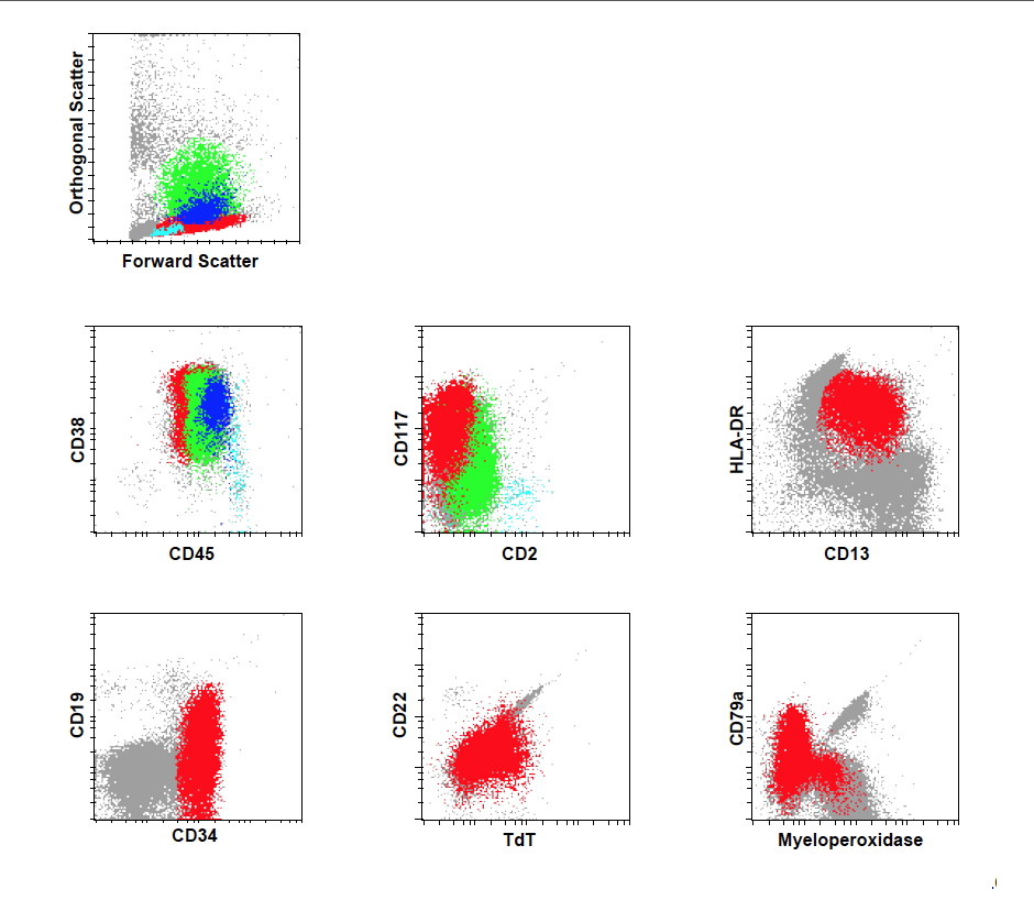 |
| 7. Karyotype | 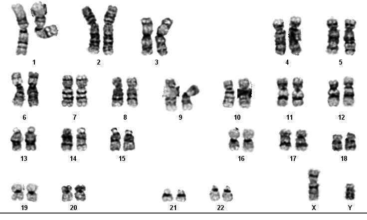 |