Institution: LAC+USC Medical Center
Additional authors:Kiran Qidwai MD, Brit Shackley MD, Imran Siddiqi MD PhD
Session: Acute leukemias of ambiguous lineage
HISTORY
The patient is a 41-year-old Asian woman with a history of iron deficiency anemia who presented with worsening fatigue, easy bruising and progressive abdominal pain. A peripheral blood smear revealed numerous blasts. The patient’s CBC results were as follows: WBC 24.4 K/cumm with 54% blasts, Hgb 8.9 g/dL, Hct 23.9%, MCV 79.7, and platelets 36 K/cumm.
DETAILS
Peripheral blood: Borderline microcytic anemia was present, and occasional nucleated red blood cells were seen. White blood cells were increased in number and included 35% blasts with variable morphology. The majority were medium to large with high nuclear-to-cytoplasmic ratios, mild nuclear irregularities, fine to granular chromatin, multiple nucleoli, and scant deeply basophilic cytoplasm with occasional vacuoles. Occasional blasts had indented nuclei, slightly more cytoplasm and rare fine azurophilic granules. Immature granulocytes were present. No overt neutrophil dysplasia was evident. Platelets were markedly decreased and largely unremarkable.
Aspirate smear: The aspirate smears contained abundantly cellular particles, mostly effaced by sheets of blasts. Blasts comprised approximately 75% of nucleated cells; most were large with mildly irregular nuclear contours, fine to granular chromatin, prominent nucleoli, and deeply basophilic cytoplasm with occasional vacuoles. Granulocytic and erythroid precursors were present, but decreased, each accounting for about 10-15% of cells, with granulocytic left shift. No significant dysplasia was evident. Megakaryocytes were decreased but largely unremarkable. The submitted iron stains on aspirate smears showed decreased stores; incorporation was increased, without an increase in ring sideroblasts. Clot and biopsy sections: The clot and biopsy sections were hypercellular (greater than 95% cellularity) and showed marrow mostly effaced by sheets of blasts. Residual hematopoiesis, including megakaryocytes, was present but decreased.IMMUNOHISTOCHEMISTRY AND FLOW CYTOMETRY
Flow cytometry detected 74% blasts with dim CD45. Blasts expressed CD34, CD13, CD14 (subset), CD15 (partial), CD33, CD38, HLA-DR, CD19 (moderate), intracellular MPO, and intracellular CD79a.
CYTOGENETIC FINDINGS
Chromosome analysis revealed an abnormal karyotype, as follows:
82-84, XXXX,-1,-2,-3,-4,-7,+8,-9,-10, t(12;17)(p13;q11.2)x2, der(12)t(12;17)(p13;q11.2),+13,-15,-16,-17,-20,-21,+22[cp7]/46,XX[13]MOLECULAR FINDINGS
The following studies were performed showing negative results, as follows:
Negative for t(9;22) BCR/ABL fusion by FISHNegative for t(8;21) AML1/ETO [RUNX1/RUNX1T1] fusion by FISHNegative for t(15;17) PML/RARA fusion by FISHNegative for RARA break apart rearrangement by FISHNegative for FLT3 ITD mutation and FLT3 D835 mutation by PCRNegative for NPM1 mutation by PCRNegative for CEBPA mutation by PCRNegative for KIT D816V mutation by PCRINTERESTING FEATURES
Flow cytometry revealed a single population of blasts (74%) co-expressing myeloid (MPO, CD13, partial CD14, partial CD15, CD33) and B lymphoid (CD19 and CD79a) lineage markers, consistent with AML with mixed phenotype (B/myeloid; WHO Classification) Cytogenetic analysis revealed a complex, near tetraploid, abnormal composite karyotype. In addition to marked aneuploidy, an apparently balanced translocation t(12;17)(p13;q11.2) was identified.
When involving the ZNF384/CIZ (chromosome 12) and TAF15(chromosome 17) genes, the t(12;17)(p13;q11) rearrangement has been described as a rare, but recurrent, abnormality predominantly in B-ALL cases (which demonstrate aberrant myeloid antigen CD13 and/or CD33 expression), less commonly in AML (which show aberrant B-cell antigen CD19 expression [see reference 1], as well as in a recent case report of a pro-B ALL which switched to an AML at relapse [see reference 2]. This abnormality has not previously been reported in cases meeting WHO criteria for mixed phenotype acute leukemia.Rarely, ambiguous B/myeloid blast phenotypes can be attributable to t(12;17)(q13;q11) translocation. The prognostic relevance of this abnormality is unclear, particularly in the background of a complex karyotype, as seen in this case. References: 1. La Starza R, et al. CIZ gene rearrangements in acute leukemia: report of a diagnostic FISH assay and clinical features of nine patients. Leukemia. 2005 Sep; 19(9): 1696-9. 2. Grammatico S, et al. Lineage Switch From Pro-B Acute Lymphoid Leukemia to Acute Myeloid Leukemia in a Case with a t(12;17)(p13;q11)/TAF15-ZNF384 Rearrangement. Leuk Lymphoma. 2013 Aug;54(8):1802-5PROPOSED DIAGNOSIS
Acute leukemia with mixed phenotype (B/myeloid) associated with t(12;17)(p13;q11.2).
CONSENSUS DIAGNOSIS
Mixed phenotype acute leukemia (B/myeloid), associated with t(12;17)(p13;q11.2).
| Numerous blasts showing no definitive lineage-specific features (aspirate smear, Wright-Giemsa, 100x) | 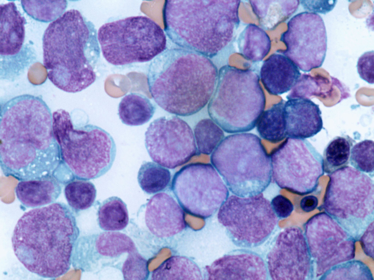 |
| Flow cytometry scatter plot showing a significant population of CD45 dim blasts. | 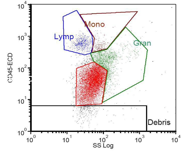 |
| Flow cytometric immunophenotyping of blasts shows co-expression of B cell (cCD79a) and myeloid (cMPO) markers. |  |