Institution: Wake Forest Baptist Health
Additional authors:Natalie Walker, Mark Pettenati, David Grier
Session: Erythroleukemia and megakaryoblastic AML and mimics
HISTORY
The patient is a 30 year-old female with a history of leukopenia and thrombocytopenia that was noted in December 2010 while being treated for pneumonia. A bone marrow biopsy in January 2011 revealed moderate erythroid and granulocytic hyperplasia and a 20q- using fluorescence in situ hybridization. The patient remained pancytopenic over the interval months. In August 2011, the patient was admitted to an outside hospital with a complaint of hip and back pain and was found to have moderate ascites as well as lytic lesions and marrow expansion on an MRI of the lumbar spine. She remained neutropenic and developed fevers and chills. She was transferred to our facility while intubated and hypotensive with concern for leukemia.
DETAILS
A bone marrow biopsy was obtained from the posterior iliac crest on 8/2011 to include cores, a clot section, a bone marrow aspirate and touch preps. The clot sections and cores were fixed in B+ fixative and the aspirates and touch preps were air dried and stained with Wright-Giemsa stain. Examination of the bone marrow biopsy cores revealed significant necrosis with focally viable cells within a background of moderate-to-severe fibrosis. Markedly pleomorphic cells with prominent nucleoli and irregular nuclear membraes were present as well as multinucleated cells. A manual differential count of the bone marrow touch preps revealed a population of atypical enlarged blasts with scant cytoplasm and occasional binucleation, consisting of 18% of the total nucleated cells. The remaining cells consisted of 49% lymphocytes, 6% plasma cells, 9% erythroid precursors, and 18% myeloid precursors. The peripheral blood had atypical mononuclear cells with scant blue cytoplasm, irregular cytoplasmic blebbing, and condensed round nuclei.
IMMUNOHISTOCHEMISTRY AND FLOW CYTOMETRY
Immunohistochemistry revealed focally positive glycophorin A and CD43 and positive CD117 and CD45 studies. The specimen was negative for CD34, MUM1, CD30, CD56, cyclin D1, CD3, S100, pancytokeratin, CD138, CD19, CD20, PAX5, and MPO.
Flow cytometry of the peripheral blood detected a population of cells comprising approximately 18% of all cells with positive expression of CD45, CD71, CD117, CD61, and dim expression of glycophorin A and HLA-DR. The cells were negative for CD3, CD5, CD7, CD138, CD10, CD19, CD20, surface kappa and lambda, CD13, CD15, CD33, CD14, CD34, CD38, and Tdt.CYTOGENETIC FINDINGS
A karyotype of the bone marrow obtained on 8/19/2011 identified 5 out of 5 mitotically active cells with 65,X,i(X)(q10),+1,+2,+3,+6,+6,+8,+8,+10,add(12)(p13),+13,+14,+15,+19,+19,+20,+21,+21,+22,+22,+22,+mar[cp5].
FISH analysis revealed no loss of chromosome 5, chromosome 7, or chromosome 20 and tetraploidy of chromosome 8.MOLECULAR FINDINGS
The patient is homozygous for c.258+2 T>C mutation in the SBDS gene establishing a molecular diagnosis of Shwachman-Diamond Syndrome (SDS) and heterozygous for c.129-3 A>G variant of unknown significance in the SBDS gene.
INTERESTING FEATURES
The case was interesting for several reasons including the severe atypia of the neoplastic cells coupled with the extensive necrosis and fibrosis of the bone marrow. The neoplastic cells expressed CD117, a hematopoietic stem cell/progenitor cell marker; CD61, a megakaryocytic marker; and glycophorin A and CD71. In addition, the patient was diagnosed with Shwachman-Diamond Syndrome via genetic testing.
PROPOSED DIAGNOSIS
The proprosed diagnosis was acute myeloid leukemia, not otherwise specified, with overlapping features of erythroid and megakaryocytic lineages.
CONSENSUS DIAGNOSIS
Acute myeloid leukemia with myelodysplasia-related changes, blasts with erythroid/megakaryocytic differentiation, arising in a patient with Shwachman-Diamond syndrome
| Low power of bone marrow showing pockets of neoplastic cells separated by extensive necrosis |  |
| Malignant cells, bone marrow (40x) |  |
| malignant cells, bone marrow touch prep (40x) |  |
| Peripheral blood (40x) | 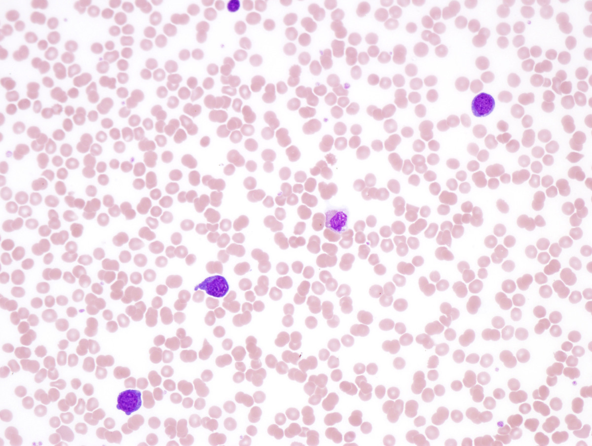 |
| CD45 immunostain, bone marrow (40x) |  |
| CD43 immunostain, bone marrow (40x) |  |
| CD117 immunostain, bone marrow (40x) | 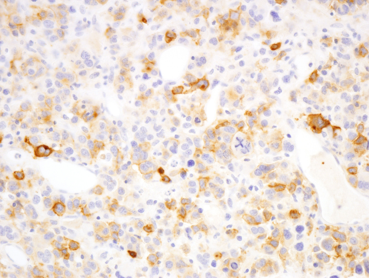 |
| Glycophorin A immunostain, bone marrow (40x) |  |
| Flow cytometry of peripheral blood; abnormal cell population gated red; expression of CD117, CD71, and CD61 | 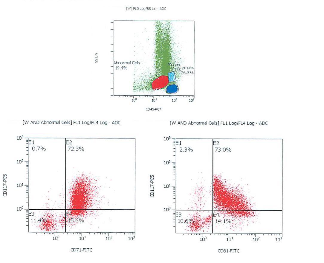 |
| karyotype |  |