Institution: Indiana University
Session: T Lymphoblastic Leukemia/Lymphoma
HISTORY
72 year-old male presented with weight loss, severe cough and hoarseness. Imaging studies showed an anterior mediastinal mass with heterogeneous enhancement suspicious for thymoma. The comparison with previous radiographs demonstrated progressive slow enlargement over a period of 14 months. CBC and peripheral blood differential count were within normal limits. The patient underwent fine needle aspiration with flow cytometric immunophenotyping followed by thymectomy and bone marrow examination.
Three years after the original presentation, the patient was admitted due to a progressive fatigue, 30 pounds weight loss and pancytopenia (WBC 2.4x10⁹/L with reported 84% lymphocytes, hemoglobin 7.6 g/dL and platelet count 100x109/L). Bone marrow examination was performed.DETAILS
Fine needle aspiration of the mediastinal mass showed immature lymphoid population with rare epithelial cells.
Thymectomy specimen demonstrated partially nodular growth pattern with diffuse areas. Aggregates of epithelial cells, a proportion with spindle, fibroblast-like cytology, were seen alongside lymphocyte-rich areas with less prominent epithelial cells. In addition, several areas which lacked epithelial cell meshworks were best visualized using cytokeratin cocktail immunohistochemical stain. Mediastinal lymph nodes accompanying thymectomy specimen showed a partial involvement by immature lymphoid cells. Concurrent bone marrow showed maturing trilineage hematopoiesis. The bone marrow biopsy at the time of the second admission was cellular with numerous blasts and markedly decreased trilineage hematopoiesis. Bone marrow aspirate smear showed predominantly blasts (89%), and rare elements of granulopoiesis and erythropoiesis. Peripheral blood smear showed frequent lymphoid blasts.IMMUNOHISTOCHEMISTRY AND FLOW CYTOMETRY
Flow cytometric immunophenotyping of fine needle aspirate of the mediastinal mass showed a predominant population of immature cells positive for low density CD45, bright CD2, bright CD7, partial CD5 and TdT. The CD34, CD10, surface CD3, CD1a, CD4 and CD8 antigens were negative.
Cytokeratin cocktail immunohistochemical stain of the mediastinal mass highlighted a prominent proliferation of epithelial cells and focal areas lacking epithelial cell meshworks. Mediastinal lymph nodes showed aggregates and sheets of TdT- and CD3-positive lymphoblasts. Cytokeratin cocktail immunostain was negative in mediastinal lymph nodes.The flow cytometric immunophenotyping of bone marrow sample procured at the time of the second admission showed a predominant population of blasts positive for cytoplasmic CD3, bright CD7 and bright CD2. There was a partial expression of CD34, HLA-DR and TdT.MOLECULAR FINDINGS
TCR gene rearrangement performed on paraffin embedded mediastinal mass and bone marrow clot section from the time of the second admission were non-contributory.
INTERESTING FEATURES
This case demonstrates a rare presentation of concurrent thymoma and T lymphoblastic lymphoma. At the time of initial diagnosis, the patient showed no evidence of bone marrow involvement. Three years post thymectomy, the patient presented with circulating blasts and prominent bone marrow involvement.
PROPOSED DIAGNOSIS
Concurrent thymoma and T lymphoblastic lymphoma.
CONSENSUS DIAGNOSIS
Concurrent thymoma and T lymphoblastic lymphoma
| Fine needle aspiration of mediastinal mass showed numerous immature lymphoid cells | 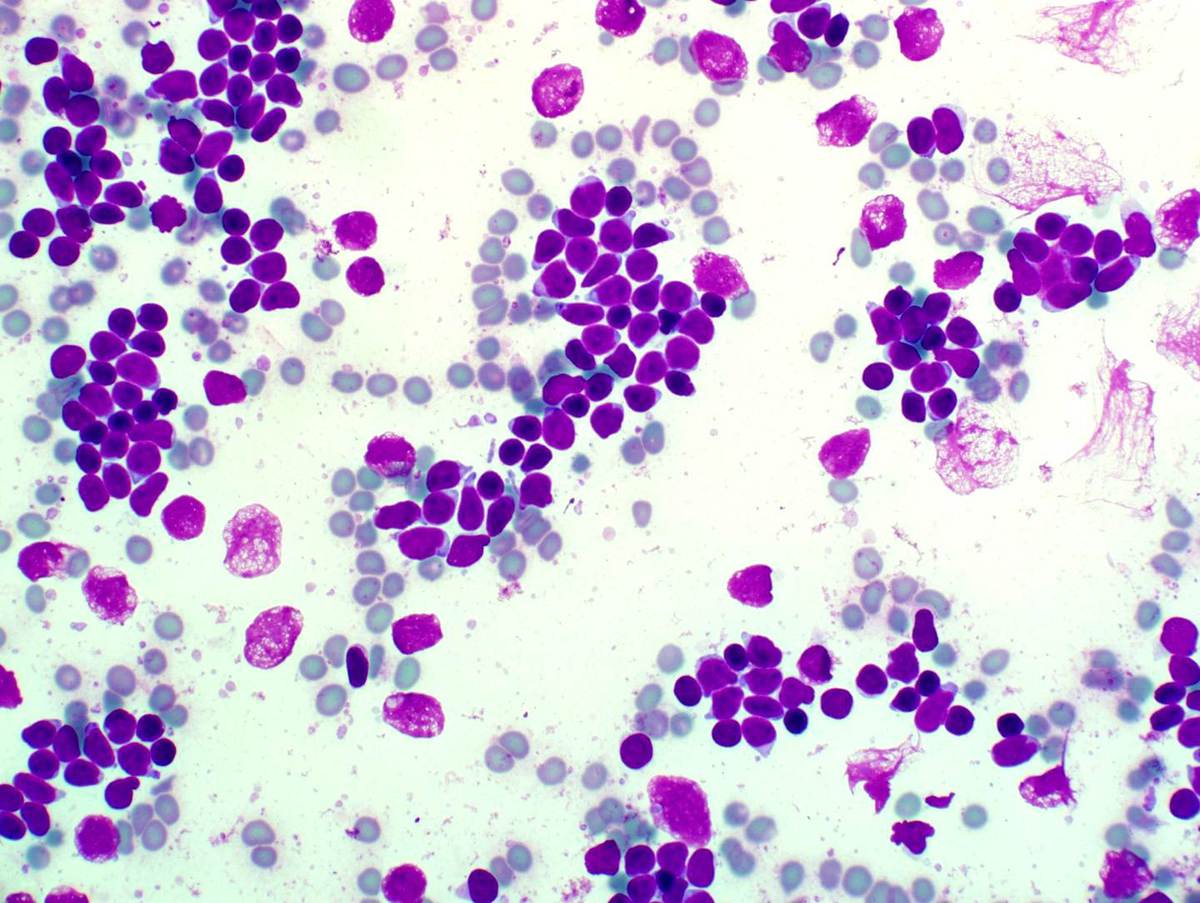 |
| Flow cytometry of mediastinal mass FNA | 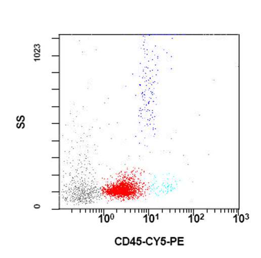 |
| Flow cytometry of mediastinal mass FNA | 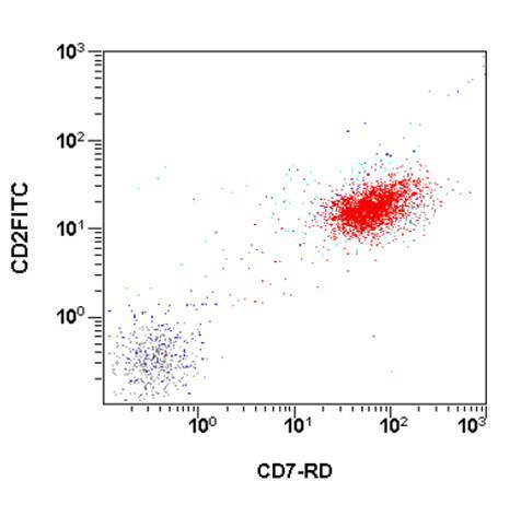 |
| Flow cytometry of mediastinal mass FNA |  |
| Mediastinal mass, epithelial cell proliferation | 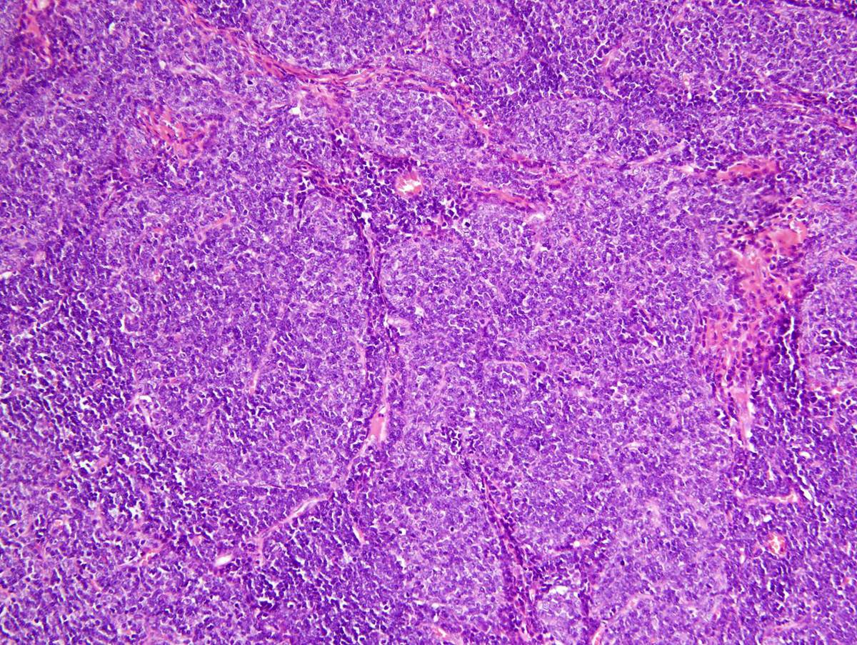 |
| Mediastinal mass, epithelial cell-rich and lymphocyte-rich areas (40x) |  |
| Mediastinal mass, epithelial cell-rich and lymphocyte-rich areas (400x) | 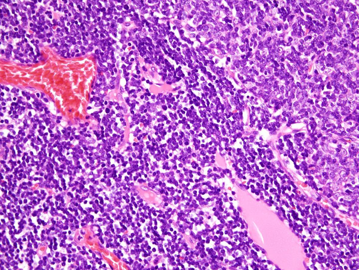 |
| Cytokeratin cocktail immunostain of mediastinal mass | 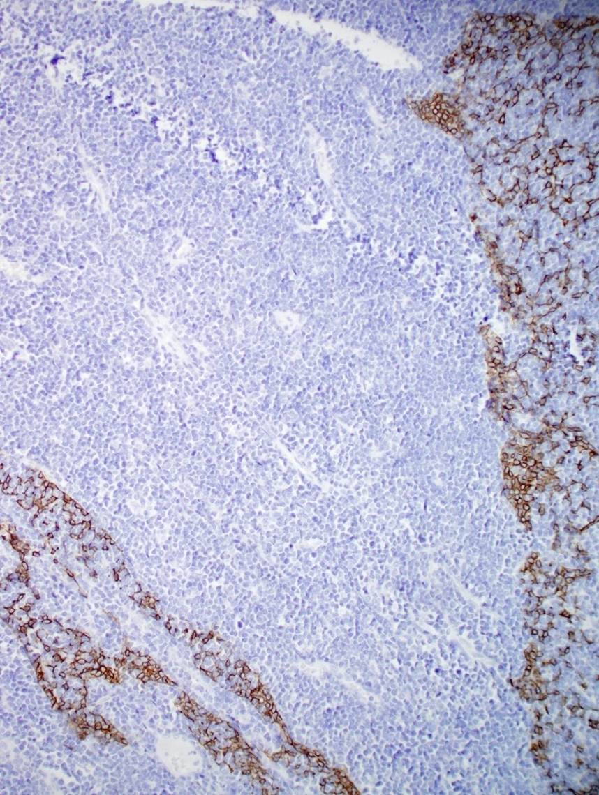 |
| Bone marrow biopsy at the time of leukemic presentation | 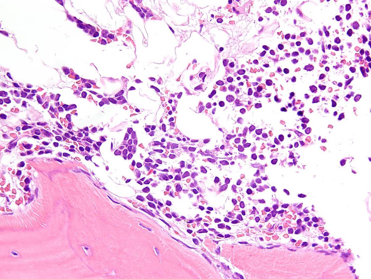 |
| Bone marrow aspirate showing numerous lymphoblasts (second admission) | 1.jpg) |
| Flow cytometry of bone marrow showing a prominent T-lymphoblast population (second admission) | 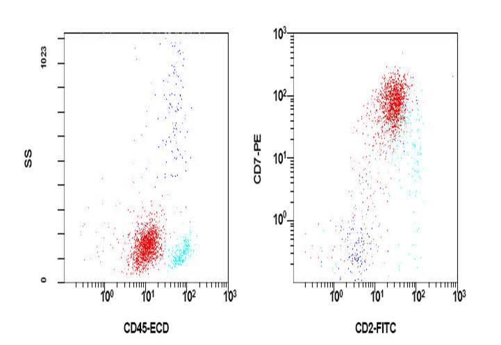 |
| Flow cytometry of bone marrow at the time of leukemic presentation-additional markers | 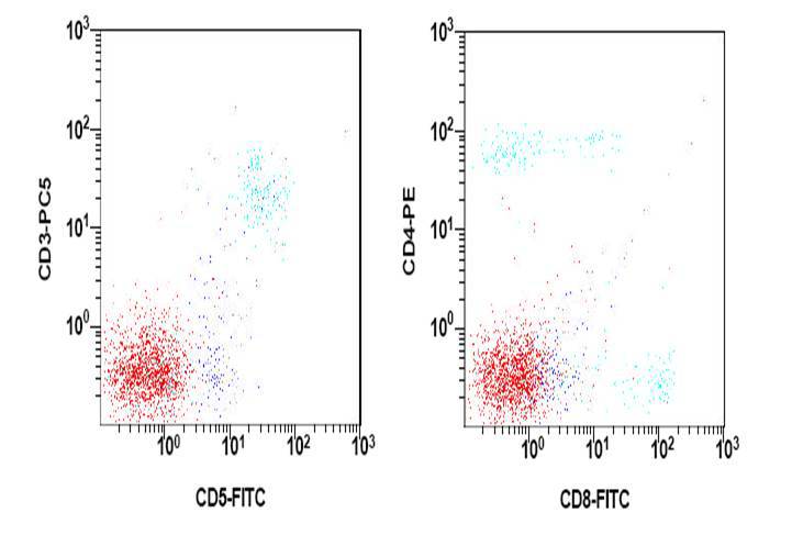 |