Institution: University of Alabama at Birmingham
Additional authors:Dara Wakefield, MD, Deniz Peker, MD
Session: AML with recurrent genetic mutations Part II
HISTORY
A 68 year-old woman with no significant past medical history sought evaluation in April 2012 for a 1 year history of constant bilateral, 8/10 facial pain located in her temporomandibular joints with associated clicking and crepitations. Radiologic studies showed severe degenerative changes in the left temporomandibular joint (TMJ) and mild to moderate changes in the right TMJ. In late September 2012, she underwent bilateral TMJ replacement. Routine pathologic evaluation of the resected TMJs revealed a hematopoietic neoplasm (CD43, CD33, and myeloperoxidase [MPO] positive) involving the bone and bone marrow space with obscured morphology secondary to specimen decalcification. Per suggestion by the hematopathologist, patient went through a bone marrow biopsy. At the time of the biopsy, complete blood count (CBC) and peripheral smear evaluation showed circulating blasts, moderate normocytic anemia and marked neutropenia. The patient was admitted to the Oncology service at the University of Alabama at Birmingham in October 2012. At the time of admission, she only stated some weight loss possibly due to being placed on a liquid diet post-operatively; she was otherwise still asymptomatic. She denied recurrent fevers, nausea, vomiting, and night sweats. Physical exam failed to reveal splenomegaly or petechiae. A bone marrow biopsy was performed and showed a hypercellular marrow (40-60% cellularity) with > 67% blasts in the background of multi-lineage dysplasia (to be discussed further). She was enrolled in the ECOG 2906 randomized clinical trial and was randomized to the 7+3 induction arm. Both day 14 and day 28 bone marrow biopsies following induction chemotherapy showed no morphologic evidence of leukemia. She has been in complete remission to date.
DETAILS
Specimen fixation details (excision bilateral mandibular condyle fragments and marrow core biopsy): The fragments of bilateral mandibular condyle submitted were fixed in buffered 10% formalin for at least 6 hours and underwent decalcification for at least 12 hours. 4-5 micron sections were stained with hematoxylin and eosin stain. The bone marrow biopsy was performed on left posterior iliac crest and was fixed in buffered 10% formalin for at least 6 hours. Brief decalcification was performed. 4-5 micron sections were stained with hematoxylin and eosin stain. Details of bilateral mandibular condyle microscopic pathology: The H&E sections illustrated an extensive monotonous infiltration of mononuclear cells involving the bone and bone marrow space. Although the morphologic exam was challenging due to marked decalcification artifact, the neoplastic cells were intermediate to large with a high nuclear:cytoplasmic ratio and irregular nuclear contours (figures 1A, 1B, and 2). There were few reactive lymphocytes and minimal normal hematopoiesis noted in the background. Of note, no overt morphology that of a degenerative joint disease was observed. Details of peripheral blood and bone marrow biopsy microscopic pathology: The peripheral blood examination was significant for marked neutropenia and moderate normocytic normochromic anemia with circulating blasts (figure 4; WBC: 0.96; HB: 9.4; MCV: 97; PLT: 142; differential count 39% neutrophils, 6% blasts). Some hypogranulated and hypolobated neutrophils noted. The aspirate smears were adequately cellular and demonstrated the predominance of large immature precursor cells with round to irregular nuclei, scant cytoplasm and open chromatin with some prominent nucleoli, compatible with myeloblasts (> 67% of cellularity). The background hematopoiesis was significant for moderate to marked dysplasia, mostly in erythroid (figure 6A) and megakaryocytic (figure 6B) lineages. The core biopsy showed a hypercellular marrow (40-60% cellularity) with > 67% of cellular elements comprised by the immature blast cell population (figure 7).
IMMUNOHISTOCHEMISTRY AND FLOW CYTOMETRY
Immunophenotype (mandibular condyle): A battery of immunostains including CD3, CD5, CD15, CD20, CD31, CD33, CD34, CD43, CD45, CD58, CD68, CD79a, CD99, CD117, CD138, pan-keratin, MPO, and S100 were performed. The neoplastic cells were diffusely positive for CD33, CD43, and MPO (figure 3) and negative for CD117, CD34, and CD45 and as well as CD4, CD5, CD15, CD31, CD58, 68, CD99, CD138, pan-keratin and S100. CD3, CD20, and CD79a highlighted the background lymphocytes. Flow Cytometry (peripheral blood and bone marrow biopsy): Flow cytometry on peripheral blood (figure 5) showed an increased aberrant myeloid lineage cell population consistent with the 14% circulating blasts with expression of CD33 and CD117 with dim CD13. CD34, HLA-DR, CD15, CD56, CD4, and CD64 were negative. Flow cytometry on bone marrow (figure 8) showed immature myeloid blasts comprising 82% of total cells with an immunophenotype identical to that detected on peripheral blood (figure 5).
CYTOGENETIC FINDINGS
Normal female karyotype.
MOLECULAR FINDINGS
PCR studies demonstrated a positive FLT3-ITD mutation, NPM1 mutation, and a single CEBPA mutation. No CKIT mutation was detected.
INTERESTING FEATURES
Interesting features of submitted case: This was an initially challenging case for the oral pathologist to work-up since AML is not common in the mandibular condyle as initial location; therefore is not high in the differential diagnosis list. Furthermore, the morphology was greatly obscured due to decalcification process and immunohistochemical markers often used diagnosing myeloid sarcoma, i.e. CD45, CD117 and CD34, were negative in this case, puzzling the general pathologist even more. In addition, the patient did not have any symptoms prior to the TMJ surgery and, per the surgeon, the pre-op CBC values performed outside were not significant (data not available). Upon hematopathology consultation, immunohistochemical stains CD43, CD33 and MPO were performed and confirmed the myeloid origin and a bone marrow biopsy followed. AML with myelodysplastic changes (AML-MDC), with no previous MDS, was rendered as the final diagnosis. Our current case did not have any cytogenetic abnormalities unlikely many AML-MDC. However, mutation analysis revealed NPM1, FLT3 and CEBPA mutations. Additional comments: AML presenting as a temporomandibular bone lesion, clinically mimicking degenerative joint disease, is very unusual. The main differential diagnosis in this region includes plasma cell neoplasms, lymphoma and metastatic carcinoma. Immunophenotypic evaluation is necessary to make the distinction, particularly in patients with no prior history. In the presence of large, discohesive, atypical cell infiltration, myeloid sarcoma should also be included in differential diagnosis. AML with myelodysplasia-related changes typically occurs in the elderly and accounts for 24-35% of AML cases. Patients often present with severe pancytopenia which was likely overlooked in this case during the pre-operative evaluation. The most common chromosome abnormalities seen in AML-MDC are similar to those seen in MDS, including -5/del(5q), -7/del(7q), and complex karyotypes; our case did not have any of the karyotypic abnormalities. In general, those affected by AML-MDC have a poorer prognosis relative to those with other subtypes of AML. As in MDS, patients with adverse cytogenetic abnormalities have an increased risk for worse outcomes. Morphology alone does not offer prognostic significance in multi-variant analysis. In our current case, the patient had a normal karyotype; however the FLT3-ITD, NPM1 and single CEBPA mutations were detected. In AML-MDC without prior MDS or MDS-related adverse cytogenetic abnormalities, the clinical significance of NPM1 or CEBPA mutations in the presence of FLT3-ITD is unclear. Furthermore, the appropriate classification of these unique situations is not yet clear. This case represents a prime example of the importance of the pathologist’s role in patient care when evaluating surgical excision specimens. It shows that life-threatening diseases, like AML, can escape detection after an extensive clinical evaluation including an in depth history review, physical exam, and advanced modality imaging. At times, there is no replacement for the information that a thorough microscopic examination can offer. In addition, this case also highlights the potential clinical impact that a routine CBC with differential can offer in the pre-operative setting.
PROPOSED DIAGNOSIS
Acute myeloid leukemia with myelodysplasia-related changes (multilineage dysplasia), bone marrow biopsy.
CONSENSUS DIAGNOSIS
Acute myeloid leukemia with myelodysplasia-related changes, NPM1 mutation, FLT3 ITD mutation, and heterozygous CEBPA mutation
| Figures 1A and 1B. Extensive involvement of bilateral maxillary condyles by an diffuse atypical cell infiltrates. | 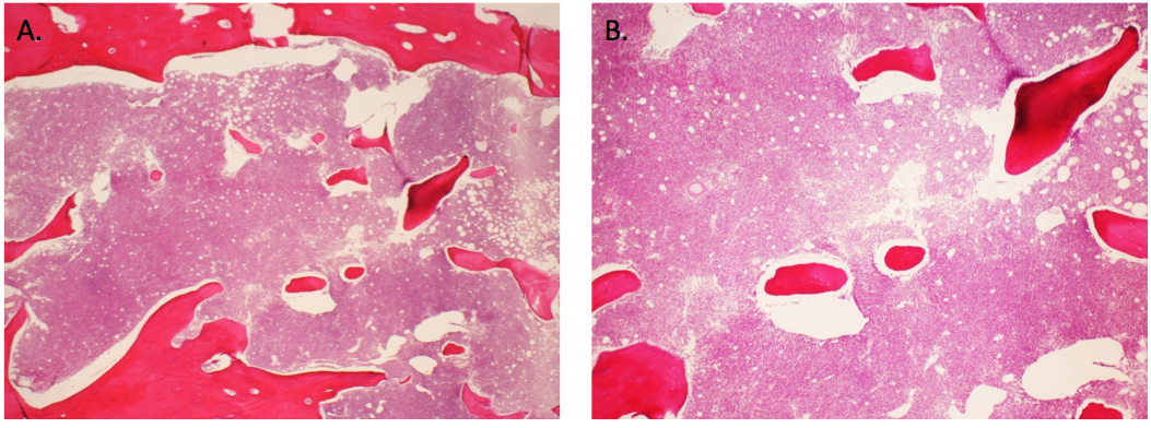 |
| Figure 2. Neoplastic cells are discohesive and mononuclear with extensive artifact, mandibular condyle. |  |
| Figure 3. Neoplastic cells are strongly positive for CD33, CD43 and MPO and negative for keratin, CD34 and CD117. |  |
| Figure 4. Peripheral blood smears displaying circulating large immature myeloid cells with open chromatin, irregular nuclear contours and some prominent nucleoli. |  |
| Figure 5. Peripheral blood flow cytometry demonstrating CD117, CD33 and dim CD13 expression in blast population with no CD34 expression. |  |
| Figures 6A and 6B. Bone marrow aspirate smear displaying (>60% of total cells) high blast count in the background of dysplastic changes in erythroid and megakaryocytic lineages. |  |
| Figure 7. Cellular bone marrow core biopsy with large clusters of blasts. |  |
| Figure 8. Flow cytometry performed on bone marrow aspirate shows similar phenotype to that of shown in peripheral blood analysis; CD33+, CD117+ and dimCD13+, CD34-. |  |