Institution: Cleveland Clinic
Additional authors:James R. Cook, M.D., Ph.D., Eric D. His, M.D.
Session: B Lymphoblastic Leukemia/Lymphoma
HISTORY
The patient was a previously healthy 63 year old man who developed sudden mid-back pain, shortness of breath, and severe constitutional symptoms. He then developed decreased output of brown-colored urine, and eventually presented in renal failure. At admission he was anemic (Hgb 11.4 g/dl) and thrombocytopenic (plt 55,000/ul) with a leukocytosis (WBC 21,400/uL); pelvic ultrasound showed hydronephrosis and a large retroperitoneal mass (8.5 x 9.5 x 13 cm). The next day 38% blasts were reported. He underwent dialysis and was transferred to our institution for management of presumed lymphoma. Upon arrival he had 34% blasts in his peripheral blood (Figure 1). His renal failure was attributed to tumor lysis syndrome, for which he was treated. Flow cytometry was performed on the peripheral blood and a bone marrow biopsy was obtained.
DETAILS
The Wright-stained aspirate smear obtained from the posterior iliac crest shows an expansion (80% of cells) of intermediate to large blasts with round nuclei, variably prominent and occasionally multiple nucleoli, and scant basophilic and often vacuolated cytoplasm (Figure 1). A zinc formalin-fixed, decalcified bone marrow core biopsy from the same site is hypercellular (80%). The majority of marrow cellularity consists of a diffuse infiltrate of blasts with only minimal residual trilineage maturation (Figures 2-3). The formalin-fixed aspirate clot section shows similar morphology.
IMMUNOHISTOCHEMISTRY AND FLOW CYTOMETRY
Flow cytometry, peripheral blood (blast gate 39% of events, Figure 4):
Positive: CD10, CD19, CD22 (dim, partial), CD38, CD45 (dim), HLA-DR, TdT
Negative: CD20, kappa and lambda surface immunoglobulin light chains, CD34, CD117, NK/T-cell markers (CD2, CD3, CD4, CD5, CD7, CD8, CD56), myeloid/monocytic markers (CD11b, CD13, CD14, CD16, CD33, CD64, CD65)
Immunohistochemistry, bone marrow core (Figure 5):
Positive: CD10, CD79a (partial), PAX5, BCL2, TdT, cMYC
Negative: CD3, CD20, CD34, CD99, BCL6
CYTOGENETIC FINDINGS
49,XY,+X,add(3)(p13),+del(7)(q22q34),t(8;22)(q24.1;q11.2),+11,t(14;18)(q32;q21.3),der(14)t(14;18)[5]/49,idem,add(16)(p13.3)[15] (Figure 7)
MOLECULAR FINDINGS
Interphase fluorescence in situ hybridization (FISH), bone marrow aspirate: Positive (Figure 6): BCL2 (18q21) translocation (dual color, break-apart probe), and MYC (8q24) translocation (dual color, break apart probe). 87% of nuclei display a gain at the telomeric portion of the BCL2 (18q21) gene locus. 64% of nuclei display a gain at the MYC (8q24) gene locus or aneusomy of chromosome 8. Negative: IGH@-MYC translocation (tri-color, dual fusion probe), t(9;22) BCR-ABL1 translocation (dual color, single fusion probe), and MLL (11q23) translocation (dual color, break apart probe). Qualitative reverse-transcription polymerase chain reaction assay for BCR-ABL1 p190 transcripts (peripheral blood): negative.
INTERESTING FEATURES
This is a case of an aggressive B-cell neoplasm presenting with both leukemic and extramedullary (retroperitoneal mass) manifestations and translocations involving BCL2 and MYC. The morphology, leukemic presentation, and immunophenotype (TdT expression with lack of CD20 and surface immunoglobulin) are consistent with a B lymphoblastic leukemia/lymphoma (B-ALL/LBL). However, the neoplasm also has a complex karyotype including BCL2 and MYC translocations, which can be seen in aggressive B-cell neoplasms (so-called “double hit” neoplasms). Given the overall findings in this case, a diagnosis of B lymphoblastic leukemia/lymphoma is favored. The patient was started on limited (due to renal failure) chemotherapy (cytoxan, vincristine, prednisone); idarubicin and pegasparagase were added after improvement of renal function with itrathecal methotrexate for CNS prophylaxis. WBC count dropped with apparent clinical improvement, but on day 22 increased blasts (26%) were again found in the periphery. The patient opted for palliative treatment and was discharged on oral 6-mercaptopurine and methotrexate. Abdominal CT 1.5 months later showed no evidence of the retroperitoneal mass. However he expired approximately four months after onset of symptoms. There are few reports of B-cell neoplasms with both BCL2 and MYC translocations presenting as de novo B-ALL/LBL; at least two presented with extramedullary involvement (abdominal masses or inguinal lymphadenopathy). Both were positive for TdT and HLA-DR with minimal or no surface immunoglobulin light chain expression. Although one case initially responded to chemotherapy, ultimately both were fatal, as in the case of our patient. TdT-positivity has also been reported in rare cases of B-cell lymphomas with both of these translocations arising from a prior low-grade follicular lymphoma. The 2008 WHO section describing the provisional entity B-cell lymphoma, unclassifiable, with features intermediate between diffuse large B-cell lymphoma and Burkitt lymphoma notes that “[i]mmunohistochemistry for TdT is required to exclude lymphoblastic lymphoma.” Given the shared molecular alterations, it is unclear whether cases such as the one submitted here, arising either de novo or in the setting of prior follicular lymphoma, should be included in this group. ____________________________________________________________________________ Mufti GJ, Hamblin TJ, Oscier DG, Johnson S. Common ALL With Pre-B-Cell Features Showing (8;14) and (14;18) Chromosone Translocations. Blood 1983; 62(5):1142-6. Thangavelu M, Olopade O, Beckman E, Vardiman JW, Larson RA, McKeithan TW, Le Beau MM, Rowley JD. Clinical, Morphologic, and Cytogenetic Characteristics of Patients With Lymphoid Malignancies Characterized by Both t(14;18)(q32;q21) and t(8;14)(q24;q32) or t(8;22)(q24;q11). Genes, Chromosomes & Cancer 1990; 2:147-58.
PROPOSED DIAGNOSIS
B lymphoblastic leukemia/lymphoma
CONSENSUS DIAGNOSIS
High-grade TdT-positive blastic B-cell leukemia/lymphoma with BCL2 (18q21) translocation and MYC (8q24) translocation
| Peripheral blood and bone marrow aspirate smear (1000x) | 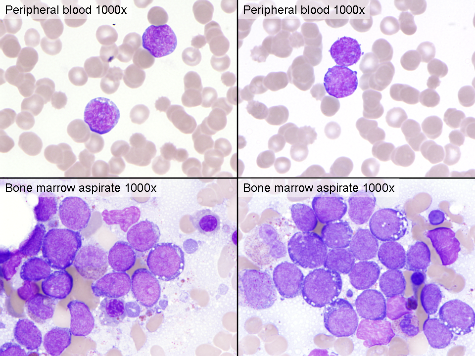 |
| Bone marrow core biopsy (100x) | 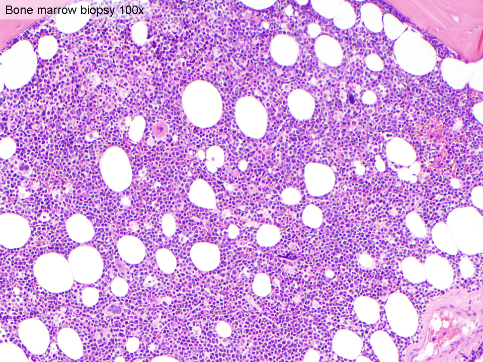 |
| Bone marrow core biopsy (500x) |  |
| Flow cytometry, peripheral blood (blast gate 39% of events) |  |
| Immunohistochemical stains, bone marrow core biopsy (200x) |  |
| Fluorescence in situ hybridization, bone marrow aspirate |  |
| Karyotype |  |