Institution: Cleveland Clinic
Additional authors:Karl S. Theil, M.D.
Session: AML secondary to myeloproliferative neoplasms and other types of disease progression in MPN
HISTORY
A 22 year-old woman was found to have leukocytosis (28,000/uL) and thrombocytosis (873,000/uL) on routine blood work performed at 12 weeks gestational age of her second pregnancy. A bone marrow biopsy showed chronic myelogenous leukemia (CML) in chronic phase with 1% blasts by aspirate count. Conventional cytogenetics showed a 46,XX,t(9;22)(q34;q11.2)[20] karyotype and molecular studies were positive for p210 BCR-ABL1 transcript. Due to the relative contraindication against tyrosine kinase inhibitors during pregnancy, she was treated with hydroxyurea for cytoreduction and heparin for prophylaxis against thrombosis and successfully carried her pregnancy to term. Five weeks after delivering a healthy boy, she presented with chest pain and shortness of breath and was found to have a pulmonary embolism. CBC and manual differential showed WBC 11,910/uL, HGB 12.6 g/dL, PLT 419,000/uL with 18% blasts, and a bone marrow biopsy confirmed blast phase CML. Nilotinib was initiated.
DETAILS
Wright-stained peripheral blood smear shows blasts which are moderate in size with scant relatively agranular cytoplasm without Auer rods (Figure 1). The right iliac crest Wright-stained bone marrow aspirate shows 66% blasts with morphology similar to those in the periphery (Figure 1). Erythropoiesis is decreased (myeloid:erythroid ratio 9.3). Megakaryocytes are present with occasional small, hypolobated forms. A cytochemical stain for myeloperoxidase is positive in a subset of blasts. A zinc-formalin-fixed, decalcified bone marrow core biopsy from the same site is hypercellular (80%) with a granulocytic left-shift and large clusters and sheets of blasts. There is scant residual hematopoiesis. Megakaryocytes are decreased with occasional small and large abnormal forms (Figures 2-3).
IMMUNOHISTOCHEMISTRY AND FLOW CYTOMETRY
Flow cytometry, peripheral blood (blast gate 26% of events; Figure 4): Positive: CD10, CD19, CD22 (dim), CD34, CD38, CD45, TdT, HLA-DR, CD13, CD33, CD117 (minor subset) Negative: CD2, CD3, CD4, CD5, CD7, CD8, CD11b, CD14, CD16, CD20, CD56, CD64, CD65 Immunohistochemistry, bone marrow core biopsy (Figure 5): subsets of blasts are positive for myeloperoxidase (arrows), CD79a, and PAX5.
CYTOGENETIC FINDINGS
46,XX,t(9;22)(q34;q11.2)[20] (Figure 5)
MOLECULAR FINDINGS
Reverse transcription polymerase chain reaction for BCR-ABL1 p210 transcripts (peripheral blood): Positive (baseline).
INTERESTING FEATURES
This case shows rapid progression to blast phase CML with an unusual mixed B/myeloid phenotype in a young (22 years) pregnant woman not previously treated with tyrosine kinase inhibitors (TKI). Although there have been reports of patients successfully treated with TKI during pregnancy without apparent harm to the fetus, in this case the disease was managed with hydroxyurea and heparin treatment only and pregnancy was successfully carried to term. The disease progressed to blast phase less than 9 months after diagnosis. This is a rapid progression, even for a patient not receiving TKI treatment, which is currently the standard of care. Although the effect of pregnant and post-partum states on disease course in CML is not well characterized, this aggressive course is in contrast to a case series of five women diagnosed with CML during pregnancy who were not treated with TKI until after delivery (1 week to 5 months later) in which all women showed molecular or cytogenetic response with none progressing to blast phase. The immunophenotype of blasts in blast phase CML is known to vary and can include myeloid, B- and T-cell differentiation. The 2008 edition of the WHO states that 25% of cases of blast phase CML fulfill criteria for mixed lineage acute leukemia, however this is based on data collected prior to the requirement of proof of myeloperoxidase expression to establish myeloid differentiation put forth in the same edition. Thus many cases previously reported as B/myeloid may not fit that designation based on current criteria. The patient’s follow-up bone marrow biopsy in August 2012 showed a hypocellular marrow with no morphologic evidence of residual/recurrent CML and no increased blasts. Although the karyotype was 46,XX[20], quantitative PCR for p210 BCR/ABL1 transcripts showed an absence of major molecular response (normalized copy number: 5.079; International Scale: 3.784). She underwent myeloablative matched-sibling HLA-identical bone marrow stem cell transplant. Initial engraftment studies showed 100% donor DNA. More recent molecular studies (January 2013) showed low-level (non-quantifiable) BCR/ABL1 p210 transcripts and 8% recipient DNA in the T-cell enriched peripheral blood fraction. Klamova H, Markova M, Moravcova J, Siskova M, Cetkovsky P, Polakova KM. Response to treatment in women with chronic myeloid leukemia during pregnancy and delivery. Leukemia Research 2009; 33:1567-9.
PROPOSED DIAGNOSIS
Mixed phenotype (B/myeloid) blast phase chronic myelogenous leukemia
CONSENSUS DIAGNOSIS
Chronic myelogenous leukemia, BCR-ABL1 positive, blast phase with mixed phenotype (B/myeloid)
| Peripheral blood and bone marrow aspirate smear (1000x) | 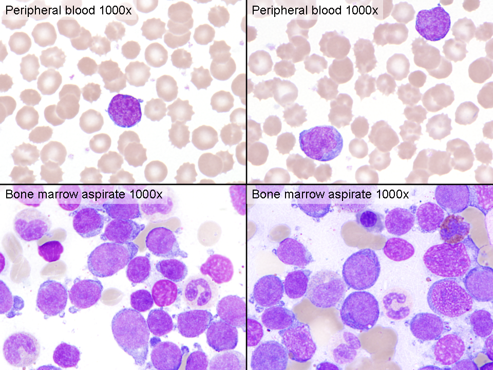 |
| Bone marrow core biopsy (100x) | 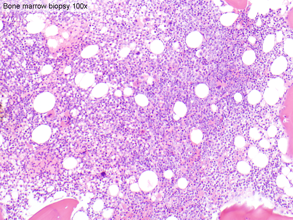 |
| Bone marrow core biopsy (200x; inset 1000x) | 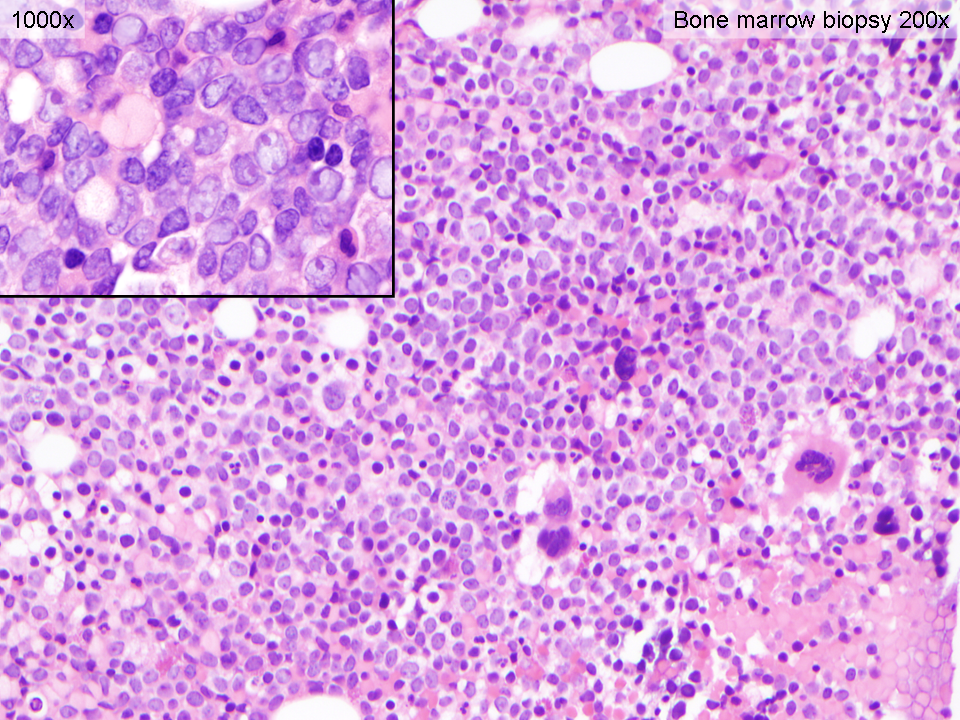 |
| Flow cytometry, peripheral blood (blast gate 26%) | 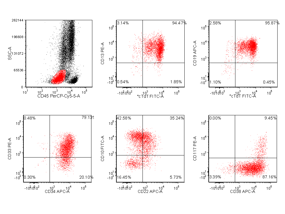 |
| Immunohistochemical stains, bone marrow core biopsy (400x) and karyotype | 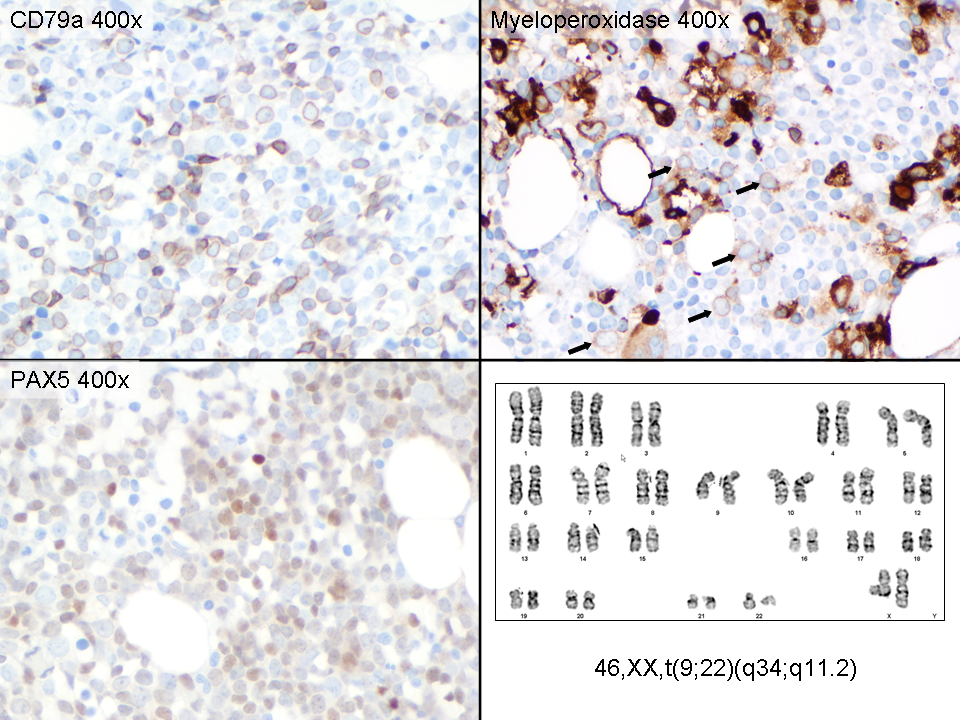 |