Institution: Toronto General Hospital/University Health Network
Session: Acute leukemias of ambiguous lineage
HISTORY
The patient is a 67 year old man who was diagnosed with MPAL (5/12) with monosomal karyotype (monosomy 7 and 20q deletion) who underwent induction chemotherapy and intensification phase of Dana Farber protocol. In January 2013 he developed features suggestive of relapse with circulating blasts of myeloid phenotype. He also developed lymphadenopathy, night sweats and skin lesions with intractable pruritus despite prednisone, antihistamines and local measures. His skin biopsy was suspicious for leukemic involvement. His significant past medical history include history of rheumatoid arthritis for which he is currently not receiving any medication. In the past he had been treated with methotrexate and ARAVA (leflunomide) almost until diagnosis of leukemia in 5/12.
DETAILS
1. Bone marrow biopsy (5/2012) Left illiac crest biopsy Fixative I.B.F at least 4 hours Decalcification in RDO decalcifier- 1 hour CBC: Hg 81 g/dl, MCV 88.7 fL, RDW 19.1%, WBC 3.9 x10⁹/L, PLT 43x109/L, Neutr A 0.7x10⁹/L, Lymphocytes A 0.31x10⁹/L, Monocytes A 0.00x10⁹/L, Blasts A 2.89x0.31x109/L (74%). Peripheral blood smear shows normal white blood cell count but abnormal differential with 74% circulating blasts and an absolute neutropenia. The blasts are mostly intermediate in size with dense chromatin, and scant cytoplasm. Neutrophils show variable morphology, including normal morphologically segmented neutrophils as well some dyspoietic forms with abnormal nuclear segmentation/hyposegmentation, chromatin hypercondensation and hypogranular cytoplasm. The red cells are normocytic but show marked anisopoikilocytosis, including macro-ovalocytes, elliptocytes, red cells fragments and occasional tear drop shaped red cells and some are hypochromic. There is also a slightly increased polychromasia. Platelets are markedly reduced in number but show normal granulation. Some large and giant platelets are noted The bone marrow biopsy is hypercellular with somewhat variable cellular distribution, averaging at 60-70% and shows evidence of underlying fibrosis, as suggested by focal streaming of the marrow cells. There is an interstitial infiltrate of blasts, which account for approximately half of the marrow cellularity. The blasts are variable in size, ranging from small to intermediate to large in size with dispersed chromatin and scant to abundant cytoplasm. Intermingled with blasts are islands of maturing erythropoiesis. Granulocytic maturation is shifted towards immaturity but progressive to segmented neutrophils. The megakaryocytes are reduced in number and show variable morphology, including normal morphologically megakaryocytes as well as some dyspoietic forms with widely separated nuclear lobes. Differential: Blasts 50%, Promyelocytes 0%, Myelocytes 3%, metamyelocytes 2%, neutrophils 17%, Lymphocytes 7%, Monocytes 1%, Erythroid precursors 20% The bone marrow aspirate smear reveals sizable spicule that are cellular and essentially reflect findings of the bone marrow biopsy. Blasts are increased, comprising 50% of the marrow cells and range in size from small to intermediate in size with scant cytoplasm to large in size with abundant agranular cytoplasm. Erythroid maturation is essentially normoblastic. Some erythroid precursors show mild megaloblastoid change and evidence of mild dyserythropoiesis. Granulocytic maturation is shifted towards immaturity but progressive to mature segmented neutrophils. Occasional neutrophils show mild dyspoietic features, however there is no evidence of significant dysgranulopoiesis. The megakaryocytes are reduced in number and only few can be appreciated on this preparation, including normal morphologically megakaryocytes as well as occasional small hypolobated dyspoietic forms. 2. Bone marrow biopsy 1/2013 Right illiac crest biopsy Fixative I.B.F. at least 4 hours Decalcification in RDO decalcifier- 1 hour CBC: Hg 126 g/dl, MCV 99 fL, RDW 22.4%, WBC 20.4 x10⁹/L, PLT 50x109/L, Neutr A 14.48x10⁹/L, Lymphocytes A 1.63x x109/L, Monocytes A 0.61 x10⁹/L, Blasts A 3.67x10⁹/L (18%). Peripheral blood smear shows moderate leukocytosis with 18% circulating blasts. The blasts are intermediate to large in size with oval to slightly irregular/indented nuclei, finely dispersed chromatin, variably distinct nucleoli and scant to moderate to abundant cytoplasm. The majority of circulating leukocytes are comprised of mature segmented neutrophils showing evidence of dysgranulopoiesis, including abnormal nuclear segmentation/hyposegmentation, chromatin hypercondensation/nuclear excrescences and hypogranular cytoplasm. The red cells are mildly macrocytic and show significant anisocytosis, including macro-ovocytes, elliptocytes, occasional tear drop shaped red cells and increased polychromasia. Platelets are markedly reduced in number The bone marrow biopsy is variably cellular, overall is hypercellular (50-70% cellular). There is an interstitial infiltrate of blasts, which account for approximately 50% of the marrow cellularity. The blasts are intermediate to large in size and show similar morphology to those described in peripheral blood smear and bone marrow aspirate smear (see below). Intermingled with blasts is residual dyspoietic hematopoiesis. Granulocytic maturation is shifted towards immaturity, however progressive to the mature segmented neutrophils. Erythroid maturation appears megaloblastoid. The megakaryocytes are reduced in number and show variable morphology, including normal morphologically megakaryocytes as well as occasional small hypolobated dyspoietic forms. Differential (500 cells): Blasts 51%, Promyelocytes 2%, Myelocytes 2%, metamyelocytes 1%, neutrophils 18%, Lymphocytes 1%, Monocytes 1%, Erythroid precursors 24% The bone marrow aspirate smear reveals increased blasts, which account for 51% of the marrow cells and show similar morphology to those described in peripheral blood smear and bone marrow biopsy: i.e. intermediate to large in size with oval to irregular/indented nuclei and variable nuclear /cytoplasmic ratio. Granulocytic maturation is shifted towards immaturity, however progressive to mature segmented neutrophils but dyspoietic. The majority of mature neutrophils are dysplastic with abnormal segmentation/ hyposegmentation, chromatin hypercondensation/nuclear excrescences and hypogranular cytoplasm. Erythroid maturation is megaloblastoid and shows evidence of dyserythropoiesis. The megakaryocytes are reduced in number and show evidence of dysmegakaryopoiesis, including small hypolobated forms and megakaryocytes with multiple widely separated nuclear lobes
IMMUNOHISTOCHEMISTRY AND FLOW CYTOMETRY
1. Bone marrow biopsy (5/2012):
BM flow cytometry analysis demonstrated that the blasts show the following phenotype: CD34+, partial CD117+, CD13+,CD33+, CD11b+, HLA-DR(-), TdT(-) and MPO(-), cyt.CD3+(dim), CD5+ and CD7+, cyt. CD79a(-), consistent with T/myeloid phenotype.
2 Bone marrow biopsy (1/2013):
BM flow cytometry analysis demonstrated that the blasts show the following phenotype: CD34+, CD117(-), CD13+, CD33+, CD11b+, partial HLA-DR+, TdT(-), partial MPO+, cyt. CD3(-), CD5(-), CD7+, cyt. CD79a-, consistent with myeloid phenotype with aberrant expression of CD7
Immunohistochemistry:
CD34 demonstrate increased blasts dispersed interstitially with variable cellular distribution, accounting overall for approximately 50% of the marrow cellularity
CYTOGENETIC FINDINGS
1. Bone marrow biopsy (5/2012): 45,XY,-7,del(20)q11.2q13.1)[4]/47,idem,+2mar[2]/46,XY[3] Six of nine metaphases analyzed showed monosomy 7 and an interstitial 20q deletion. Two of those abnormal metaphases also showed two small unidentified marker chromosomes. The remaining metaphases had a normal male karyotype. 2. Bone marrow biopsy (1/201313): 46, XY, del(20)q11.2)[3]/45,idem,-7[7]/46,XY[10] Ten of twenty metaphases analyzed showed 20q deletion. Seven of those metaphases also bore monosomy 7. Both of these abnormalities were identified in the previous diagnostic sample from 5/2012. The subclone with two unidentified marker chromosomes was not seen in the current sample. No additional abnormalities were identified in the current sample
MOLECULAR FINDINGS
1.Bone marrow biopsy (5/2012): N/A 2 Bone marrow biopsy (1/2013): N/A
INTERESTING FEATURES
1. The case demonstrates a clonal evolution of MPAL (T/Myeloid) towards predominantly myeloid lineage with cytogenetic evidence of a clonal selection/evolution. 2. Exemplifies association of autoimmune diseases (RA), with an increased risk of myeloid leukemia / MDS and address the role of disease modifying anti-rheumatic drugs (DMARDs) and immune response modulators (methotrexate, leflunomide) in potentiation of leukemogenesis risk. 3. Address common mechanisms underlying autoimmune diseases and cancer pathogenesis 4. Shows an association of MPAL with a complex cytogenetic abnormalities and monosomies commonly seen in therapy related AML and multilineage dysplasia. 5. Address a problem in classification of leukemia (MPAL/AML) in the context of DMARDs therapy for autoimmune disease (RA) as therapy related leukemia.
PROPOSED DIAGNOSIS
Acute leukemia with mixed phenotype (T/Myeloid), in a patient with history of rheumatoid arthritis treated with disease modifying agents (DMARDs), showing clonal evolution towards predominantly myeloid phenotype.
CONSENSUS DIAGNOSIS
Mixed phenotype acute leukemia (T/myeloid), with relapse as acute myeloid leukemia
| Bone marrow biopsy diagnsotic 5.2012 | 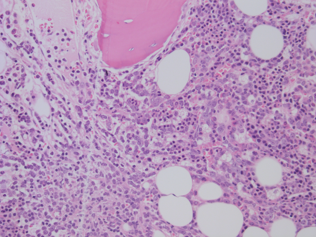 |
| Bone marrow aspirate diagnostic 5.2012 | 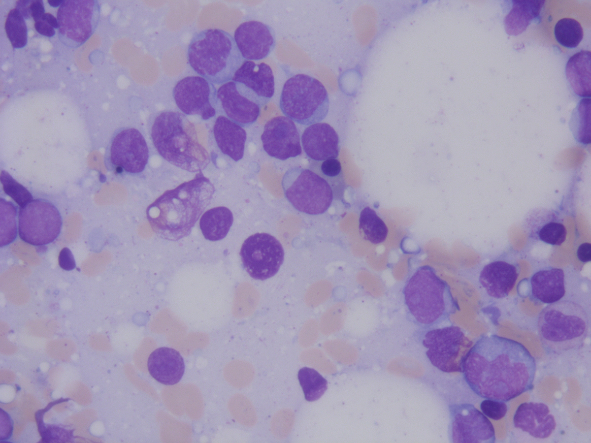 |
| Bone marrow biopsy relapse 1.2013 | 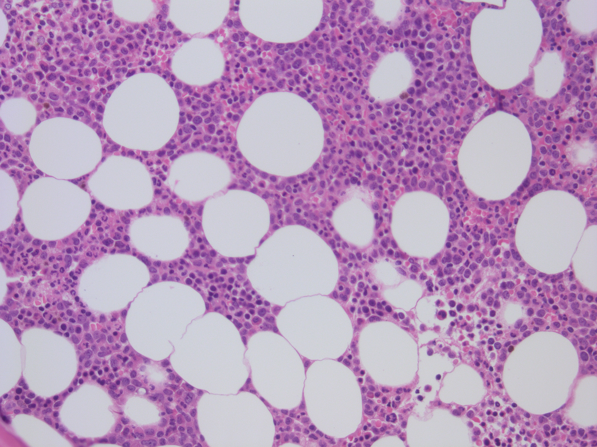 |
| Bone marrow aspirate relapse 1.2013 | 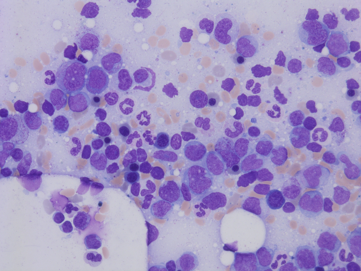 |