Institution: UCLA
Additional authors:Sheeja T. Pullarkat
Session: Therapy-related myeloid neoplasms
HISTORY
This is a 48-year-old Asian female with a past medical history of stage 1 leiomyosarcoma of uterus diagnosed in 2006, status post salpingo-oophorectomy and adjuvant chemotherapy with Taxol (paclitaxel) for 12 months.
In May 2012, patient started to feel fatigued and shortness of breath. Other symptoms include joint stiffness, lightheadedness, and tachycardia. Her symptoms worsened, and she was admitted on June 2012. At the time of admission, she had the following blood studies: white blood cell count was 14.4, hemoglobin was 4.8, hematocrit of 13.1, and platelets were 10,000. A bone marrow biopsy (06/08/12) was performed and patient was diagnosed with AML with t(8;21) (q22;q22); RUNX1-RUNX1T1. Patient received chemotherapy with ARA-C and Idarubicin and was in remission 6 months after therapy. However, recent bone marrow biopsy performed in January 29th of this year showed relapseDETAILS
PERIPHERAL BLOOD SMEAR
The peripheral smear shows normochromic, normocytic erythrocytes. Polychromatic red cells are not easily detectable. The platelet count is markedly decreased. The white cell count is increased with a differential of 64 blasts, 4 promyelocytes, 16 myelocytes, 2 metamyelocytes, 1 band, 1 neutrophil, 1 eosinophil, 0 basophils, 10 lymphocytes, and 1 monocyte. BONE MARROW SMEARS AND TOUCH PREPARATIONSBone marrow smears and touch preparations reveal left shifted myeloid maturation with markedly increased myeloblasts. These blasts are large in size and have large nuclei with irregular nuclear contours, prominent nucleoli and cytoplasm containing salmon colored granules. Other hematopoietic elements are markedly reduced. Megakaryocytes are reduced. Scattered lymphocytes and plasma cells are present. There is no evidence of eosinophilia or basophilia. Iron stores are present per the iron stain.BONE MARROW BIOPSY AND CLOT SECTIONSThe decalcified bone marrow biopsy sections and the aspirate clot sections show a hypercellular marrow with an approximate cellularity of 80-90%. The marrow shows left shifted myeloid maturation with markedly increased myeloblasts. Mature myeloid cells are inconspicuous. Other hematopoietic elements are markedly reduced. Iron stores are increased per the iron stain.IMMUNOHISTOCHEMISTRY AND FLOW CYTOMETRY
The blast gated population comprises 32% of the total cells and contain excess and abnormal blasts (30% of the total cells) that express CD34, CD117(partial), CD13, CD15 (partial), CD19 (partial), CD38, CD71a (partial), and HLA-DR.
CYTOGENETIC FINDINGS
45, X, -X, t(8:21)(q22;q22)
MOLECULAR FINDINGS
C-KIT mutation analysis is pending.
INTERESTING FEATURES
The morphologic features with abundant azurophilic granules ( salmon colored granules) and immunophenotype positive CD34, CD13, and CD19 expression in this case is classic for a t(8:21)(q22:q22) AML. Also, a loss of a sex chromosome is a common additional chromosomal abnormality. It is interesting to note how quickly patient’s disease relapsed after chemotherapy since AML with t(8:21)(q22:q22) is usually associated with a good response to therapy and high complete remission rate. It will be interesting to correlate analysis of KITmutation result since a positive mutation confers poorer prognosis. Other factors to consider is her previous history of therapy especially with her relapse post chemotherapy. Paclitaxel (Taxol), which is an antitubulin agent, is implicated in therapy-related hematologic neoplasms. However, balance translocations such as t(8:21) like this case are most commonly associated with use of topoisomerase II inhibitors due to its mechanism of action.
PROPOSED DIAGNOSIS
Acute myeloid leukemia with t(8;21)(q22;q22); RUNX1-RUNX1T1
CONSENSUS DIAGNOSIS
Therapy-related myeloid neoplasm, acute myeloid leukemia with t(8;21)(q22;q22); RUNX1-RUNX1T1
| Bone marrow biopsy core at low power | 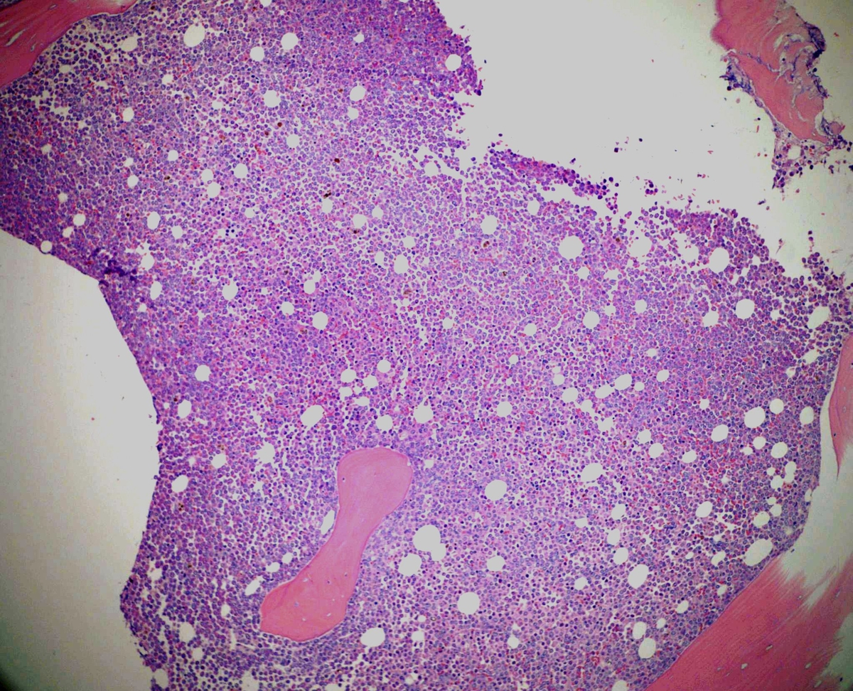 |
| Blasts with abundant azurophilic granules | 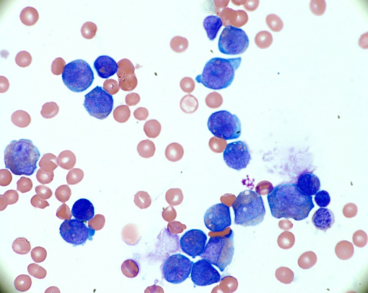 |
| Blasts with abundant azurophilic granules | 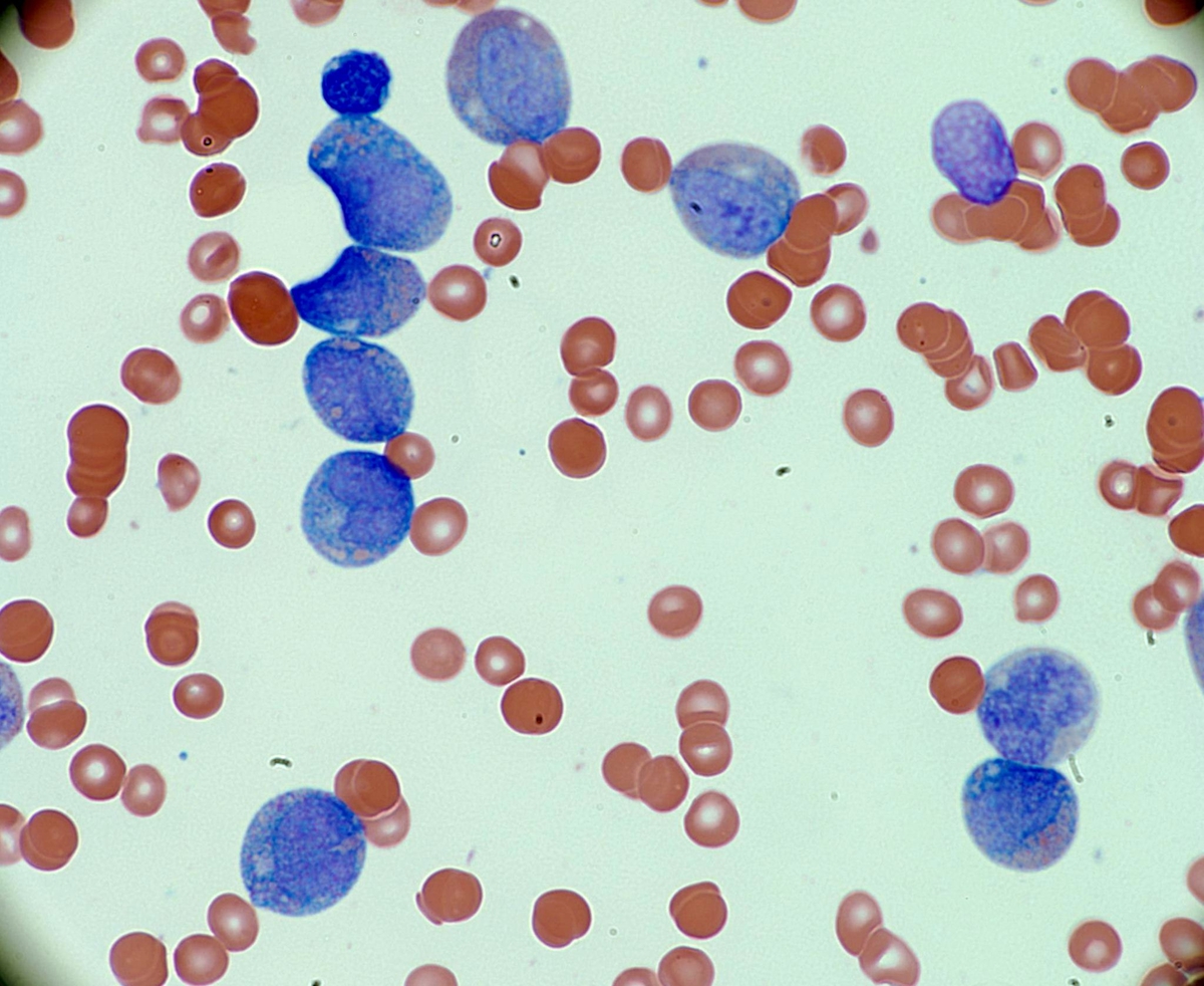 |
| Circulating blasts, peripheral smear | 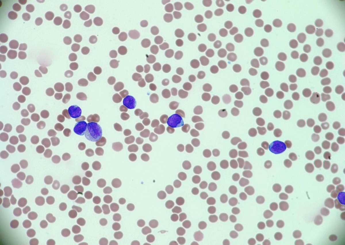 |
| Flow Cytometry | 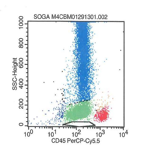 |
| Flow cytometry shows excess blasts | 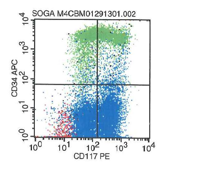 |
| Aberrant CD19 expression |  |
| CD13 expression | 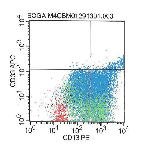 |