Institution: Massachusetts General Hospital
Additional authors:Salil Garg, Michelle E. DeLelys, Howard J. Weinstein, Robert P. Hasserjian
Session: Acute leukemias of ambiguous lineage
HISTORY
The patient is an 11-year-old girl with fevers, night sweats and fatigue. Past medical history significant for asthma and recent diagnosis of goiter and hypothyroidism, treated with levothyroxine. Found to have pancytopenia on routine CBC analysis: WBC 1,400/ul (manual differential: 8% polys, 76% lymphs, 12% atypical lymphs, 4% eos), Hgb 10.4 g/dl, HCT 29.0%, MCV 98 fl, PLT 66,000/ul. Reticulocyte count: 1.3%. Normal chemistries, calcium, ferritin, B12, folate and LDH.
DETAILS
B-plus fixed and decalcified bone marrow biopsy from right iliac crest, and air-dried Wright-Giemsa stained bone marrow aspirate and peripheral blood smears. Bone marrow core biopsy: The marrow cellularity is variable (5-90%), overall approximately 50%. Among maturing hemopoietic elements, there is a relative increase in erythroid elements with left-shift and decreased megakaryocytes with overall normal morphology. Primitive cells with irregular and folded nuclei, consistent with blasts, occur in large clusters and comprise about 20% of the cellularity. Bone marrow aspirate smear: Differential (based on 400 cell count): 15% neutrophils and precursors, 2% promyelocytes, 30% erythroid precursors, 24% lymphocytes, 2% monocytes, 1% eosinophils, 1% basophils, 22% blasts and 3% plasma cells. Blasts are medium-sized to large cells with scant to moderately abundant cytoplasm, irregular or clefted nuclei and prominent nucleoli. No Auer rods are seen. Mild erythroid dysplasia is seen in maturing hemopoietic elements. Scattered hemophagocytic cells are also seen. Peripheral blood smear: Occasional circulating blasts identified.
IMMUNOHISTOCHEMISTRY AND FLOW CYTOMETRY
Bone marrow core biopsy immunohistochemistry: Blasts are positive for CD34, with a subset showing cytoplasmic CD3 and CD117 positivity, while another subset shows staining for Pax5 and TdT. Lysozyme and myeloperoxidase (MPO) highlight scattered maturing myeloid precursors, but are negative on the blasts, as are CD20 and CD56. Bone marrow aspirate smear: Blasts are negative for MPO and non-specific esterase by enzyme cytochemistry. Bone marrow aspirate flow cytometry (gating on lymphocyte and blast populations based on light scatter and CD45 expression): Two immunophenotypically distinct blast populations, each comprising about 10% of all cells. Population #1: CD45dim+, CD19+, CD20-/dim+, CD10+, TdT+, CD34dim+, CD117-, MPO-, CD13-, CD33-, CD38+ (consistent with B lymphoblasts). Population #2: cytoplasmic CD3+, surface CD3-, CD7+, CD34+, CD117+, HLA-DR+, CD38+, CD19-, CD20-, CD10-, TdT-, MPO-, CD13+, CD33- (consistent with T lymphoblasts). Both populations were negative for monocytic markers (CD14, CD11c and CD64).
CYTOGENETIC FINDINGS
Karyotype: 47,XX,+11[19]/46,XX[1].
Interphase FISH shows no evidence of monosomy 7 or for BCR-ABL1 or ETV6-RUNX1 (TEL-AML1) rearrangements. FISH for MLL confirms trisomy 11, but shows no rearrangement involving MLL.
MOLECULAR FINDINGS
Not performed.
INTERESTING FEATURES
This is an unusual example of a mixed phenotype acute leukemia (MPAL) with two immunophenotypically distinct blast populations exhibiting B lymphoid (CD19, CD10, Pax5) and T lymphoid (cytoplasmic CD3) phenotype. B/T MPAL is estimated to comprise <5% of MPAL in one large series (Matutes E et al. Blood 2011;117:3163). The T lymphoblast population in this case expresses both stem cell (CD117 and CD34) and myeloid (CD13) antigens, consistent with an early T-cell precursor leukemia phenotype (Coustan-Smith E et al. Lancet Oncol 2009;10:147). The absence of MPO or any other evidence of myeloid differentiation on this population is against a diagnosis of B/myeloid MPAL. Interesting morphological aspects of this case include the patchy nature of the leukemic infiltrate, variable marrow cellularity with some areas of relatively preserved hemopoiesis showing mild erythroid dysplasia, and presence of hemophagocytosis. Both the erythroid dysplasia and hemophagocytosis may represent a reaction to the neoplastic infiltrate. Alternatively, the erythroid dysplasia may reflect an underlying early stem cell defect manifesting in a non-lymphoid marrow lineage.
PROPOSED DIAGNOSIS
Mixed phenotype acute lymphoblastic leukemia (B/T).
CONSENSUS GROUP: ADDITIONAL INFORMATION/STUDIES
Additional immunostains performed by the conference consensus group:
CD3: Positive in blasts
CD79a: Positive in blasts
CONSENSUS DIAGNOSIS
Mixed phenotype acute leukemia (B/T)
| Bone marrow core biopsy showing large cluster of blasts with admixed hemopoietic elements (H&E, 40x). |  |
| High power view of aggregate of primitive-appearing blasts in core biopsy (Giemsa, 100x). | 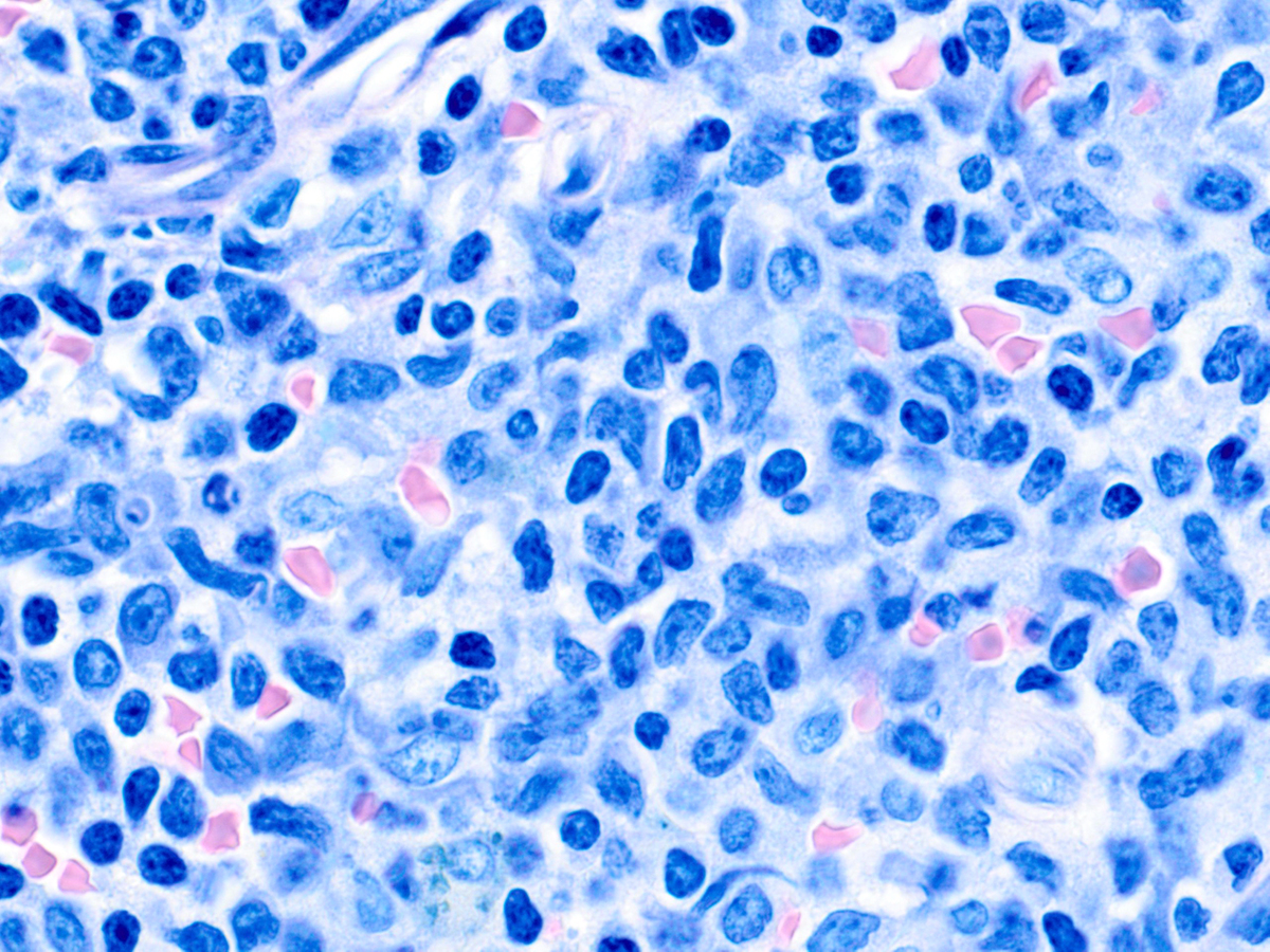 |
| Area of relatively preserved hemopoiesis in core biopsy with hemophagocytic cell (arrow, H&E, 40x). |  |
| CD34 immunohistochemical stain of core biopsy highlighting blasts (20x). | 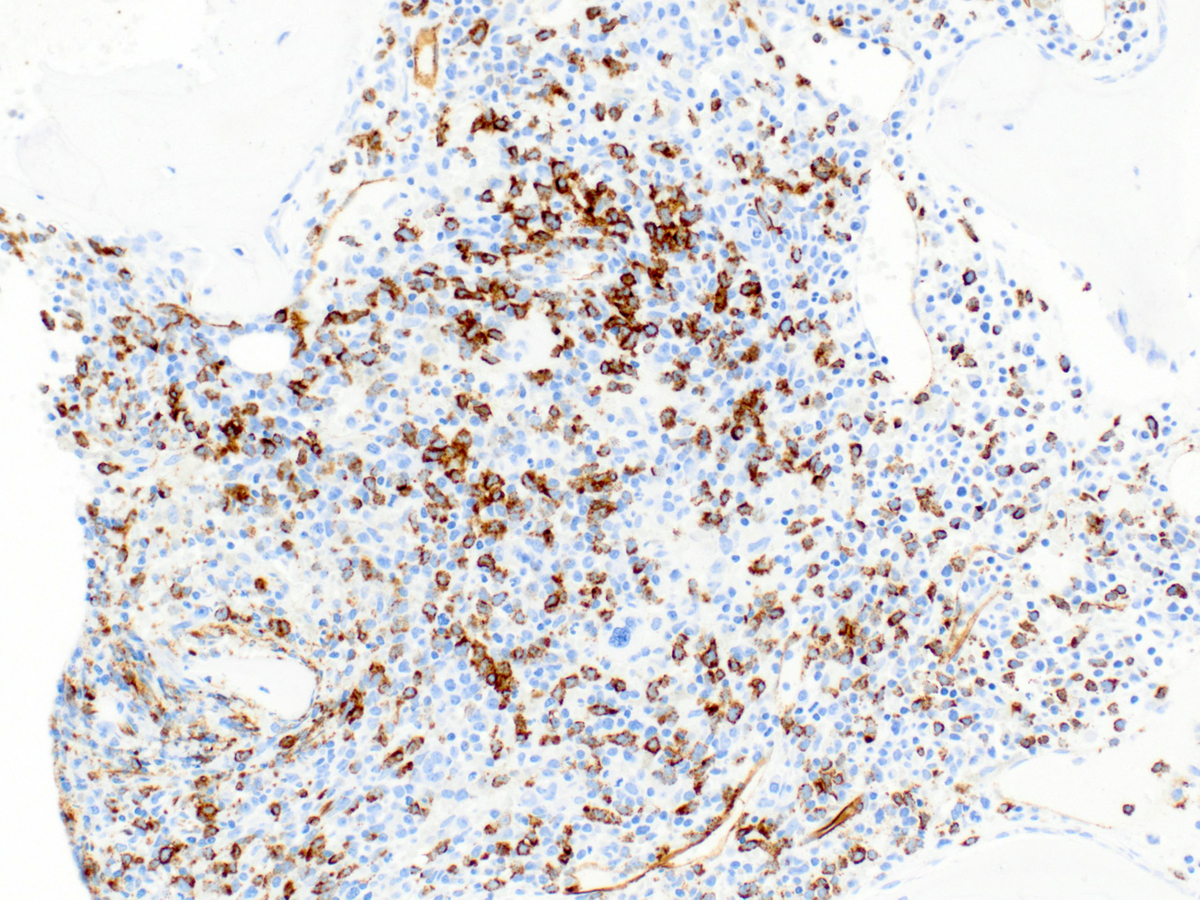 |
| CD3 staining of subset of blasts. Weak staining of some of the larger cells is consistent with cytoplasmic staining (40x). | 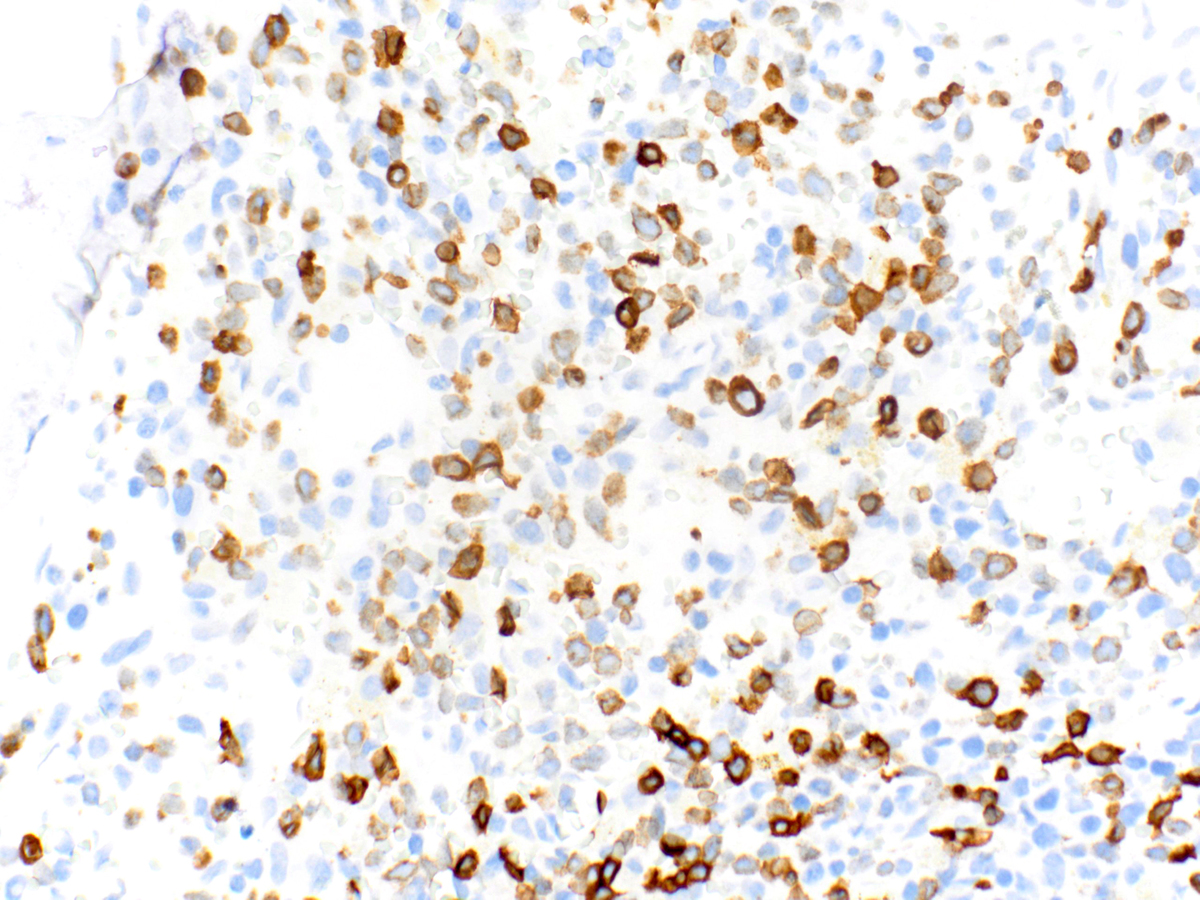 |
| CD117 staining of subset of blasts (40x). | 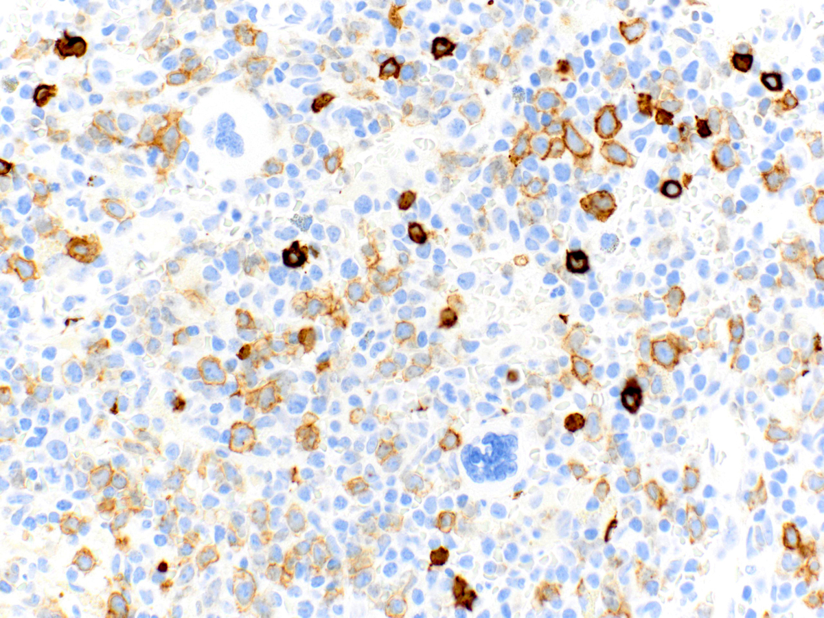 |
| Pax5 staining of subset of blasts (40x). |  |
| TdT staining of subset of blasts (40x). | 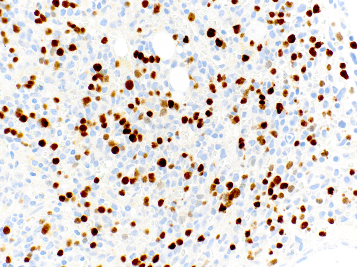 |
| Blasts are negative for MPO by immunohistochemistry (40x). | 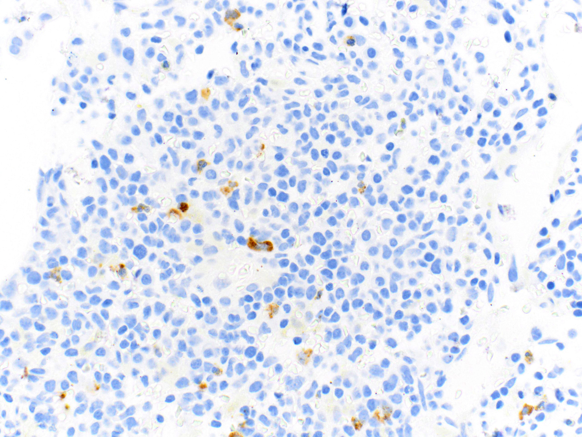 |
| Bone marrow aspirate smear showing morphology of blasts in comparison to mature lymphocytes (Wright-Giemsa, 100x). | 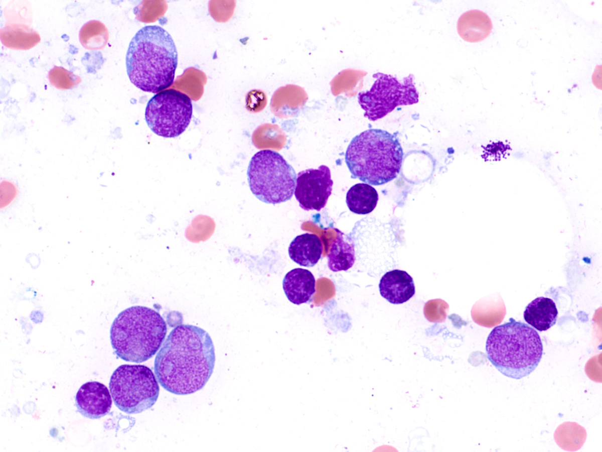 |
| Bone marrow aspirate smear showing hemophagocytic cells (Wright-Giemsa, 100x). |  |
| Bone marrow aspirate smear showing mild nuclear-cytoplasmic asynchrony of maturing erythroid precursors (arrow, Wright-Giemsa, 100x). |  |
| 8-color flow cytometry showing two distinct CD45dim+ blast populations. One population (gold) expresses CD19, CD10, TdT, CD34 (dim), and CD38. The second population (green) expresses CD34 (bright) and CD38, and is negative for CD19, CD10 and TdT. |  |
| Additional analysis for B-cell antigens shows that the B-lymphoid population (gold) expresses very dim CD20 and is negative for surface kappa and lambda immunoglobulin light chains. Background mature polytypic B cells are present (blue). | 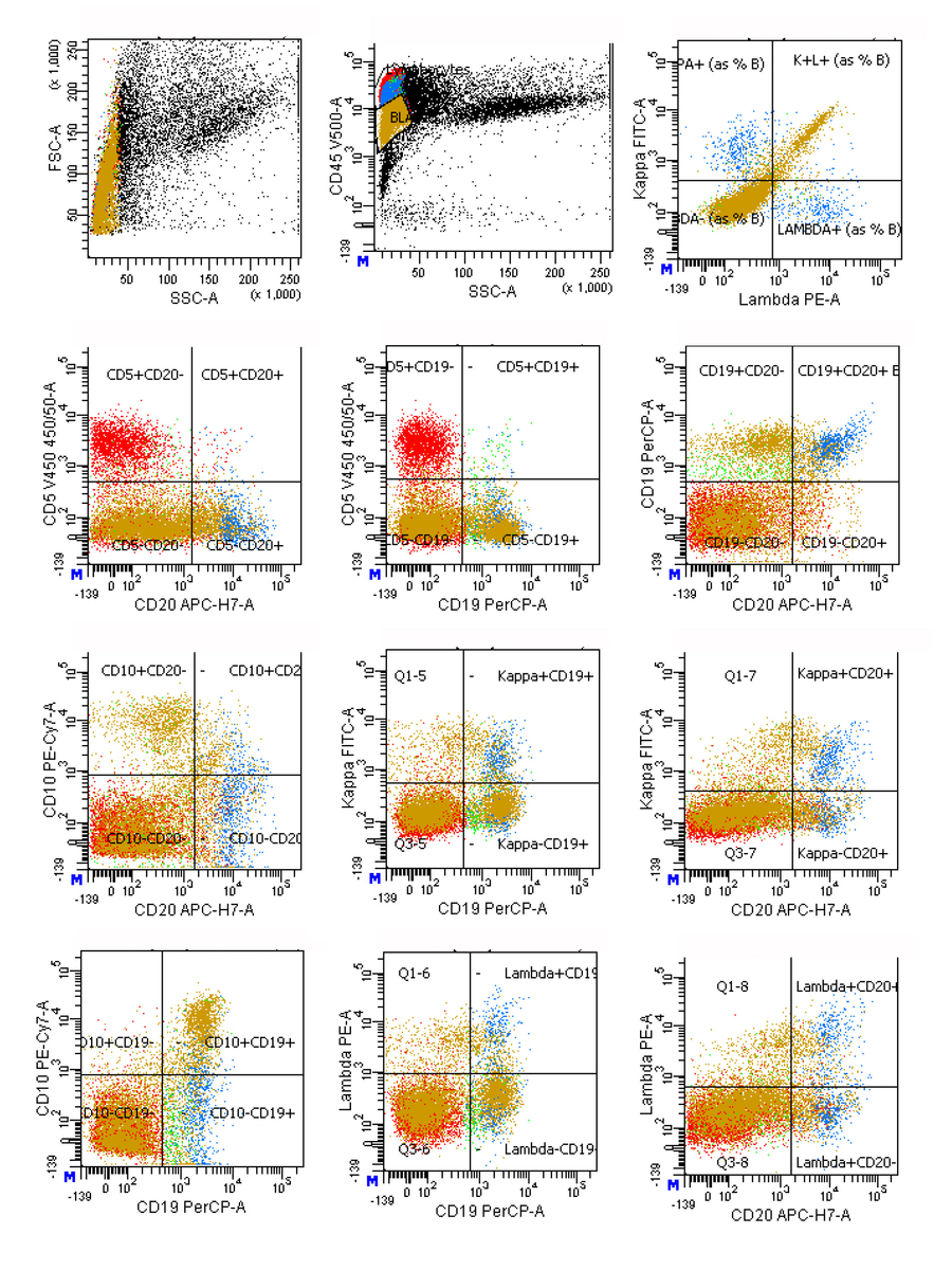 |
| Additional analysis for cytoplasmic CD3 and stem cell antigens shows that the brightest CD34+ cells (green) co-express cytoplasmic CD3 and CD117. | 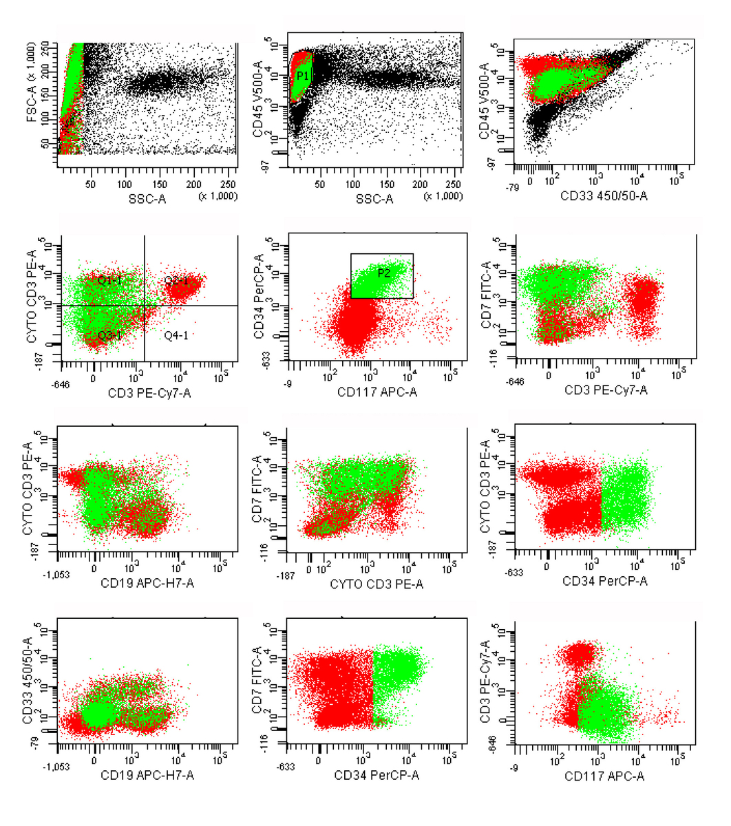 |
| Additional analysis for myeloid and stem cell antigens shows that the CD34+, CD117+ cells (gold) co-express CD13 and HLA-DR. | 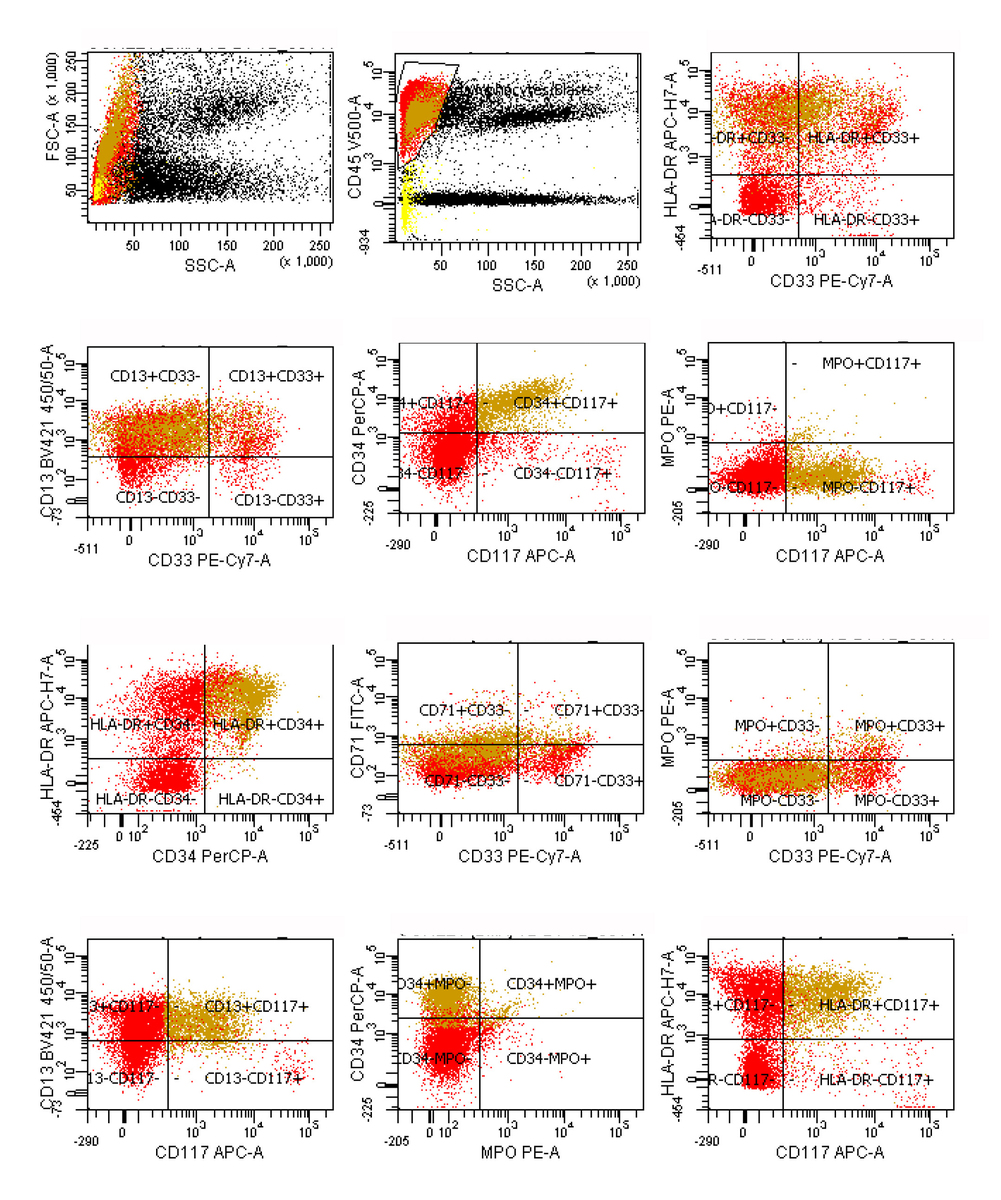 |
| Additional analysis for T-cell antigens shows that a subset of the CD45dim+ blasts (gold) is positive for CD7, and negative for surface CD3, CD4 and CD8. Lower right inset shows light scatter and CD45 analysis (FSC: forward scatter, SSC: side scatter). |  |