Institution: University of New Mexico Health Sciences
Session: B Lymphoblastic Leukemia/Lymphoma
HISTORY
A 31-year-old Native American male with Down syndrome presented with 2 months history of fatigue, cough, and weight loss. Examination of a peripheral blood and bone marrow biopsy with flow cytometric and cytogenetic work-up was diagnostic of a Philadelphia chromosome-positive precursor B- lymphoblastic leukemia. He was initially treated with single agent dasatinib after discussion with family as patient is primarily non-verbal. The patient’s disease relapsed 6 months after initial presentation and was treated with Hyper-CVAD chemotherapy with delayed administration of second cycle of chemotherapy due to persistent pancytopenia. Examination of a cerebrospinal fluid (CSF) specimen revealed extensive CNS involvement. He was treated with intrathecal chemotherapy and all subsequent CSF samples became negative. Review of a peripheral blood smear, prior to administration of the last cycle of Hyper-CVAD revealed no circulating blasts and quantitative PCR on a peripheral blood sample showed no detectable BCR-ABL1 transcripts. Four months after completion of chemotherapy, the patient developed CNS relapse and died in late 2012.
DETAILS
A complete blood count performed at admission revealed WBC count of 104X10^9/L, hemoglobin of 12.7 g/dL, hematocrit of 38.5%, MCV of 95 fL, and a platelet count of 788 X10^9/L. Manual differential count on peripheral blood includes: 21% neutrophils, 4% lymphocytes, 1% monocytes, 1% metamyelocytes, 2% basophils, and 71% blasts.
Wright-stained bone marrow aspirate smears reveal decreased erythropoiesis, decreased neutrophilic myelopoiesis and greater than 60% blasts. Blasts contain intermediate to large oval to indented nuclei with fine chromatin, frequent distinct nucleoli and moderate amounts of pale blue cytoplasm without appreciable granules. The H&E-stained clot and trephine biopsy sections show a hypercellular bone marrow with sheets of blasts estimated to account for greater than 80% of marrow cellularity. Megakaryocytes are increased in number with occasional loose clustering and include small hypolobated forms as well as subset with separate nuclei. Residual erythropoiesis and neutrophilic myelopoiesis is present but significantly decreased. The peripheral blood and bone marrow biopsy at the time of disease relapse revealed morphologic findings similar to those noted at the time of initial admission except for decreased numbers of megakaryocytes.IMMUNOHISTOCHEMISTRY AND FLOW CYTOMETRY
Flow cytometric analysis performed on peripheral blood revealed an increased population of B lymphoblasts (55% of total events) expressing CD19, subset CD10, CD34, CD13, dim CD33, CD79A, HLA-DR and TdT and lacking megakaryocytic antigens.
The blasts were also negative for myeloperoxidase as assessed by a cytochemical stain.CYTOGENETIC FINDINGS
Cytogenetic analysis performed at the time of admission on a peripheral blood sample revealed: 47,XY,t(9;22)(q34;q11.2),+21[20].
MOLECULAR FINDINGS
Quantitative real time PCR analysis performed on a peripheral blood sample, 2 months after treatment for relapse disease, identified a b2-b3/a2 type BCR-ABL1 transcripts with a BCR-ABL1/ABL1 ratio of 0.0075. A subsequent follow up quantitative PCR (4 months post therapy after disease relapse) revealed no detectable BCR-ABL1 transcripts.
INTERESTING FEATURES
Children with Down syndrome (DS) show an increased incidence of transient myeloproliferative disease, acute myeloid leukemia of megakaryocytic type, and acute lymphoblastic leukemia (ALL). The most common cytogenetic abnormality reported in DS-associated ALL includes additional copy of chromosome X. Additional cytogenetic abnormalities that may be seen more frequently in DS-ALL include t(8;14) and del(9p). Most recently, it has been shown that about 20% of DS-ALL demonstrates JAK2 mutations that are different from those commonly observed in myeloproliferative neoplasm. However, the occurrence of ALL with recurrent 9;22 translocation in adult patients with DS is rare.
This case is a rare example of an adult onset DS-associated ALL with a recurrent BCR-ABL1 fusion gene that is uncommon in the setting of DS-associated hematologic ma;ignancies.PROPOSED DIAGNOSIS
Down syndrome-associated B lymphoblastic leukemia with recurrent t(9;22)(q34;q11.2)/BCR-ABL1
CONSENSUS DIAGNOSIS
B-acute lymphoblastic leukemia with t(9;22)(q34;q11.2); BCR-ABL1 in a patient with Down syndrome
| Wright-stained peripheral blood smear demonstrates circulating blasts and thrombocytosis | 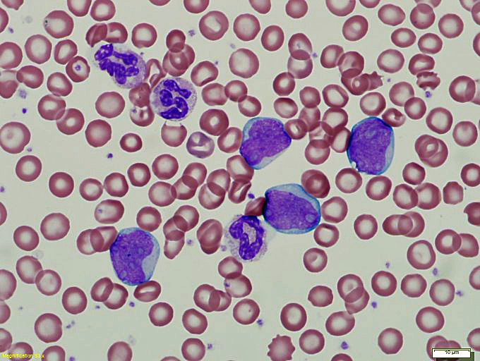 |
| Wright-stained bone marrow aspirate smear demonstrates increased blasts and residual maturing hematopoietic cells | 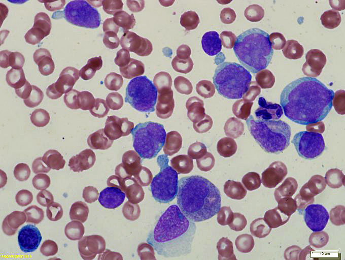 |
| H&E-stained bone marrow trephine biopsy shows a hypercellular marrow with numerous blasts and increased atypical appearing megakaryocytes | 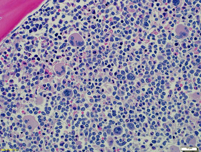 |
| Representative histogram of flow cytometry showing blasts with expression of TdT and dim CD79A | 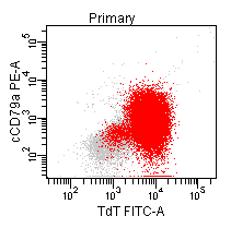 |
| Post treatment quantitative real time PCR at the time of disease relapse detects b2-b3 type BCR-ABL1 transcripts (green-colored amplification curves) with a BCR-ABL1/ABL1 ratio of 0.0075 | 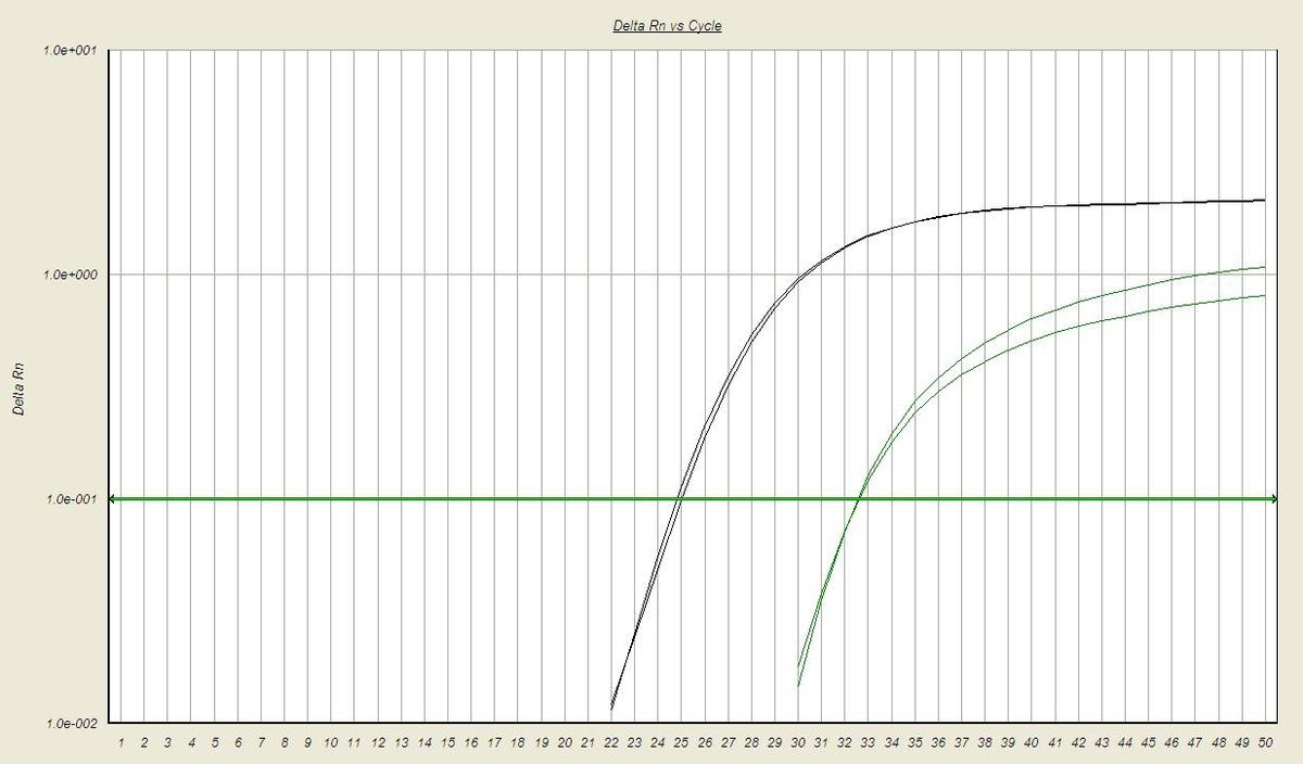 |