Institution: University of Tuebingen, Germany
Additional authors:Falko Fend
Session: Extramedullary manifestations of myeloid neoplasms
HISTORY
A 56 year-old man presented in September 2011 with bilateral pain in the elbows. Physical examination revealed subcutaneous lesions involving rather symmetrically both elbows, left supraescapular region and right arm. An MRT revealed intramuscular haematoma with rupture of the deltoid muscle. The blood picture at the time was normal. In August 2012 the patient sought again medical attention because of persistence of his symptoms. A new MRT revealed a 12.5 cm subcutaneous tumor on the right arm with bone destruction and a 7.5 cm tumor on the left arm involving only the dermis and subcutaneous tissue. Biopsies of both elbow lesions were performed. The lesions were classified as indeterminate dendritic cell tumor. Because of the diagnosis a conservative approach was decided.
In October 2012 the patient presented again with general malaise, weight loss (14 kg), and fever. A peripheral blood picture showed Hb: 11.4 g/dl, platelets: 16K/uL, leukocytes: 53K/uL with 48% monocytoid cells. A bone marrow biopsy was performed. The patient started treatment with ICE+ATRA (AML-SG 07/04 study). The patient showed initially a good response with partial remission; however, he developed sepsis and liver failure. A new bone marrow biopsy in December demonstrated a 30% infiltration. The patient died in January 2013.DETAILS
Subcutaneous lesions (Formalin fixed and paraffin embedded): There is a diffuse infiltration of the dermis and subcutaneous tissue with histiocytic-appearing cells. The cells are rather large with blastic open chromatin, irregular nuclei and sometimes grooves and clefts. The nucleoli are eosinophilic rather inconspicuous. The cytoplasm is abundant and eosinophilic. There is clear spindling of the cells, sometimes with whorls. Mitosis are abundant, as well as apoptosis.
Bone marrow biopsy (Formalin fixed and paraffin embedded): The bone marrow showed diffuse infiltration with large atypical cells with similar morphology as in the skin lesions. Chloroacetate esterase was negative in the tumor cells. The silver stain showed a fibrosis grade 1 up to 2 in some areas. Due to the fibrosis the BM aspirate was a "dry-tap"IMMUNOHISTOCHEMISTRY AND FLOW CYTOMETRY
Subcutaneous lesion:
S100+, CD1a+/-, CD56+, CD4+, CD163+, CD45+, lysozyme +/-, CD68+/-, CD35-, CD23-, CD21-, Langerin-, BRAF-Bone marrow: CD34-, CD117-, MPO-, Lysozyme++, S100+/-, CD56+, CD1a+/-, CD33+, CD14+/-, CD45+, CD163+, CD43+, CD4+Flow cytometry of peripheral blood cells showed that 60% of the cells were CD34-, CD117-, CD15 low, CD14 +, CD33+, MPO-, HLADR+, sCD11c+, sCD11b+, TDT-, and aberrant expression of CD1a+ and CD56+ similar to the skin tumors. Around 20% of tumor cells coexpressed CD1a and CD14. B- and T- cell markers were negative.CYTOGENETIC FINDINGS
Not performed
MOLECULAR FINDINGS
Subcutaneous tissue: NPM1 type A mutation was demonstrated in the skin. BRAF mutation negative.
Peripheral blood: NPM1 type A mutation was demonstrated. MLLT3/MLL t(9;11) negative; CEBPA Wildtype;PML/RARA t(15;17) negative; RUNX1/RUNX1 T1 t(8;21) negative; CBFB/MYH11 inv(16) negative, FLT3-ITD negative; FLT3-TKD WildtypeINTERESTING FEATURES
This is a myeloid leukemia with mutated NPM1. The interesting feature of this case is the aberrant phenotype with S100, CD1a and CD56. Although AML with NPM1 mutation usually shows monocytic features and extramedullar involvement, especially of the skin, the presentation as an indeterminate dendritic cell tumor preceding the development of acute leukemia for one year is rather unique. Also of interest is the demonstration of the NPM1 mutation in the skin tumors supporting the clonal relationship of both tumors. Of note is the change in phenotype from the skin lesion to the BM infiltrating cells. In the skin lesion the tumor cells were strongly S100+ and weak and partially lysozyme +, whereas in the BM was the opposite. In both lesions there was only a partial positivity for CD1a that with flow cytometry was estimated to be 20% of the neoplastic population. In the literature there is one case reported similar to this one (Br J of Dermatology 2007;156:1357)
PROPOSED DIAGNOSIS
Acute myeloid leukemia with mutated NPM1 with aberrant phenotype presenting initially as an indeterminate dendritic cell tumor.
CONSENSUS GROUP: ADDITIONAL INFORMATION/STUDIES
Additional immunostains performed by consensus group:
NPM1: Cytoplasmic positivity
CONSENSUS DIAGNOSIS
Myeloid sarcoma (monocytic/dendritic differentiation) with mutated NPM1 involving soft tissue of elbow; subsequent acute myeloid leukemia 1 year later
| Bone marrow morphology |  |
| Bone marrow immunostains |  |
| Flow cytometric analysis of peripheral blood | 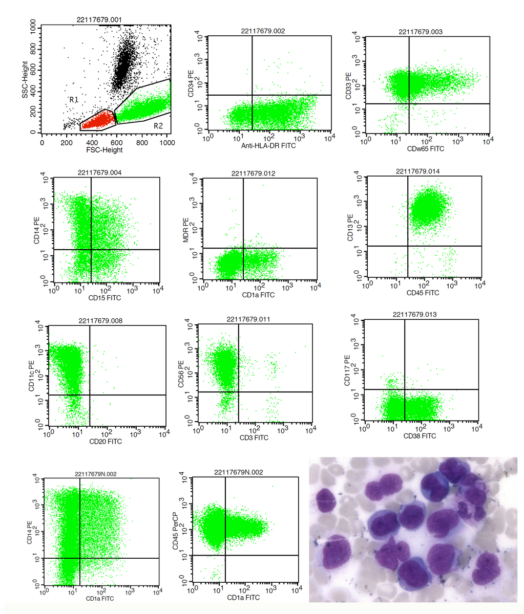 |
| Morphology of the indeterminate dendritic tumor | 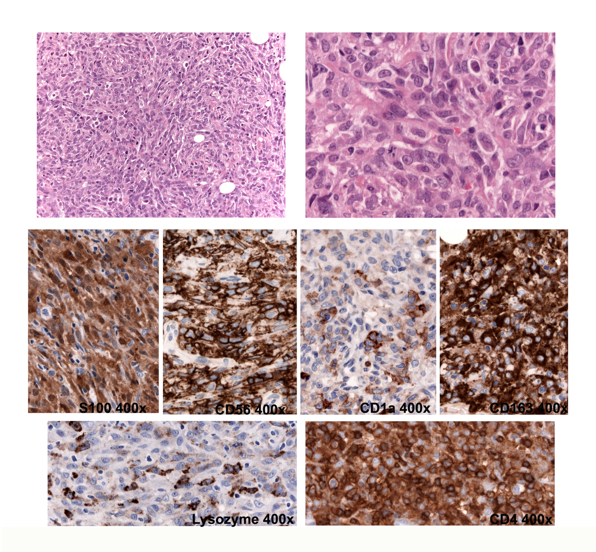 |