Institution: Roswell Park Cancer Institute
Additional authors:Asangi Kumarapeli, MD (1), Sheila Sait, PhD (2), Amy M Sands, MD (1) 1. University of Buffalo 2. Roswell Park Cancer Institute
Session: Acute leukemias of ambiguous lineage
HISTORY
A 90-year-old Caucasian woman lived independently before presenting to her physician for intractable fatigue for past two months. Past medical history included hypertension, dyslipidemia, hypothyroidism, and osteoporosis. Physical examination was unremarkable. CBC at presentation: WBC 1.3 x 10⁹/L, HGB 11.5g/dL, HCT 33.8 %, platelets 109 x 109/L, ANC 0.28 x 10⁹/L. A bone marrow biopsy was performed but no aspirate could be obtained. The patient was admitted at a second hospital where a bone marrow aspiration was performed, material submitted for ancillary studies and both samples concurrently reviewed.
DETAILS
The zinc formalin–fixed, paraffin-embedded fragmented biopsy showed diffuse marrow space replacement by blasts. Peripheral blood film review demonstrated pancytopenia with severe neutropenia and 24% circulating blasts with large round-ovoid nuclei, many with nuclear angulations and variably condensed nuclear chromatin, prominent central nucleoli, basophilic cytoplasm with numerous vacuoles and occasional coarse eosinophilic granules. Aspirate smears showed 85% blasts with similar morphology. By cytochemistry (CC), many cytoplasmic vacuoles were PAS-positive while the eosinophilic granules were myeloperoxidase-negative. Myeloperoxidase, alpha-naphthyl butyrate esterase, chloroacetate esterase and dual CAE/ANBE stains were all negative (<3% of blasts positive).
IMMUNOHISTOCHEMISTRY AND FLOW CYTOMETRY
Flow cytometry (FCM) in one laboratory detected CD14dim, CD15, CD19dim, CD45dim, CD56dim, CD71dim, and CD34 expression in the blast population, lacking CD13, CD20 or CD33. In the second laboratory the blasts phenotype by FCM was: CD15het, CD19, sCD22 dim, CD34, CD32, CD71het, CD45dim and HLA-DR while negative for immunoglobulin light chains, CD10, CD13, CD14, CD33, CD38 and CD117. cCD79a, but not cMPO or cCD3 expression, was detected by FCM. By immunohistochemistry (IHC) the blasts were strongly and diffusely positive for PAX5 and TdT, focally positive for CD79a and CD22, and negative for lysozyme, CD10, CD20, CD42b, CD61 and hemoglobin. MPO IHC was strongly, diffusely positive in one lab (Dako polyclonal rabbit, predilute) and faintly positive (originally interpreted as background staining) in the second lab (Dako polyclonal rabbit; 1:20,000), both using no pretreatment and same incubation time but different detection systems (Dako; EnVision + Dual Link System-HRP and Rabbit EnVision, respectively). The same sample was tested in a third lab where a positive IHC MPO reaction with intermediate staining intensity and extent between the 2 previous results was detected using a polyclonal Ab from another vendor (Ventana, rabbit polyclonal).
CYTOGENETIC FINDINGS
Conventional cytogenetics:
35,XX,-3,-4,-5,-7,-8,-9,-13,-15,-16,-17,-20[9]46,XX,?t(5;6)(q23;q24)[1]46,XX[10]Fluorescence in situ hybridization:nuc ish(ABL1 x 1,BCR x 2)[154/200]/(ABL1,BCR) x 1[12/200]nuc ish(MLL x 2)[190/200]/(MLL x 4)[10/200]MOLECULAR FINDINGS
Negative for NPM1, FLT3 mutation. B cell clonality testing (IGH FR1-3, IGK PCR) performed in a subsequent sample with persistent disease was negative.
INTERESTING FEATURES
Interesting Feature(s) of Submitted Case:
A. Aberrant versus genuine antigen expression of essential lineage-specific markers (MPO, CD79a, PAX5)B. Conflicting results of critical markers (MPO, CD79a) tested with different modalities (CC, FCM, IHC)C. Interlaboratory variability even when using the same reagentThis leukemia is difficult to classify since CD19bright PAX5+ sCD22+/- cCD79a+/- profile would support B-lineage (B-ALL) whereas CD15 and IHC MPO+ expression support myeloid lineage, fulfilling thus the criteria for the current WHO classification of mixed phenotype acute leukemia (MPAL), B-myeloid (1).Of note, if only FCM immunophenotyping had been used, this case would have been classified as B-ALL in both EGIL (2) and WHO 2008 systems. Expression of IHC MPO (polyclonal antibody) varied in extent and intensity among 3 labs. Despite its absence by FCM (monoclonal antibody) or CC–both more specific methods–, the IHC MPO expression may be sufficient for MPAL diagnosis in the current WHO proposal. Aberrant immunohistochemical expression of myeloperoxidase was reported in adult B-ALL by one group (3) but discordance between various methods can be seen in up to 21% of AML (4). cCD79a was convincingly positive by FCM (clone HM47) but less clear by IHC (clone HM57). PAX5 is the most sensitive and specific B-cell marker, usually diffusely and strongly expressed in B-ALL and only rarely, focally and weakly expressed in AML except RUNX1-RUNXT1 cases. However, PAX5 is not included in the current WHO MPAL schema. While widely used in paraffin-embedded tissue, PAX5 expression can also be assessed by FCM using a commercially available antibody with excellent sensitivity and specificity (5).This case provides a good illustration of inter-laboratory variability even when using the same antibody with adequate controls but different dilutions and detection systems, and raises the issue of polyclonal antibodies specificity for lineage assignment in the absence of or with only minimal expression of other lineage-supporting markers. MPO or CD3 expression is sufficient to determine myeloid or T lineage, respectively, in the new MPAL proposal. Validation, verification and rigorous QA are very important; however, inter-methodology discordant results or inter-laboratory variability, possibly due to pre-, intra- or post-analytical differences (e.g., fixative, dilution, detection system) may play a significant role. Standardization of critical assays is desirable but currently not widely implemented (6). In select cases of acute leukemias with ambiguous differentiation, employing multiple testing modalities for one marker and integrating all information, including molecular-genetic findings, may help resolve these discrepancies.1. Borowitz MJ et al in Swerdlow et al (eds). WHO classification of haematopoietic and lymphoid tumors. IARC 2008 pp 150-155.2. Bene MC et all. Leukemia 1995; 9: 1783- 1786.3. Arber DA et al. AJCP 2001;116: 25-334. Saravanan L, Juneja S. Int J Lab Hematol 2010; 32:e132-6.5. Merzianu M et al. Mod Path 2013;26 S2:347-348A6. Goldstein NS et al. Appl Immunohistochem Mol Morphol. 2007;15:124-33PROPOSED DIAGNOSIS
B-ALL with aberrant expression of myeloperoxidase by immunohistochemistry
CONSENSUS GROUP: ADDITIONAL INFORMATION/STUDIES
Additional immunostaining performed by the conference consensus group:
MPO: Strongly positive in blasts
CONSENSUS DIAGNOSIS
Mixed phenotype acute leukemia, B/myeloid (with hyperdiploidy)
| Blood with pancytopenia and blasts |  |
| Blast with cytoplasmic vacuoles and condensed chromatin |  |
| Marrow aspirate blasts WG |  |
| Marrow aspirate blasts PAS | 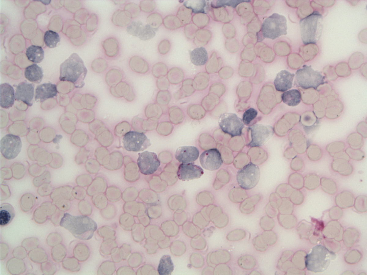 |
| Marrow aspirate blasts MPO |  |
| Marrow aspirate blasts-detail: cytoplasmic granules are MPO negative |  |
| Blast with cytoplasmic vacuoles and eosinophilic granules and condensed chromatin WG |  |
| CAE/ANBE dual staining-blasts are negative |  |
| HE 20x |  |
| HE 60x |  |
| PAX5 |  |
| TdT |  |
| CD22 |  |
| CD79a Lab1 |  |
| CD79a Lab2 | 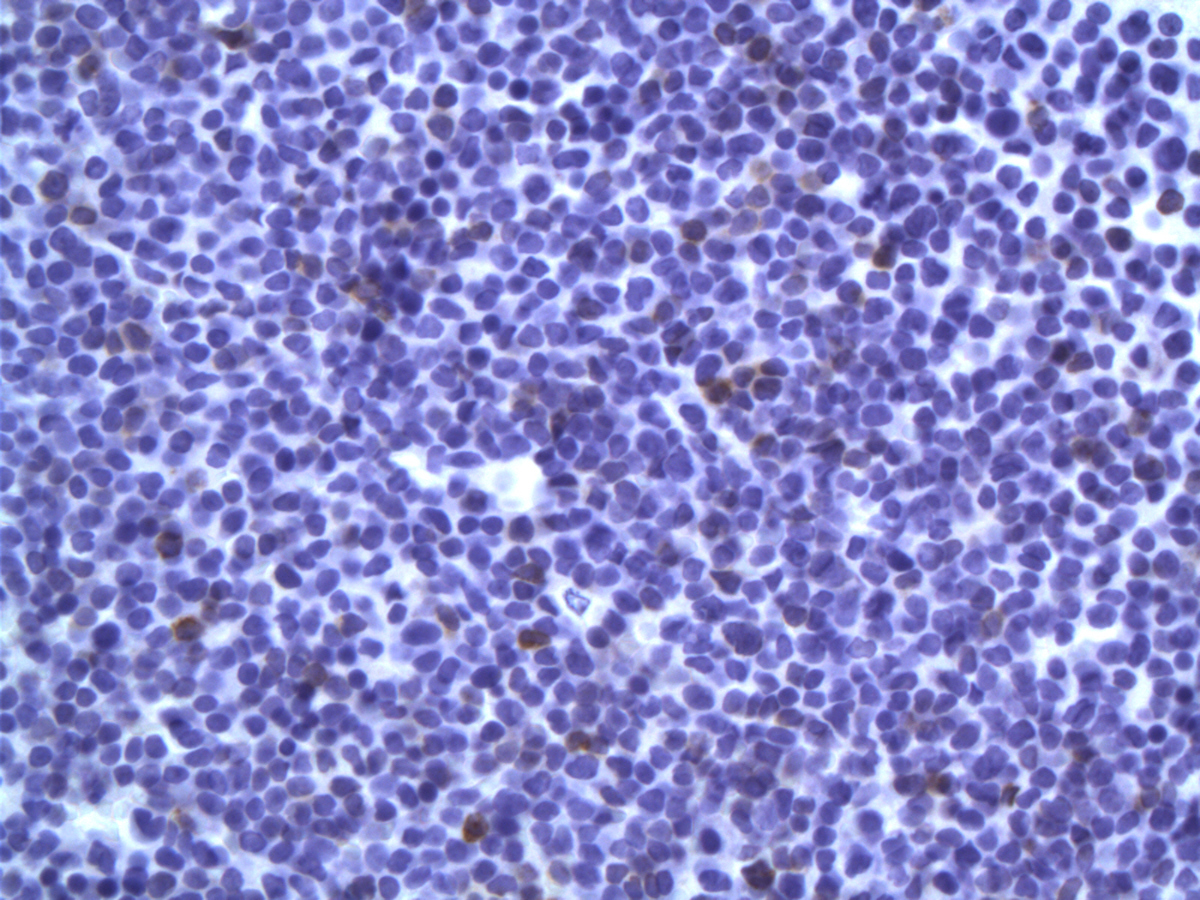 |
| MPO IHC in 3 different laboratories |  |
| Lysozyme |  |
| Hemoglobin |  |
| CD61 |  |
| CD68 | 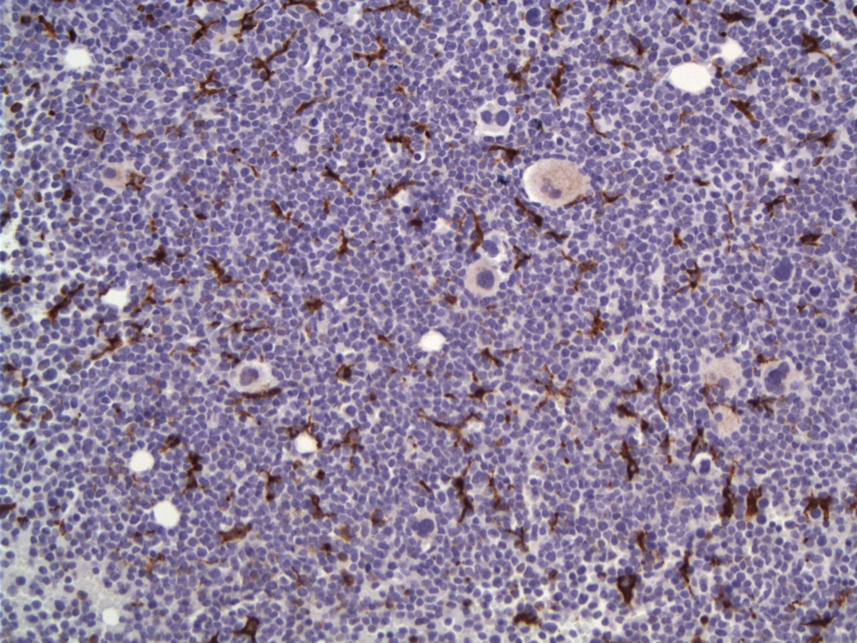 |
| Flow cytometry marrow aspirate in Lab2 | 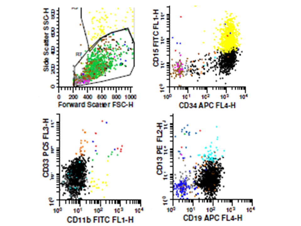 |
| Flow cytometry marrow aspirate in Lab2 |  |
| Flow cytometry marrow aspirate in Lab2 | 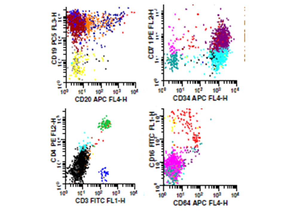 |
| Flow cytometry marrow aspirate in Lab2 |  |