Institution: University of Pittsburgh School of Medicine
Additional authors:Raymond Felgar, M.D., Ph.D.
Session: Erythroleukemia and megakaryoblastic AML and mimics
HISTORY
The patient is a 7 month old, previously healthy female infant who initially presented with fever and irritability, hepatosplenomegaly, anemia, thrombocytopenia, and leukocytosis including 25% circulating blasts. At the time of initial bone marrow biopsy, CBC data revealed the following: Patient Normal Ranges for Laboratory WBC 26.5 x10E+9/L [H] ( 6.0 – 17.5) RBC 2.74 x10E+12/L [L] ( 3.70 – 5.30) Hgb 8.1 g/dl [L] ( 10.5 – 13.5) Hct 23.5 % [L] ( 33.0 – 39.0) MCV 85.7 fL ( 70.0 – 86.0) MCH 29.6 pg [H] ( 27.0 – 33.0) MCHC 34.6 gm/dL ( 31.0 – 35.0) RDW 17.5 % [H] ( 11.5 – 15.0) PLT 20 x10E+9/L [L] ( 150 – 450) POLYS 7.0 % (1.86 ) ( 1.00 – 8.50) BANDS 5.5 % (1.46) [H] ( 0.00 – 1.00) LYMPHS 16.5 % (4.37 ) ( 4.00 –13.50) MONOS 1.0 % (0.26) ( 0.05 – 0.70) EOS 1.5 % (0.40) ( 0.05 – 0.70) BLASTS 66.5% (17.62) META 2.0 % (0.53)
DETAILS
Blood smear review demonstrated leukocytosis with numerous blasts, some of which exhibited prominent cytoplasmic blebbing. (Images 1 and 2). Significant drying artifact precluded adequate morphologic characterization of the other peripheral blood elements. Bone marrow biopsy was very limited in size. (Image 3). Most cells appeared to be intermediate and large sized blasts (88% by counts on the biopsy touch imprint). (Image 4). A reticulin stain demonstrates minimally increased reticulin deposition but no overt fibrosis (Image 5). Limited immunohistologic studies performed on the core biopsy demonstrated CD34 positive staining on the blasts (Image 6).
IMMUNOHISTOCHEMISTRY AND FLOW CYTOMETRY
Flow cytometric evaluation of bone marrow aspirate showed 66-75% of blasts marking as follows: Dim CD45+, CD14-, CD56+, CD34+, CD13/33+, partial CD19+, partial CD10+, CD36-, CD64-, CD117+, partial CD15+, CD33+, partial/weak CD7+, dim CD13+, CD123+, CD41a+, CD61 (partial/subset+), HLA-DR (mostly negative). (image 7)
CYTOGENETIC FINDINGS
Karytotype: 46,XX [20/20 metaphases] (Image 8)
MOLECULAR FINDINGS
PCR negative for FLT3 internal tandem duplication. PCR negative for FLT3 D835 mutation. PCR negative for NPM1 mutation.
INTERESTING FEATURES
This case illustrates morphologic and other findings typical of involvement by AML, M7 type, and would fall within the category of Acute Myeloid Leukemia, not otherwise specified, in the 2008 WHO blue book. M7 AML has a bimodal incidence pattern with the first peak occurring in early childhood (<3 years old) and the second peak in middle-aged adults, and typically carries a poor prognosis. Although AML-M7 in children is much more common in Down syndrome, the presence of a normal female karyotype makes this unlikely, and the patient did not have any other evidence of Down syndrome. The patient underwent 2 cycles of induction chemotherapy followed by allogeneic matched sibling donor progenitor cell transplantation, and patient remains free of disease, 7 months following.
PROPOSED DIAGNOSIS
Acute myeloid leukemia with megakaryoblastic features (FAB AML M7).
Per 2008 WHO: Acute Myeloid Leukemia, NOS.
CONSENSUS DIAGNOSIS
Acute megakaryoblastic leukemia
| Peripheral blood blasts. |  |
| Peripheral blood blast with cytoplasmic blebbing. |  |
| Low power view showing limited bone marrow on biopsy. |  |
| Biopsy touch imprint shows numerous blasts. |  |
| Biopsy reticulin stain. | 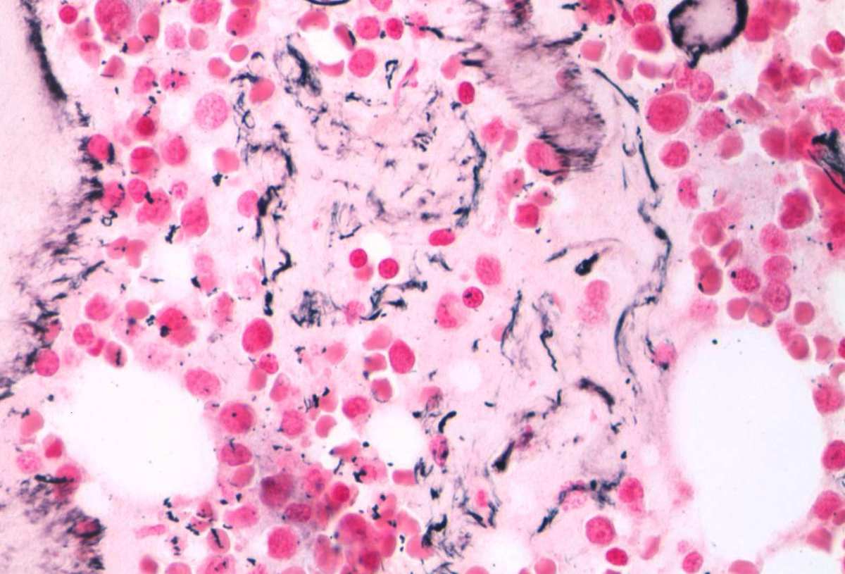 |
| CD34 immunohistochemical stain on marrow biopsy. |  |
| Representative flow cytometric scatter plots. |  |
| Representative karyotype (20/20 metaphases). |  |