Institution: H. Lee Moffitt Cancer Center
Additional authors:Ling Zhang, MD
Session: AML secondary to myeloproliferative neoplasms and other types of disease progression in MPN
HISTORY
The patient is a 34-year-old female presented with leukocytosis and diagnosed with chronic myelogenous leukemia in 06/2010. Bone marrow biopsy at that time revealed 100% cellularity with a karyotype of 46,XX,inv(2)(p11.2q13),t(9;22)(q34;q11.2). Fluorescence in situ hybridization (FISH) showed BCR-ABL1 fusion signals in all 200 cells. She was treated with nilotinib for 1 year and achieved hematologic remission. In 04/2011, the patient presented with an increased white blood cell count (53.4 x 199/L) with 82% circulating lymphoblasts confirmed by flow cytometry immunophenotyping and was initiated on chemotherapy with hyper-CVAD (cyclophosphamide, vincristine, Adriamycin and dexamethasone) and dasatinib (arm 2B until 6/2011). During the chemotherapy the patient was found to have an episode of leptomeningeal involvement by lymphoblasts in cerebrospinal fluid and underwent intrathecal chemotherapy with a full response. She subsequently received an allogeneic matched unrelated bone marrow transplant on 8/17/2011. Post-transplant dasatinib (100 mg daily) was given without side effects. Beginning in 12/2012 the patient experienced right-sided facial numbness/tingling and diplopia. MRI showed a left temporal lobe intra-axial enhancing mass (3.2cm x 2.2cm) (Figure 1). Cytology of CSF and immediate bone marrow biopsy were reviewed with the following interpretation.
DETAILS
A lumbar puncture was performed and CSF was collected for cytology evaluation and flow cytometry analysis. The CSF cytospin showed abundant immature cells that are small to medium in size with fine chromatin, prominent nucleoli, high N:C ratio and small amount of cytoplasm (Figure 2 and 3). Posterior Iliac crest bone marrow aspiration and biopsies were subsequently performed and showed normocellular marrow with trilineal hematopoiesis. The M:E ratio was 1.5:1 and there was no increase in blasts (Figure 4 and 5).
IMMUNOHISTOCHEMISTRY AND FLOW CYTOMETRY
Flow cytometry on the CSF detected a large population of immature cells (69% of total events), phenotypically consistent with lymphoblasts. The cells expressed dim CD45, CD19, CD10, CD13 and CD34 (subset) and they were negative for CD3, TdT, CD11b, CD11c, CD117 and CD33.
CYTOGENETIC FINDINGS
In bone marrow, cytogenetics reported male donor karyotype (46,XY[20]); FISH for BCR-ABL1 was negative. FISH studies were performed on CSF, and 95% of cells showed two to four BCR-ABL fusion signals (Figure 6).
MOLECULAR FINDINGS
BCR-ABL1 p210 transcripts were not detected by real-time RT-PCR in peripheral blood and bone marrow.
INTERESTING FEATURES
This is a rare case with central nervous system lymphoblastic relapse of chronic myeloid leukemia in hematologic and molecular remission 500 days post allogeneic matched unrelated hematopoietic stem cell transplant. The patient presented with a solitary mass lesion in CNS consistent with extramedullary B-lymphoblastic blast crisis while her peripheral blood and the donor-derived marrow retained a molecular remission status.
PROPOSED DIAGNOSIS
Chronic myelogenous leukemia, B lymphoblastic blast crisis, extramedullary relapse in central nervous system
CONSENSUS DIAGNOSIS
Chronic myelogenous leukemia, BCR-ABL1 positive, blast phase – B lymphoblastic, with central nervous system relapse
| Figure 1. MRI scan of the brain showed a left temporal lobe intra-axial enhancing mass. | .jpg) |
| Figure 2. Cerebrospinal fluid cytospin showed abundant immature cells. | 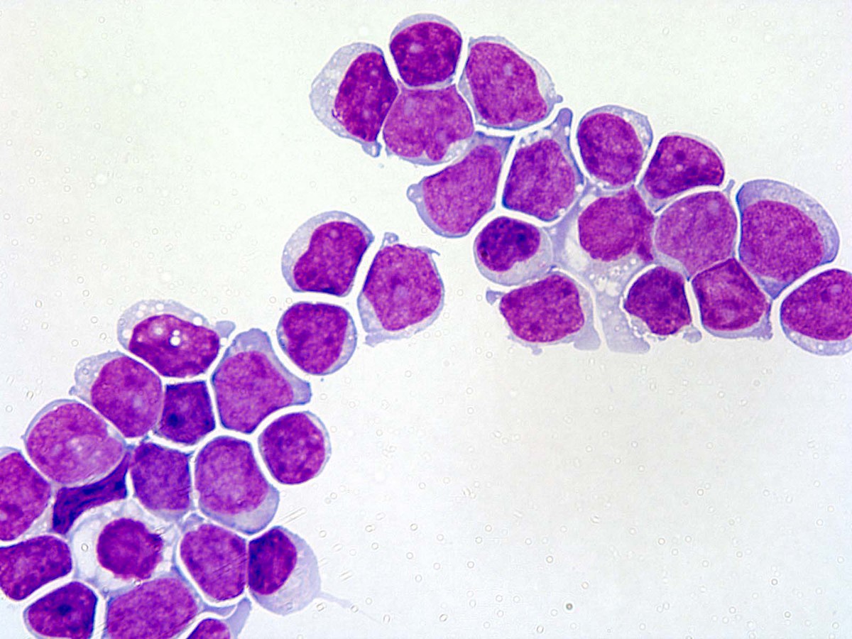 |
| Figure 3. Cerebrospinal fluid cytospin showed abundant immature cells. | 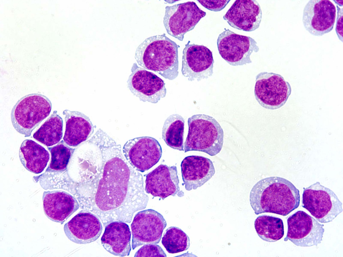 |
| Figure 4. Bone marrow biopsy showed normocellular marrow with trilineal hematopoiesis. | 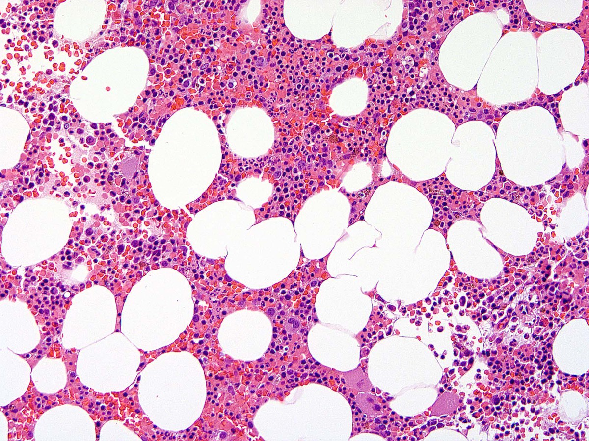 |
| Figure 5. Bone marrow aspiration showed trilineal hematopoiesis with no increase in blasts. |  |
| Figure 6. FISH studies on CSF showed 95% of cells have two to four BCR-ABL fusion signals. |  |