Institution: The University of Texas MD Anderson Cancer Center
Additional authors:Pei Lin, MD; Jeffrey L. Jorgensen MD, PhD; Sa Wang MD, PhD
Session: Acute leukemias of ambiguous lineage
HISTORY
A 35-year-old man who initially presented to the local Emergency Room for management of fatigue and discomfort in the perianal area. Laboratory evaluation showed abnormal complete blood count (CBC) and the patient was referred to a local hematologist. At that time, a bone marrow biopsy was performed on 12/05/2012 and the patient was diagnosed with acute myeloid leukemia. He received one cycle of induction chemotherapy with cytarabine and idarubicin (3+7 regimen). Conventional cytogenetic studies performed on the bone marrow specimen were reported to be positive for the presence of Philadelphia chromosome. He was subsequently transferred to our institution for further evaluation regarding treatment options. At the time of admission a CBC count showed: WBC: 1.1 k/uL, Hb 9.3 g/dL, MCV: 88, and platelet 147 k/uL. A repeat bone marrow biopsy was performed on 01/14/2013 that showed mixed phenotype acute leukemia, with t(9;22), myeloid and B lineage. the initial bone marrow biopsy was also obtained for review. The patient was started on chemotherapy with hyper-CVAD and dasatinib.
DETAILS
1. Specimen 1: Bone marrow biopsy, clot, aspirate smears and peripheral blood smears prior induction chemotherapy (12/05/2012): • Peripheral blood smears showed marked leukocytosis with numerous blasts, large in size with scant to moderate cytoplasm and some with fine azurophilic granules. No Auer rods were identified. Some of the blasts had slightly folded nuclear contours. Maturing myeloid elements showed dysgranulopoiesis. Basophils and eosinophils were not increased (Figure 1A). • Bone marrow differential count based on 200 cells revealed 76% blasts, 0% promyelocytes, 2% neutrophils, 1% eosinophils, 1% monocytes, 18% lymphocytes, 2% erythroid precursors. • Bone marrow aspirate smears and touch imprint contained good bone marrow particles for assessment. Megakaryocytes were reduced in numbers, including some small hypolobated forms. Blasts were markedly increased and ranged from small, to medium and large, containing scant to moderate cytoplasm. Many of the blasts had slightly folded and nuclear contours and conspicuous nucleoli. No Auer rods were identified. Maturing myeloid elements and erythroid precursors were markedly reduced. Basophils and eosinophils were not increased (Figure 1B). • Bone marrow biopsy and clot section showed 95% cellularity. Megakaryocytes were reduced in number including small and hypolobated forms. Blasts were markedly increased and large, with scant cytoplasm. There was no significant fibrosis present (Figure 1C and D). 2. Specimen 2: Bone marrow biopsy, clot and aspirate smears after one cycle of induction chemotherapy and prior hyper-CVAD and dasatinib (01/14/2013): • Bone marrow differential count based on 500 cells revealed 29% blasts, 1% progranulocytes, 3% granulocytes, 1% eosinophils, 3% monocytes, 18% lymphocytes, 38% erythroid precursors. • Bone marrow smears were adequate for evaluation and showed increased blasts raging from small to intermediate, some with cytoplasmic granules. Rare megakatyocytes were seen. Maturing myeloid elements and erythroid precursors were markedly reduced with relative erythroid predominance (Figure 3A and B). • Bone marrow biopsy and clot section showed a cellularity of 30-40% with interstitial clusters of immature cells. Megakaryocytes were adequate in number with unremarkable morphology (Figure 1D).
IMMUNOHISTOCHEMISTRY AND FLOW CYTOMETRY
1. Specimen 1: Bone marrow clot:
Immunohistochemical studies showed the blasts to be positive for PAX5 (80-90%), CD19 (50%), TdT (90%) and a subset for myeloperoxidase (30%). The blasts were negative for CD117 and CD20 (Figure 2).
2. Specimen 2: Bone marrow aspirate:
Flow cytometric analysis showed 13% blasts that were positive for CD34, CD117 large subset, CD33, CD13 partial, CD15 subset, CD19 moderate, CD22 partial, cytoplasmic CD79a dim, CD25 partial, and TdT, MPO small subset. The blasts were negative for CD10, CD66c, CD2, CD5, CD7 and CD56 (Figure 4).
Special Stains: Specimen 2; Bone marrow aspirate:
Cytochemical stains were performed at our institution showed that myeloperoxidase (MPO) was weakly positive in approximately 20% of blasts when compared to the strong cytoplasmic stain of granulocytes. Blasts were negative for butyrate esterase (Figure 3C).
CYTOGENETIC FINDINGS
Specimen 2; Bone marrow aspirate: Conventional cytogenetic studies were positive for the presence of two Philadelphia positive clones: 46,XY,t(9;22)(q34;q11.2) [15] and 46,idem, del(20)(q11.2q13.3)[5].
MOLECULAR FINDINGS
Specimen 2; Bone marrow aspirate: Molecular studies performed at our institution showed the presence of e1a2 BCR-ABL fusion transcript coding for the 190kDa BCR-ABL detected by real-time PCR.
INTERESTING FEATURES
• We are presenting a case of mixed phenotype acute leukemia with t(9;22) translocation. They are the most common subgroup of acute leukemias of ambiguous lineage with the most usual immunophenotype including B and myeloid lineage, but they represent less than 1% of acute leukemias. These cases are challenging and extensive immunophenotyping is necessary to confirm that the blasts belong to two different lineages or co-express differentiation antigens of more than one lineage.
• The diagnosis of mixed phenotype acute leukemia can be particularly difficult when only paraffin sections are available, as in our case for specimen 1, and co-expression of markers on the blasts can only be evaluated by immunohistochemistry. We observed that on the clot sections from specimen 1, the MPO and CD19 stains are positive for a subset of the blast population for each marker and this may also reflect the presence of two different clones identified by conventional cytogenetic stuies in the specimen 2. However, considering the results from the flow cytometric analysis on specimen 2 with residual acute leukemia, two distinct blast populations could not be definitely identified.
• A discrepancy was observed regarding the CD117 expression on blasts, being negative by immunohistochemistry on specimen 1, but highlighting a subset of blast on specimen 2 by flow cytometry. These findings indicate that, when different methods are used for the evaluation of the expression of cell antigens, a discrepancy on the results may be observed.
• Importantly, in the WHO 2008, the guidelines are not specific regarding criteria for the evaluation of the expression of the cell specific markers in order to assign a certain lineage. The latter is especially problematic for the assessment of myelperoxidase in the blast population and it is not clear whether a cytochemical, immunohistochemical or flow cytometric analysis should be used. This has important implications regarding treatment decisions, since currently there is no evidence that patients with mixed lineage phenotypic leukemia can be benefit by AML or ALL based chemotherapy protocols.
• Additional studies for evaluation of the current WHO 2008 guidelines regarding lineage assignment and definition of mixed phenotype acute leukemia are important, since this can have significant implications in therapeutic decision making.
PROPOSED DIAGNOSIS
Philadelphia chromosome positive acute leukemia of ambiguous lineage, B/myeloid.
CONSENSUS DIAGNOSIS
Mixed phenotype acute leukemia, B/myeloid, with t(9;22)(q34;q11.2)
| Figure 1. Review of microscopic findings from specimen 1. (A) Peripheral blood smear with numerous blasts, large in size with scant to moderate cytoplasm and some with fine azurophilic granules. No Auer rods were identified. Some of the blasts had slightly folded nuclear contours (500x). (B) Bone marrow smears showed markedly increased blasts with similar morphologic findings to that described in the peripheral blood smear (1000x). (C) and (D) Bone marrow biopsy showed 95% cellularity with sheets of large blasts with scant cytoplasm (C 200x; D 1000x). | 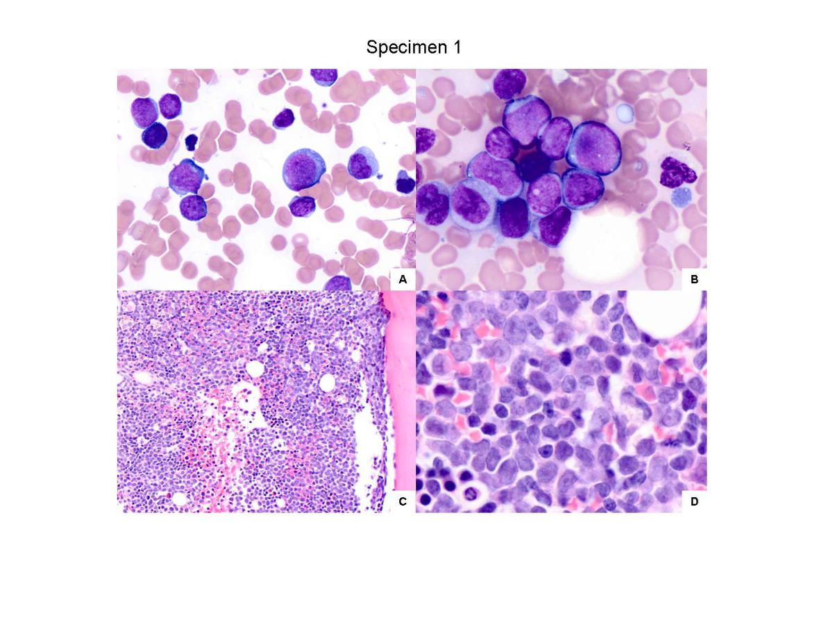 |
| Figure 2. Review of immunohistochemistry results from specimen 1. (A) Bone marrow clot section showed 95% cellularity and sheets of large blasts. Immunohistochemical studies were performed on clot sections that showed the blasts to be positive for CD19 approximately 50% (B), a subset for myeloperoxidase approximately 30% (C), PAX5 (D), and TdT (E). The blasts were negative for CD117 (F) (A-F, 400x). |  |
| Figure 3. Review of microscopic findings from specimen 2. (A) and (B) Bone marrow smears showed increased blasts raging from small to intermediate, some with cytoplasmic granules. Maturing myeloid elements and erythroid precursors were markedly reduced with relative erythroid predominance (A, 500x; B, 1000x). (C) Myeloperoxidase (MPO) was weakly positive in approximately 20% of blasts when compared to the strong cytoplasmic stain of granulocytes. (D) Bone marrow biopsy showed a cellularity of 30-40% with interstitial clusters of immature cells (400x). | 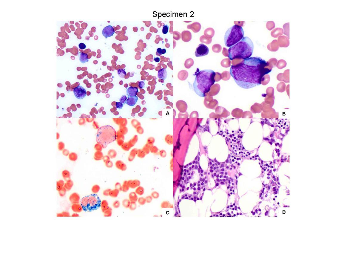 |
| Figure 4. Review of flow cytometric results on specimen 2 that showed 13% blasts (dim CD45) that were positive for CD34, CD123, CD13 partial, CD117 subset, CD33, CD19 moderate, CD22 partial, cytoplasmic CD79a dim, MPO small subset and TdT. | 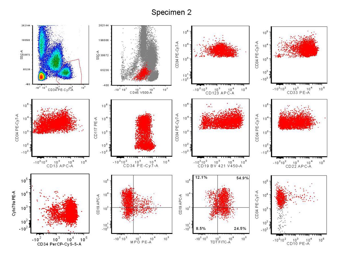 |