Institution: Stanford University Medical Center, Department of Pathology
Additional authors:Dita Gratzinger, M.D., PhD. Stanford University Medical Center, Department of Pathology; Athena Cherry, PhD. Stanford University Medical Center, Department of Pathology
Session: Therapy-related myeloid neoplasms
HISTORY
The patient is a 61-year-old female with a history of an inflammatory myopathy managed with azathioprine and monthly IVIG who presented initially with fatigue, fevers, and chills for two days. Initial blood work was significant for a marked anemia (Hgb 6.2 g/dL), a thrombocytopenia (14K/uL), and an absolute neutropenia (210/uL). Workup at that time revealed multilineage dysplasia, 8% blasts, as well as monosomy 7. The patient was then managed initially on azacytidine and given red cell and platelet transfusion as needed. Her condition continued to decline, and when she was re-evaluated for progression of disease 4 months later, a diagnosis of leukemia was made. The patient was subsequently managed on azacytidine and lenalidomide, but her condition rapidly declined. The patient died of complications related to febrile neutropenia and presumed sepsis only 2 months after her diagnosis of leukemia.
DETAILS
The peripheral blood at initial diagnosis showed leukopenia, anemia with mild to moderate anisopoikilocytosis including dacrocytes and microcytes. The air-dried methanol-fixed Wright Giemsa stained slides at the patient’s initial presentation showed hypercellular marrow with overt multilineage dysplasia. Erythroids demonstrate megaloblastoid changes and dyspoietic forms with irregular nuclear contours are readily identified. Megakaryocytes show many dysplastic forms with micromegakaryocytes and megakaryocytes with separated nuclear lobes. The Bouin’s fixed, rapid decalcified bone marrow core biopsy shows a hypercellular marrow (80%) with increased myeloids and erythroids and occasional micromegakaryocytes present.
At the patients diagnosis of more advanced disease, the peripheral blood showed 10% circulating blasts with monocytic morphologic features. An absolute basophilia and monocytosis were present. Basophils showed hypolobation and increased chromatin condensation. The bone marrow aspirate slides showed similar megaloblastoid and dyspoietic morphology as had previously been noted. Megakaryocytes also demonstrated small megakaryocyte forms and multinucleated forms. Blasts comprised 29% of nucleated cells. The bone marrow core was hypercellular (90%) and show patches of immature blasts with a background of myeloid predominance and megakaryocyte dysplasia.IMMUNOHISTOCHEMISTRY AND FLOW CYTOMETRY
Flow cytometry of the aspirate at initial presentation was significant for a CD34+ population comprising 6% of CD45 positive events. Cytoplasmic markers were not run as part of the panel.
At the patient’s diagnosis of acute leukemia, the blast population showed a myelomonocytic phenotype with expression of MPO, CD117, CD33, CD14, with partial CD34, partial CD38, partial CD13, partial CD64, partial CD4, partial CD15, as well as partial aberrant CD7 and CD2 but not surface CD3. A subset of events within the blast and monocyte gates were cytoplasmic CD3 positive, and this finding was confirmed by backgating for cytoplasmic CD3. Immunohistochemistry was not performed on this case.CYTOGENETIC FINDINGS
At initial presentation, karyotype analysis revealed monosomy 7, confirming a clonal neoplastic process. At the subsequent diagnosis of leukemia, bone marrow aspirate culture again revealed the finding of monosomy 7.
MOLECULAR FINDINGS
The bone marrow aspirate at the diagnosis of leukemia was negative for FLT3 internal tandem duplication and D835 mutation, NPM1, CEBPA, and KIT. The SNaPshot cancer mutation panel was also performed on this case which was positive for the NRAS 182A>G (Q61R) mutation.
INTERESTING FEATURES
The salient features of this case include the initial presentation of therapy-related myelodysplastic syndrome related to azathioprine therapy for inflammatory myopathy, rapidly followed by evolution to acute leukemia with partial cytoplasmic CD3 (as well as CD2 and CD7) in addition to a myelomonocytic immunophenotype. Dysplasia in both the erythroid and megakaryocytic lineages as well as monosomy 7 was consistent at both timepoints. Given the persistence of monosomy 7 and multilineage dysplasia at both timepoints, as well as the clinical course, the findings were felt to be most consistent with therapy-related myelodysplastic syndrome and subsequent AML despite the cytoplasmic CD3.
Although mutations within NRAS have recently been described, the prognostic significance of this finding is still not known but under investigation (Dunlap J, et al. Hum Pathol 2012 Dec).PROPOSED DIAGNOSIS
Therapy-related myelodysplastic syndrome evolving to acute myeloid leukemia with aberrant cytoplasmic CD3 expression.
CONSENSUS DIAGNOSIS
Acute myeloid leukemia, possibly therapy-related following azacytidine and azathioprine
| Peripheral blood, Wright Giemsa, 20x. A pancytopenia with moderate anisopoikilocytosis was evident at initial diagnosis. | 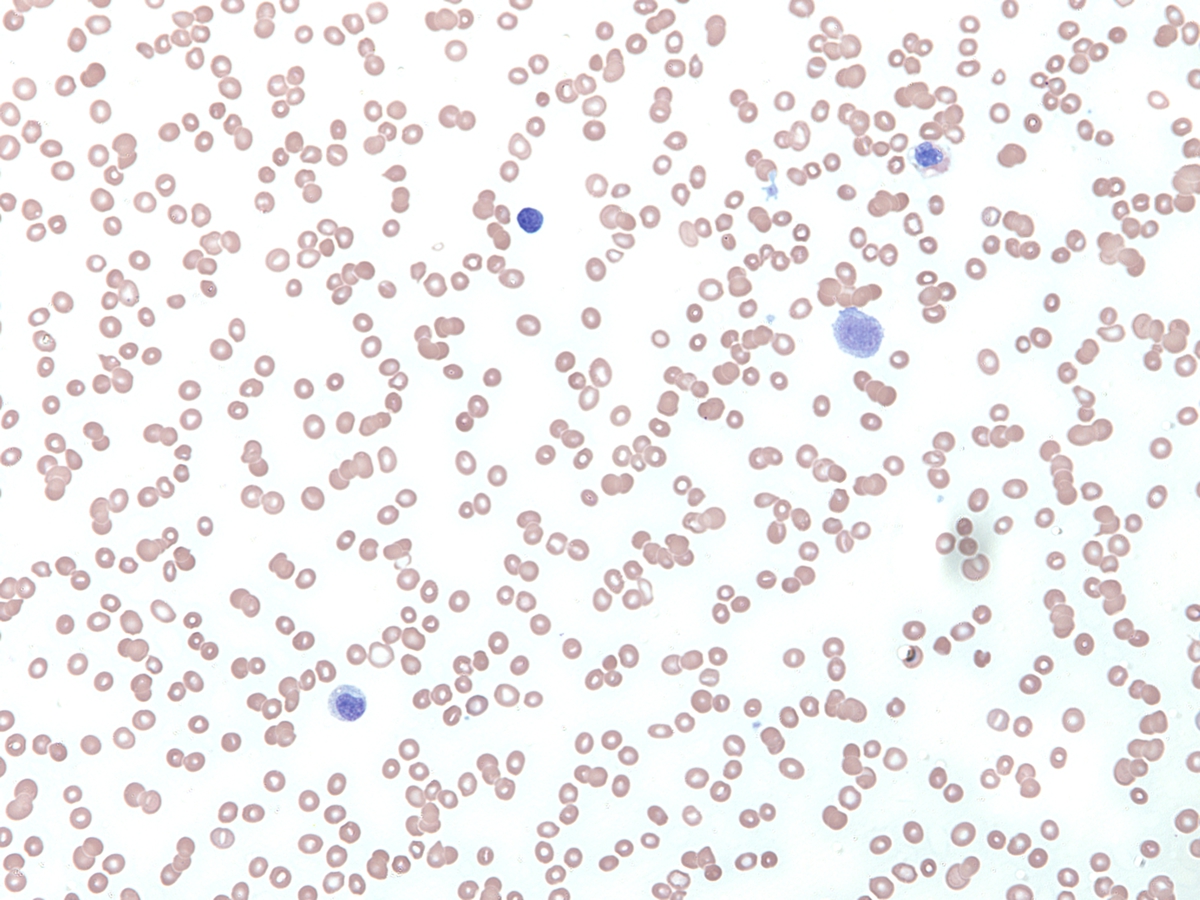 |
| Bone marrow aspirate, Wright-Giemsa, 60x. At initial diagnosis, dysplasia in both the erythroid and megakaryocytic lineages was evident. | 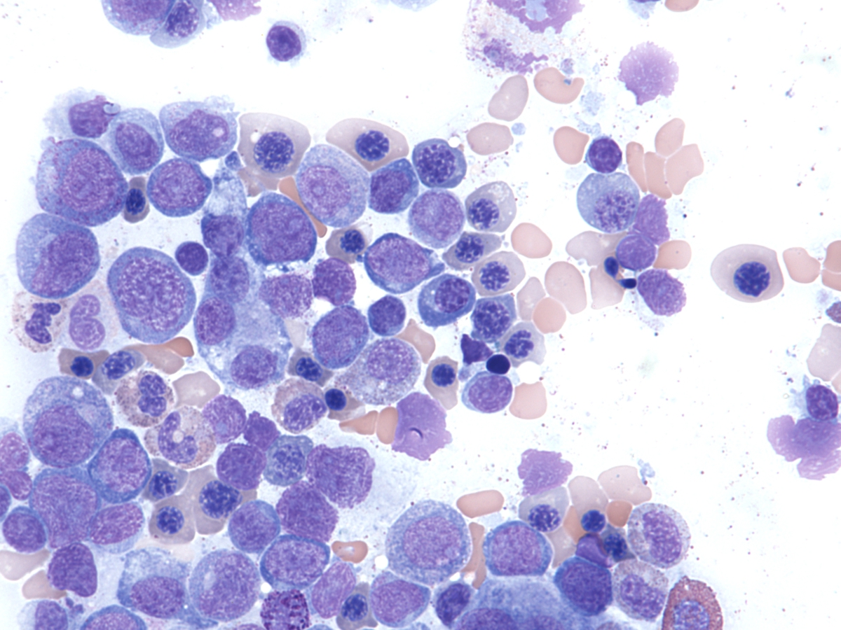 |
| Bone marrow aspirate image #2, Wright-Giemsa, 60x. At initial diagnosis, other areas show more overt erythroid dysplastic forms with abnormal nuclear contour. |  |
| Bone marrow core biopsy, H&E, 20x. The marrow at initial diagnosis is hypercellular for age. | 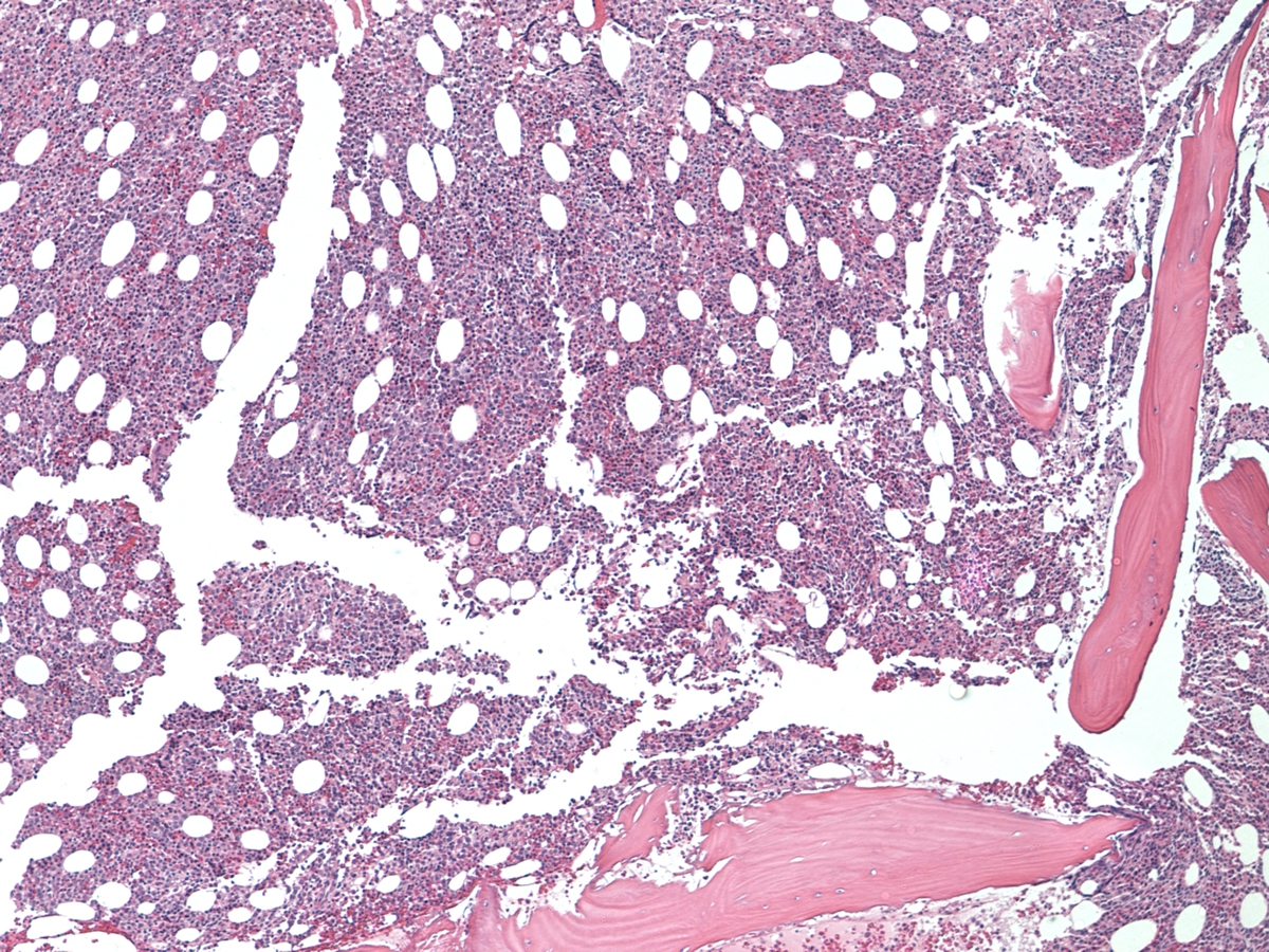 |
| FISH probe analysis at initial diagnosis demonstrating the presence of only one chromosome #7 signal (red); karyotype analysis revealed monosomy #7. | 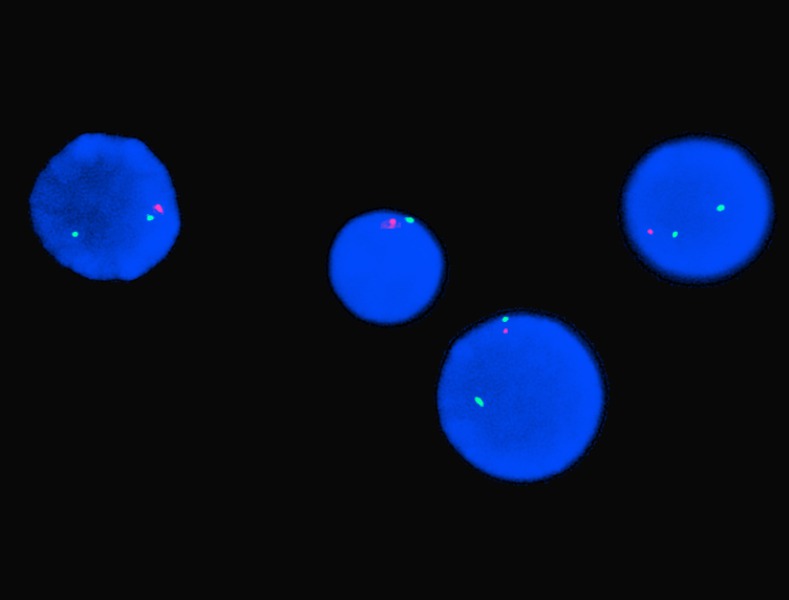 |
| Peripheral blood, Wright Giemsa, 40x. At the more advanced stage of disease circulating blasts (10%) with monocytic morphologic features and an absolute monocytosis and basophilia are present. | 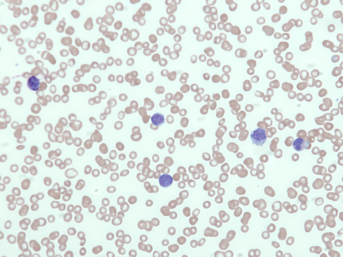 |
| Bone marrow aspirate image #1, Wright-Giemsa, 40x. At the more advanced stage of disease, increased numbers of blasts (29%) and similar multilineage dysplasia present. | 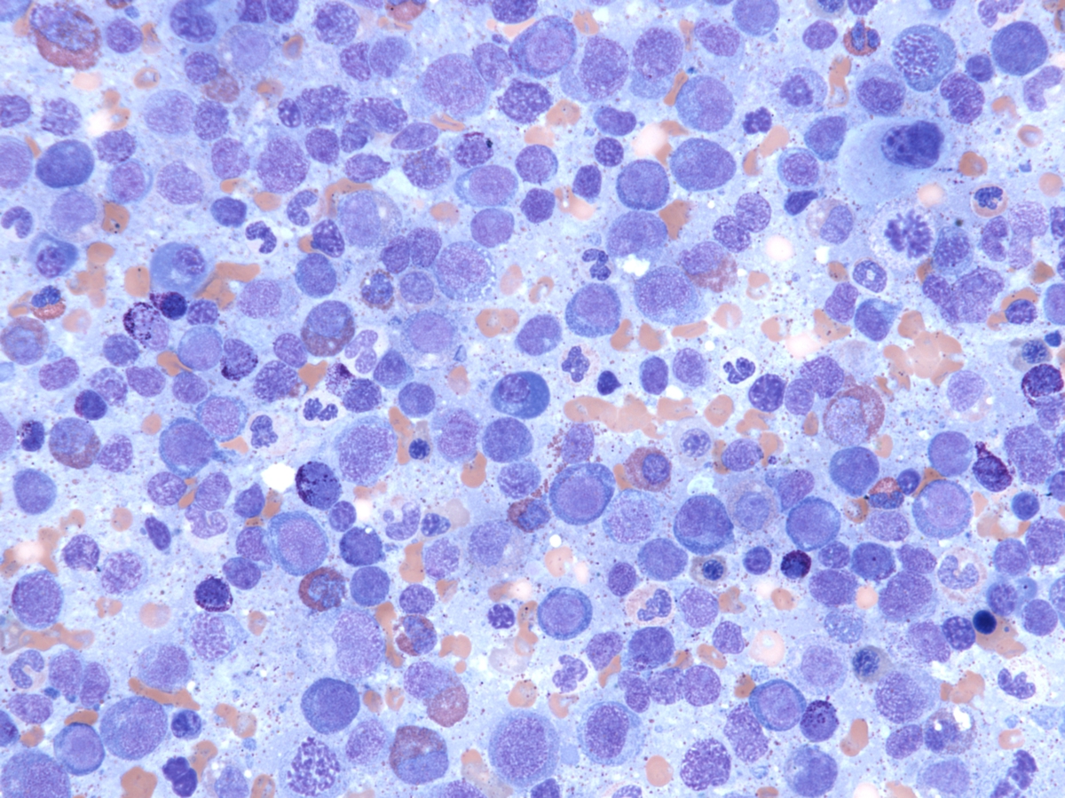 |
| Bone marrow aspirate image #2, Wright-Giemsa, 40x. At the more advanced stage of disease, increased numbers of blasts (29%) and similar multilineage dysplasia present. |  |
| Flow cytometry findings: At the more advanced stage of disease, flow cytometry revealed the blast population to express CD33, CD117, partial CD34, as well as aberrant partial CD7 and CD2. A small subset of blasts expressed cytoplasmic CD3, which was confirmed on back-gating of CD3 positive events. |  |
| Karyotype analysis at the more advanced stage of disease was again noteworthy for monosomy #7. |  |