Institution: Medical University of South Carolina
Additional authors:Sarah Brooks MD, Nicole Miller DO, Ana Maria Medina MD, Daynna Wolff PhD, John Lazarchick MD
Session: B Lymphoblastic Leukemia/Lymphoma
HISTORY
A 66 year old Caucasian man with past medical history of hypertension and type II diabetes mellitus presented to an outside hospital with chief complaint of abdominal fullness and pain. He noted that the pain was worst in his lower back. He was treated initially with diuretics and pain management. When this failed to resolve his symptoms a CT of the abdomen and pelvis was obtained. At that time he was diagnosed with an ‘abdominal mass’ per the patient. A bone marrow biopsy was then performed; these slides were made available at MUSC for review and our findings are detailed below. He was then transferred to our institution where a repeat marrow was attempted; the findings are described below. The patient was started on Gemcitabine therapy and shortly thereafter developed tumor lysis syndrome. He was admitted to the hospital for further chemotherapy and management of tumor lysis.
DETAILS
In B + solution followed by 10% formalin fixation of iliac crest bone marrow biopsy Outside Hospital Bone Marrow Biopsy: The bone marrow aspirate revealed increased numbers of blasts with scant cytoplasm, prominent nucleoli and cytoplasmic vacuoles. Myeloid and erythroid lineages were decreased in numbers with no morphologic abnormalities; megakaryocytes were present in adequate numbers with no morphologic abnormalities. The Bone marrow biopsy confirmed the presence of markedly increased numbers of blasts with scant cytoplasm, prominent nucleoli and cytoplasmic vacuoles. In House Bone Marrow Biopsy: There were no significant findings within the peripheral blood smear. The bone marrow aspirate was hemodilute and aspicular. Evaluation of marrow elements could not be performed. The bone marrow biopsy was extremely superficial and composed predominantly of cartilage. No marrow elements were present.
IMMUNOHISTOCHEMISTRY AND FLOW CYTOMETRY
Outside Hospital Bone Marrow Biopsy:
Flow cytometric analysis revealed a 70% blast population positive for CD19, CD10, CD79a, HLA-DR, cTdT, CD22 and CD38.
Immunohistochemical staining performed on the bone marrow core biopsy revealed the blast population to be CD19, TdT and PAX-5 positive. CD3 stain highlighted scattered background small mature T-cells. CD-20 highlighted scattered background small mature B-cells. Myeloperoxidase staining revealed scattered myeloid marrow elements (markedly decreased). CD34 and CD117 was negative in the blast population.
In house bone marrow biopsy:
Flow cytometric analysis revealed a 25% population of cells within the monocytoid region which expressed HLA-DR, CD19, CD10, CD22, CD38 and cytoplasmic CD79a. They were surface immunoglobulin negative.
The flow cytometric analysis report from the patient’s initial diagnosis was available for review. It reported an 85% blast population which were positive for CD19, CD10, CD79a and surface immunoglobulin negative. The cells were TdT positive. There was no mention of whether the blasts were located in the monocytoid region.
CYTOGENETIC FINDINGS
Cytogenetic analysis revealed a complex phenotype: t(14;18), possible deletion 2q, abnormal 8q, loss of 10 and 15, additional material of 17p1 (possible loss of Tp53) additional material of 19p and Xp.
MOLECULAR FINDINGS
FISH analysis confirmed the presence of t(14;18).
INTERESTING FEATURES
The entity of de novo acute lymphocytic leukemia with a t(14;18) has been very rarely described in the literature. Available information suggests that these cases tend to have complex karyotypic abnormalities, which is confirmed by our findings in this case. Additionally, the blast location within the monocytoid region is highly unusual and, as far as our research efforts have revealed, not described in the literature. This diagnosis is reported to carry a poor prognosis and highly aggressive clinical course.
PROPOSED DIAGNOSIS
Acute B Lymphoblastic Leukemia, t(14;18) positive
CONSENSUS DIAGNOSIS
High-grade TdT-positive blastic B-cell lymphoma/leukemia with t(14;18)
| Bone Marrow Touch Prep, 40X |  |
| Bone Marrow Touch Prep, 60X |  |
| Bone Marrow Biopsy, 4X | 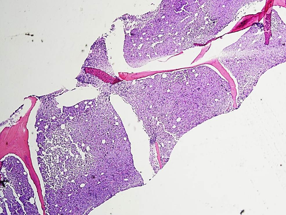 |
| Bone Marrow Biopsy, 60X | 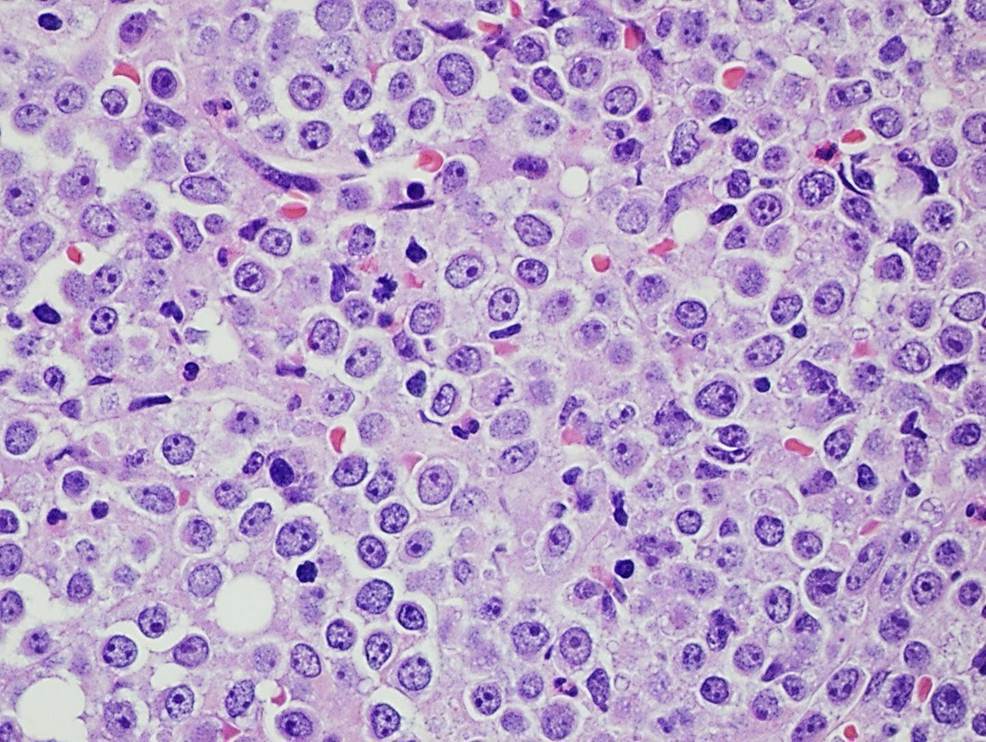 |
| Bone Marrow Biopsy, 60X |  |
| Bone Marrow Biopsy, CD19 |  |
| Bone Marrow Biopsy, PAX-5 |  |
| Flow Cytometry |  |
| Flow Cytometry | 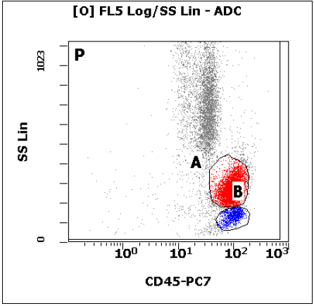 |
| Flow Cytometry | 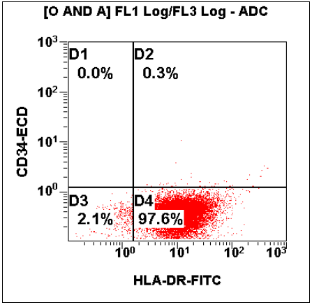 |
| Flow Cytometry |  |
| Flow Cytometry |  |
| Bone Marrow Biopsy, TdT |  |