Institution: Medical University of South Carolina
Additional authors:Ana Maria Medina MD, Sarah Brooks MD, Nicole Miller DO, Luciano Costa MD, John Lazarchick MD
Session: AML with myelodysplasia-related changes
HISTORY
A 64 year old man with past medical history of hypothyroidism, colon cancer and atrial fibrillation presented leukocytosis and thrombocytopenia noted by his family physician. The patient reported similar lab values for the prior six months with no resolution after antibiotic therapy. He reported a 25 pound weight loss over the prior 18 months and persistent fatigue. At that time, the outside institution performed a bone marrow biopsy which demonstrated findings consistent with myeloproliferative disorder concerning for evolving acute myeloid leukemia. Two weeks after presentation he was admitted to our facility and a bone marrow biopsy was obtained. Findings are described below. Physical exam revealed 14 cm splenomegaly. A serum tryptase (obtained after diagnosis) was elevated at 36 ug/L (normal= 0.4 to 10.9 ug/L). Since diagnosis the patient has been treated with a watch a wait philosophy given that he is largely asymptomatic and functioning well.
DETAILS
B + solution followed by 10% formalin fixation of iliac crest bone marrow biopsy specimen. Of significance in the peripheral blood, the white blood cell count was 20,000/CUMM with a differential cell count revealing 14% monocytes (2,800/CUMM absolute monocyte count) and no blasts. Examination of the aspirate revealed no morphologic abnormalities of the erythroid or myeloid precursors. Megakaryocytes were also morphologically normal. There was morphologic evidence of an increased blast count; a differential cell count revealed a 7% population of blasts as well as a 6% population of monocytes. The bone marrow biopsy was hypercellular (80-90%). Megakaryocytic dysplasia was identified in the form of mummified nuclei, clustering and unilobation. Two discreet foci of spindled cells were identified. Immunohistochemistry was utilized to further characterize these foci and the findings are described below. Aside from a myeloid left shift the myeloid and erythroid series were unremarkable.
IMMUNOHISTOCHEMISTRY AND FLOW CYTOMETRY
Flow cytometric analysis of the bone marrow revealed a 1% blast population which expressed CD34, HLA-DR, CD33, CD117, CD13 and CD38. The previously described spindled cell clusters within the bone marrow core biopsy were CD117 positive by immunohistochemistry (mast cells). A CD34 stain highlighted scattered blasts comprising less than 10% of the marrow elements. Special Stain for Iron: Present with no ring sideroblasts
CYTOGENETIC FINDINGS
Normal male karyotype (46, XY)
MOLECULAR FINDINGS
PCR for C-KIT (D816V) mutation: detected
INTERESTING FEATURES
This is an interesting case of systemic mastocytosis and associated chronic myelomonocytic leukemia (CMML). This entity’s incidence is likely underestimated because it can be extremely difficult to identify to identify the cytologic and histologic features of systemic mastocytosis due to an obscuring effect by the associated malignancy. In this case the mastocytosis was an incidental finding upon close bone marrow biopsy examination.
PROPOSED DIAGNOSIS
Systemic mastocytosis with associated clonal hematologic, non-mast cell lineage disease (chronic myelomonocytic leukemia-1)
CONSENSUS DIAGNOSIS
Systemic mastocytosis with associated clonal hematologic, non-mast cell lineage disease (chronic myelomonocytic leukemia-1)
| Peripheral Blood | 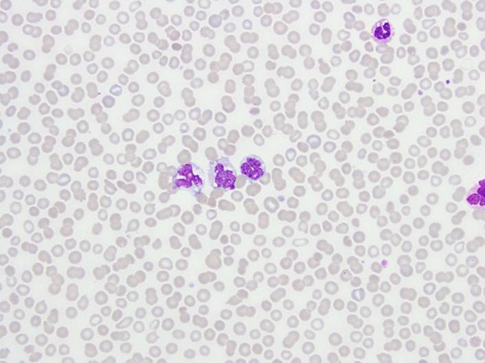 |
| Peripheral Blood | 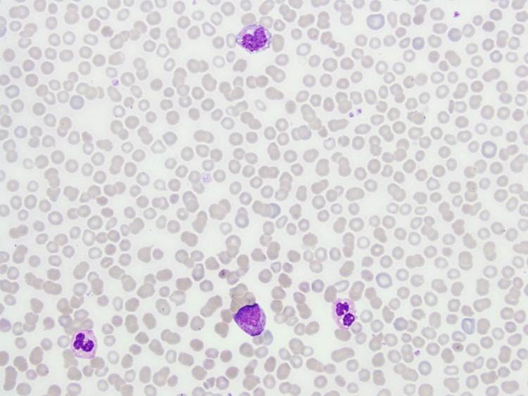 |
| Bone Marrow Biopsy, 4X |  |
| Bone Marrow Biopsy, 20X |  |
| Bone Marrow Biopsy, 20X |  |
| Bone Marrow Biopsy, CD117 | 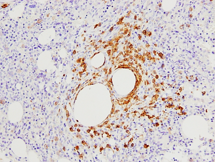 |
| Bone Marrow Biopsy, CD117 |  |