Institution: Texas Childrens Hospital, Houston, TX; Baylor College of Medicine, Houston, TX
Additional authors:Choladda V. Curry, MD Andrea M. Sheehan, MD
Session: Myeloid and lymphoid neoplasms with eosinophilia and abnormalities of PDGFRA, PDGFRB, or FGFR1
HISTORY
A 17 year old boy with past medical history of exposure to tuberculosis, status post nine months of treatment two years prior to presentation, presented with bilateral cervical and supraclavicular lymphadenopathy, right greater than left. Presenting CBC demonstrated normal counts with mild relative but not absolute monocytosis (10.6%) and eosinophilia (4.4%).
DETAILS
Multiple right neck lymph nodes were received fresh for tissue triage. Touch preparations were performed. Portions of the tissue were submitted in RPMI for flow cytometry and cytogenetics. Additional tissue was sent for microbiology and frozen for possible future studies. The remainder of the specimen was submitted for permanent sections.
Cut sections demonstrated a homogenous tan cut surface. Diff-Quik and H&E stained touch preparations demonstrated a monotonous population of small to intermediate sized immature lymphoid cells with scant cytoplasm, finely dispersed chromatin with inconspicuous nucleoli. H&E stained sections demonstrated near complete effacement of the lymph node architecture by a paracortical infiltrate of monomorphic immature lymphoid cells with finely dispersed chromatin, scant cytoplasm, irregular nuclear contours, and finely dispersed chromatin with indistinct to small nucleoli. There was brisk mitotic activity and occasional apoptotic bodies seen. There was prominent vascularity. No eosinophilic infiltrates or immature myeloid populations were identified.Bone marrow biopsies, clot and unstained aspirate smears from bilateral anterior iliac crests were received. The biopsies were decalcified and submitted for permanent sections. The clots were submitted for permanent sections. The aspirate smears were Giemsa-stained.Staging bone marrow biopsy was negative for lymphomatous infiltrates or acute leukemia, but demonstrated an abnormal chromosome analysis. At the time of bone marrow biopsy a peripheral absolute monocytosis and absolute neutrophilia was present.IMMUNOHISTOCHEMISTRY AND FLOW CYTOMETRY
Flow cytometry of the right neck lymph node demonstrated an immature T cell population that expressed CD3 (cytoplasmic only), CD2, CD5, CD7, CD1a, CD10 (partial), CD43, TdT, CD45R0, CD52, CD25 (partial), CD33 (dim partial) and CD38. Surface CD3, CD8, and other myeloid markers were negative. Immunohistochemistry was not performed.
Flow cytometry of the staging bone marrow biopsy was negative for an abnormal clone.CYTOGENETIC FINDINGS
Both the right neck lymph node and staging bone marrow biopsy demonstrated an abnormal male karyotype: 46,XY,t(8;13)(p12;q12).
Right neck lymph node: 46,XY,t(8;13)(p12;q12)[12]/46,XY[8]Staging bone marrow: 46,XY,t(8;13)(p12;q12)[18]/46,XY[2]MOLECULAR FINDINGS
Reverse transcription polymerase chain reaction (RT-PCR) performed on RNA obtained from the paraffin embedded right neck lymph node biopsy was positive for the presence of the FGFR1-ZNF198 fusion transcript, which results from the t(8;13) translocation.
INTERESTING FEATURES
The bone marrow biopsy, lymph node excision and peripheral blood did not show eosinophilia, findings typical of this entity. The peripheral blood did however have an absolute monocytosis and absolute neutrophilia at the time of biopsy.
PROPOSED DIAGNOSIS
Leukemia/Lymphoma associated with FGFR1 rearrangement, T-Lymphoblastic Lymphoma
CONSENSUS DIAGNOSIS
Myeloid and lymphoid neoplasm with t(8;13)(p12;q12); FGFR1-ZMYM2, presenting with T-lymphoblastic leukemia, myeloproliferative neoplasm, and eosinophilia
| H&E stained sections of the right neck lymph node demonstrated near complete effacement of the lymph node architecture by a paracortical infiltrate of monomorphic immature lymphoid cells with finely dispersed chromatin, scant cytoplasm, irregular nuclear contours, and finely dispersed chromatin with indistinct to small nucleoli. No eosinophilic infiltrates or immature myeloid populations were identified. | 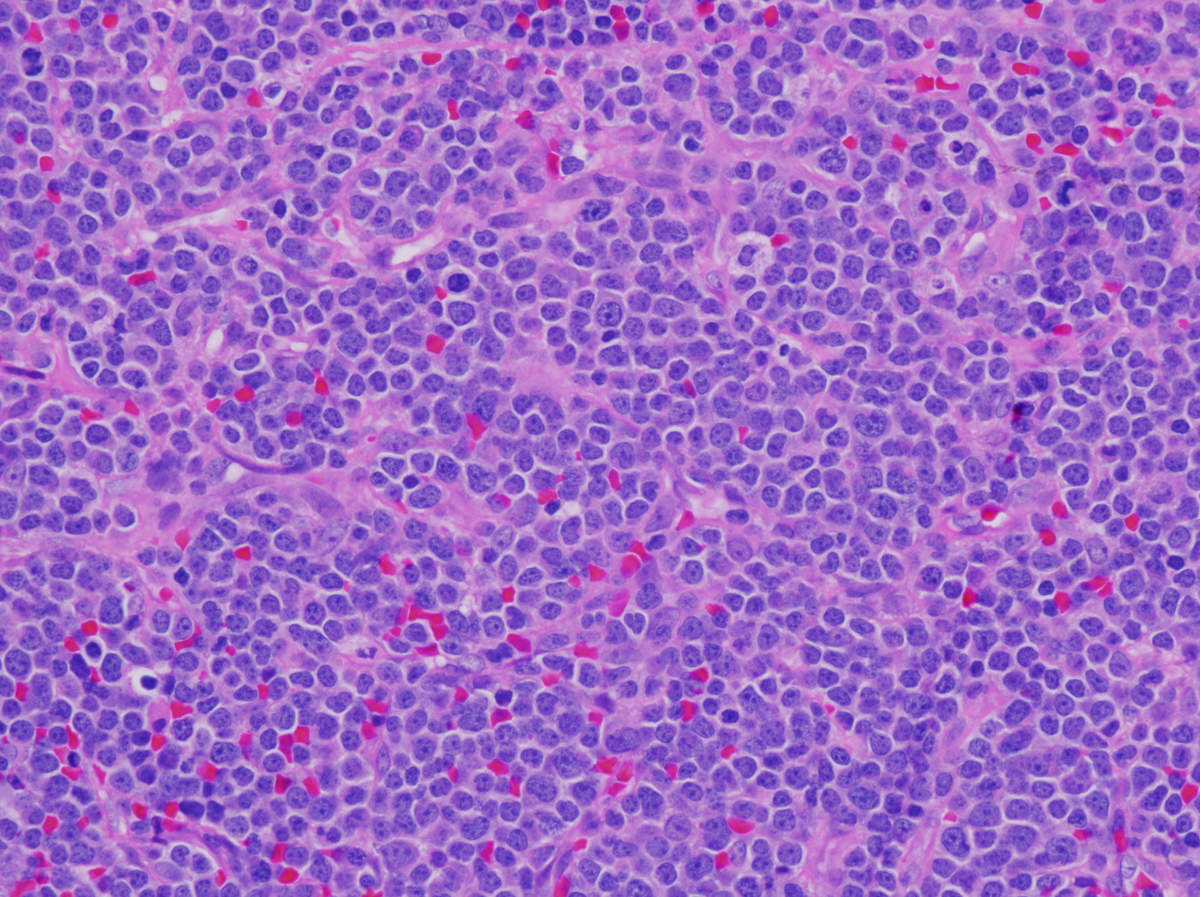 |
| The staging bone marrow biopsy was negative for any lymphomatous infiltrates or acute leukemia. H&E stained sections of the bone marrow core biopsy showed focal expansion of granulocytic cuffs. | 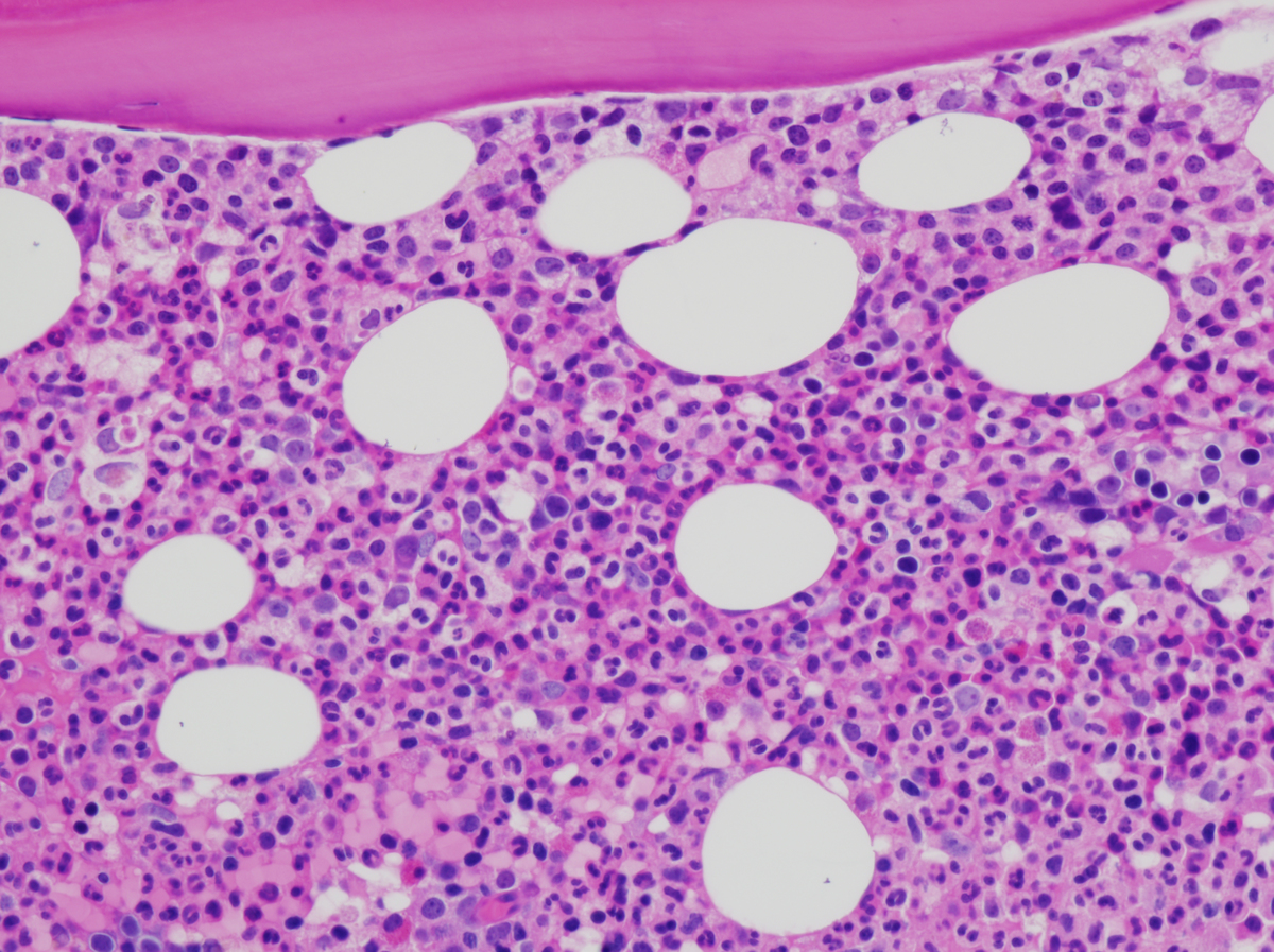 |