Institution: University of Utah/ARUP Laboratories, Salt Lake City, UT
Additional authors:Todd Kelley MD, Mohamed Salama MD, Sherrie Perkins MD, PhD
Session: Acute leukemias of ambiguous lineage
HISTORY
The patient is a 7 month old male who presented with a neck and tongue mass, base of skull lesions and circulating blasts.
DETAILS
A biopsy of a right neck mass was originally submitted. It showed partial infiltration of the lymph node by a monomorphic cellular infiltrate. No definitive cohesive growth pattern or rosettes were identified within the neoplastic cells. A bone marrow biopsy was received the next day. The WBC count was 4.2 K/MCL. The peripheral blood contained 13% circulating blasts by differential count and a mild absolute eosinophilia. The blasts were characterized by variably abundant cytoplasm, fine chromatin and occasional prominent nucleoli. Auer rods were not identified. The bone marrow aspirate was richly cellular and contained 28% blasts by differential count. Dysplasia was not identified. The bone marrow core biopsy was hypercellular at 90-95%. Large sheets and aggregates of immature monomorphic cells consistent with blasts were identified.
IMMUNOHISTOCHEMISTRY AND FLOW CYTOMETRY
Flow cytometry performed on the right neck mass biopsy revealed 47% blasts of ambiguous lineage expressing cytoplasmic CD3, CD13 (dim), CD33, CD34, CD56 (bright) and CD117. The blasts were negative for CD1a, CD2, surface CD3, CD5, CD4, CD7, CD8, CD11b, CD11c, CD14 (Mo2 and My4), CD16, CD41, CD61, CD64, glycophorin A, HLA-DR, myeloperoxidase and TdT.
Flow cytometry performed on the bone marrow revealed 31% blasts of ambiguous lineage with an antigen expression pattern similar to that seen within the right neck mass biopsy.
Immunohistochemical stains performed on the right neck mass revealed the neoplastic cells were positive for CD34, CD33, CD56, CD117 (variable), and CD43 but negative for CD1a, CD2, CD3, CD4, CD5, CD8, CD10, CD15, CD19, CD99, TdT, myeloperoxidase and PAX-5. CD45 was very dim to negative. In situ hybridization studies for EBER were negative.
The neoplastic cells retained nuclear expression of BAF-47 and were also negative for desmin, MYF4, NSE and synaptophysin .
CYTOGENETIC FINDINGS
ALL and AML FISH studies were negative. The karyotype revealed a normal karyotype, 46, XY [21].
MOLECULAR FINDINGS
Not performed.
INTERESTING FEATURES
The presentation in this case including occasional skull lesions was unusual. In addition, the immunophenotype was unusual in that the neoplastic cells expressed cytoplasmic CD3 as well as CD13 (dim), CD33, CD34, CD56(bright) and CD117. Solid tumor markers including desmin, NSE, synaptophysin were all negative.
PROPOSED DIAGNOSIS
Poorly differentiated acute leukemia. The differential diagnosis would include an early T-cell precursor leukemia, acute myeloid leukemia with aberrant cytoplasmic CD3 expression or a mixed phenotype acute leukemia (T/myeloid) but there did not appear to be sufficient lineage associated markers to allow for definitive lineage assignment.
CONSENSUS DIAGNOSIS
Acute myeloid leukemia with minimal differentiation
| Peripheral blood containing occasional circulating blast |  |
| Bone marrow aspirate with 28% blasts by differential count |  |
| Lymph node with mononuclear infiltrate |  |
| Higher power of lymph node containing sheets of mononuclear cells | 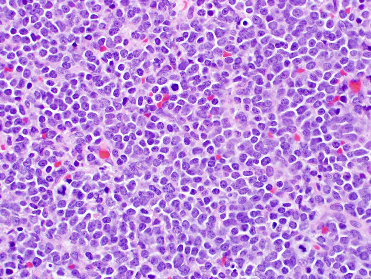 |
| CD33 staining of the neoplastic cells in the lymph node biopsy |  |
| CD34 staining of the neoplastic cells in the lymph node biopsy | 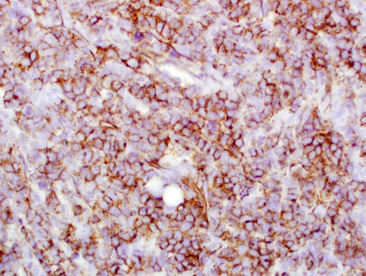 |
| CD43 staining of the neoplastic cells in the lymph node biopsy |  |
| CD56 staining of the neoplastic cells in the lymph node biopsy |  |
| CD117 staining of the neoplastic cells in the lymph node biopsy | 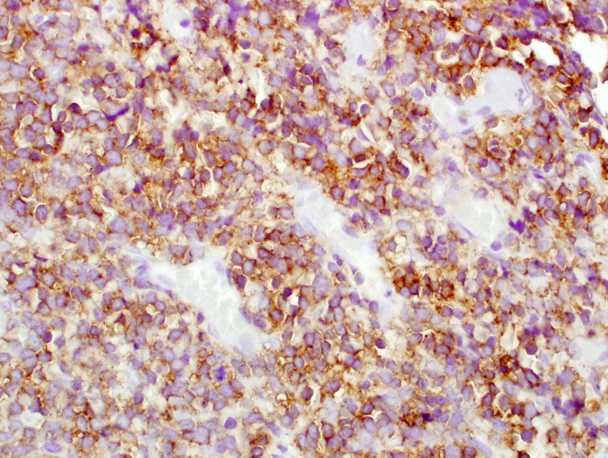 |