Institution: UT MD Anderson Cancer Center
Additional authors:Xiaohong I. Wang, Keyur Patel, Mark Routbort, Zhuang Zuo, Pei Lin, Carlos E. Bueso-Ramos, Raja Luthra, L. Jeffrey Medeiros, C. Cameron Yin
Session: Extramedullary manifestations of myeloid neoplasms
HISTORY
The patient is a 71-year-old woman who was diagnosed with myeloid sarcoma (MS) of breast and skin without bone marrow (BM) involvement in January 2001. She was treated with idarubicin and cytarabine and achieved complete remission. She relapsed with a large abdominal mass that was proven to be MS in March 2008. Bone marrow was not involved. The patient was then treated with idarubicin, cytarabine, fludarabine and mylotarg, and achieved a second complete remission. She was on maintenance therapy with decitabine till December 2009. The patient recently developed neutropenia since December 2012. A complete blood cell count showed white blood cell 4.2 K/uL, hemoglobin 11.8 g/dL, and platelet count 71 K/uL, with 5% circulating blasts. A BM biopsy performed on January 8, 2013 showed 21% blasts of myeloid immunophenotype consistent with acute myeloid leukemia (AML). She was treated with idarubicin, cytarabine and clofarabine, and achieved complete remission on February 7, 2013.
DETAILS
Histologic sections of the breast and skin specimens collected in 2001 and abdominal mass collected in 2008 showed similar morphology findings. There was an extensive infiltration of immature cells with fine chromatin, distinct nucleoli and moderate amount of cytoplasm. Mitotic figures were easily identified.
The BM biopsy and aspirate specimens showed normocellular (30-40%) bone morrow with megakaryocytic and myeloid hypoplasia, dysmegakaryopoiesis, dyserythropoiesis and increased blasts (21%).IMMUNOHISTOCHEMISTRY AND FLOW CYTOMETRY
Immunohistochemical stains performed on the breast specimen showed that the neoplastic cells were positive for myeloperoxidase, CD68, CD43, CD45, and were negative for CD3, CD20, CD34, CD79a, TdT and cytokeratin.
Flow cytometry immunophenotypic analysis of the BM aspirate material revealed a population of blasts that were positive for CD13, CD33, CD34, CD38, CD49d, CD117, CD123 and HLA-DR, and were negative for CD2, CD3, CD4, CD7, CD10, CD15, CD19, CD22, CD36, CD41, CD56, CD64, CD184, TdT and myeloperoxidase, consistent with myeloid immunophenotype.CYTOGENETIC FINDINGS
Conventional cytogenetic analysis showed 46,XX[20].
FISH analyses were negative for the presence of a clone with a RUNX1/RUNX1T1 or CBFB gene rearrangement.MOLECULAR FINDINGS
A next generation sequencing-based analysis for the detection of frequently reported point mutations in a total of 53 genes was performed on DNA extracted from the BM aspirate sample.
Mutations in IDH2 (R140Q), TP53 (S241F) and GNAS (R201H) were detected. No mutation in FLT3 or NPM1 were detected.INTERESTING FEATURES
Myeloid sarcoma usually occurs in a patient with a history of AML or in association with an antecedent myeloproliferative disorder or myelodysplastic syndrome. Less commonly, MS can manifest initially as an isolated mass; in many of these patients, AML subsequently develops. Although its clinicopathologic features have been described in case series, its molecular aberrations have rarely been reported.
We report a patient with de novo MS who subsequently developed AML with diploid karyotype. We assessed mutations in a total of 53 genes, and detected mutations in 3 genes: IDH2, TP53 and GNAS.PROPOSED DIAGNOSIS
De novo myeloid sarcoma with subsequent AML development, diploid karyotype and mutations in IDH2, TP53 and GNAS.
CONSENSUS DIAGNOSIS
Myeloid sarcomas involving breast and skin, relapse with abdominal mass, second relapse with acute myeloid leukemia, diploid karyotype with mutations in IDH2, TP53 and GNAS, involving bone marrow
| Histologic sections of the breast show an extensive infiltration of immature cells with fine chromatin, distinct nucleoli and moderate amount of cytoplasm (H&E, 400x). | 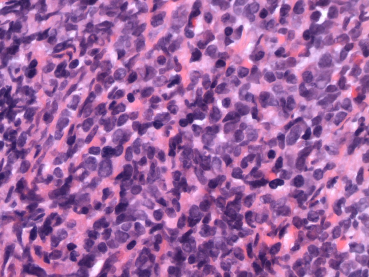 |
| Immunohistochemical stain shows that the neoplastic cells are strongly positive for CD43 (400x). | 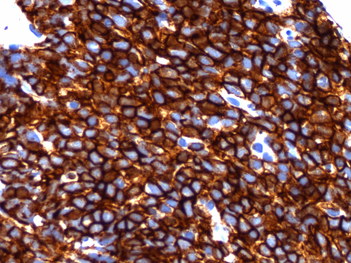 |
| Immunohistochemical stain shows that the neoplastic cells are positive for CD68 (400x). | 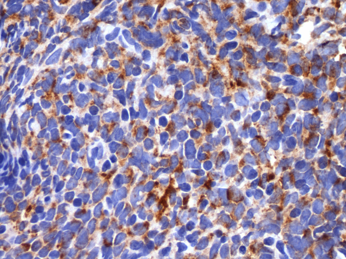 |
| Immunohistochemical stain shows that the neoplastic cells are positive for myeloperoxidase (400x). |  |
| The bone marrow biopsy shows normocellular (30-40%) bone morrow with increased immature cells (H&E, 400x). | 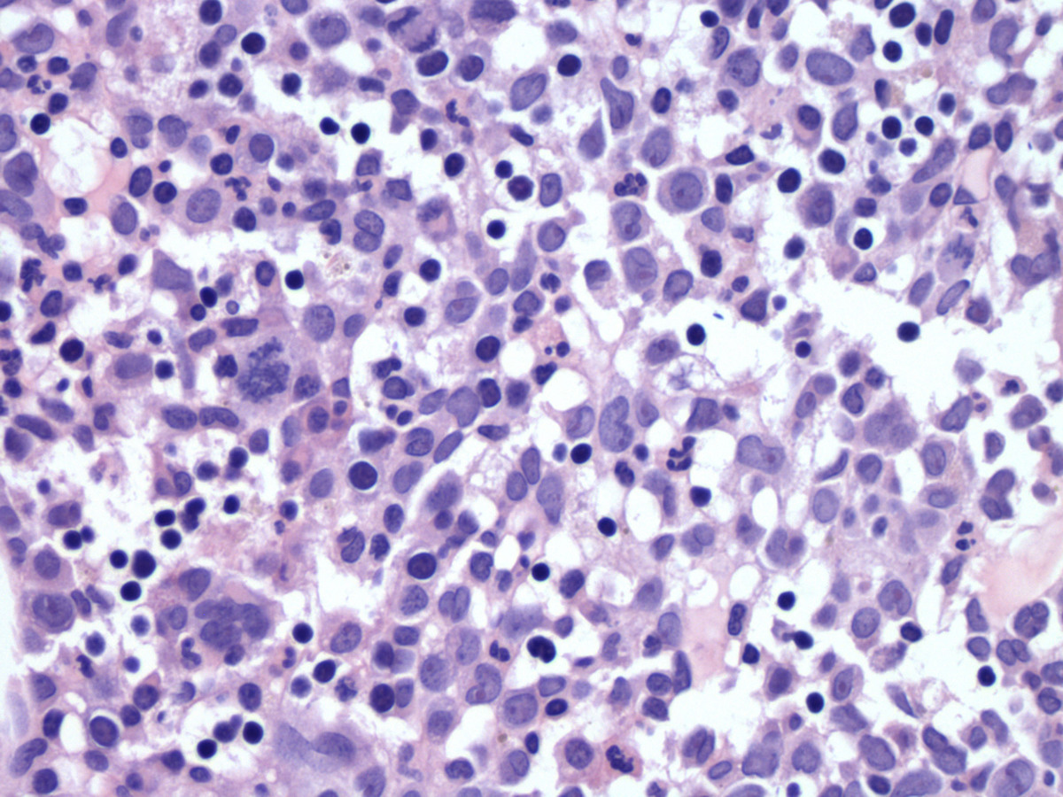 |
| The aspirate smear shows increased blasts. Some blasts have monocytic differentiation (Wright-Giemsa, 1000x). | 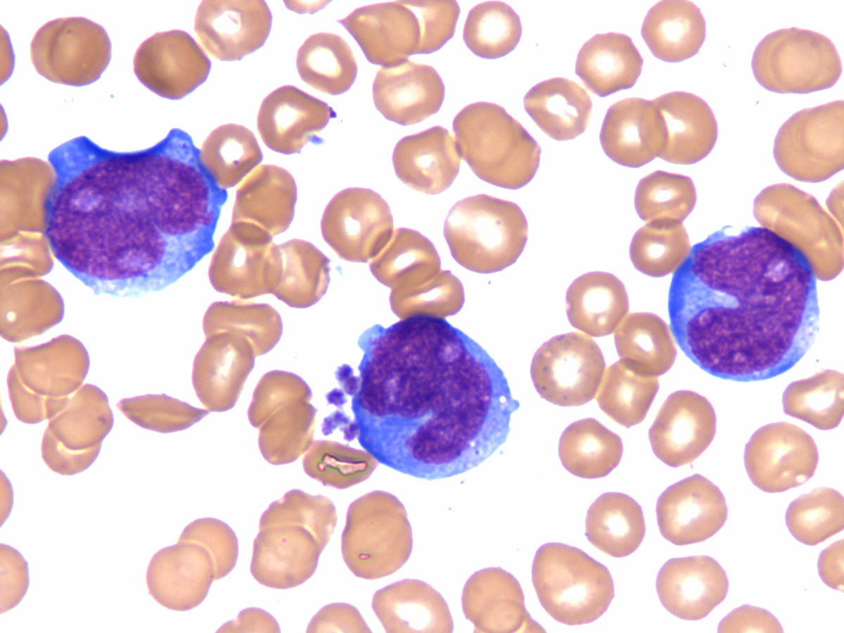 |