Institution: Mount Sinai Medical Center
Additional authors:Vathany Sriganeshan
Session: Erythroleukemia and megakaryoblastic AML and mimics
HISTORY
A 65 year old male with a history of CLL,SLL, and diagnosed in 2004 with bone marrow involvement. At that time, cytogenetics was normal and FISH was positive for trisomy 12, subsequently treated with rituxan, fludarabine, and cyclophosphamide and achieved complete remission after treatment. In 2006, the patient was diagnosed with acute promyelocytic leukemia, cytogenetics and FISH proven, subsequently treated with induction, consolidation, and maintenance, and achieved complete response. The patient represented in April 2012 with severe fatigue and a hemoglobin of 8. Bone marrow biopsy at that time was performed and was submitted for diagnostic consideration. The cytogenetics showed a complex karyotype. CLL panel did not show a trisomy 12 but did show P53 deletion in 50% of the cells. Diagnosis of therapy-related MDS was favored. The patient received 2 cycles of vidaza and follow-up marrow was performed. This is also submitted with possibility of erythroleukemia raised given tremendous erythroid proliferation.
His CBC showed: WBC: 4.2, RBC: 2.1, HGB: 6.1, HCT: 17.5 %, MCV: 83.7, MCH: 29.4, MCHC: 39.2, RDW: 14.9, PLTS: 85, NEUT: 66%, LYMPH: 19%, ATYPICAL LYMPHOCYTES: 14%, BLASTS: 1%DETAILS
A left posterior iliac crest bone marrow biopsy was fixed in 10% buffered formalin. Aspirate smear slides were stained with Giemsa, along with a peripheral blood smear.
The peripheral blood smear showed decreased platelets, mild anisocytosis, poikilocytosis, atypical lymphocytes, rare myelocytes, pseudo Pelger-Huet like cells and rare blasts. The aspirate smears showed mild dyserythropoiesis, mild megaloblastoid changes and normal granulocytic maturation. Few erythroid precursors showed cytoplasmic vacuoles. Hypolobated megakaryocytes were seen. The differential count on the smears showed: blasts: 2%, myelocytes: 3%, maturing granulocytes: 20%, erythroid: 69%, lymphocytes: 5% and eosinophils: 1%. The bone marrow biopsy showed hypercellular marrow (60% cellular) with trilineage hematopoiesis and erythroid hyperplasia. A small lymphoid aggregate is present. The Prussian blue stain of the biopsy revealed increased iron storage.IMMUNOHISTOCHEMISTRY AND FLOW CYTOMETRY
Stains for CD34 and CD117 do not show any increase in blasts. CD117 stains many of the erythroid precursors as is Hemoglobin A. CD3 and CD20 stains revealed that the lymphoid aggregate is composed predominantly of small B cells. A reticulin stain showed marked increase in reticulin fibrosis. A PAS stain showed intense cytoplasmic staining in some of the erythroid precursors.
Flow cytometry of the bone marrow showed persistent chronic lymphocytic leukemia (5% of total cells) and increase in immature hematopoietic cells (12% of total cells) showing co-expressing of CD 117 with bright CD71, raising the possibility of an erythroproliferative processCYTOGENETIC FINDINGS
FISH study showed deletion of chromosome 13 (D13S319) in 65.5% cells and deletion of chromosome 17 (TP53) in 74.5% of cells
INTERESTING FEATURES
This study is a classic example of therapy related myelodysplastic syndrome/acute myeloid leukemia with del (13q) and del 17.
PROPOSED DIAGNOSIS
Therapy related myelodysplastic syndrome/ therapy-related acute myeloid (erythroid) leukemia (t-MDS/AML)
CONSENSUS GROUP: ADDITIONAL INFORMATION/STUDIES
1. Bone marrow in April: blast= 3%, erythroid = 69%, and left-shift of the erythroid lineage was not appreciated.
2. After Vidaza treatment, there were 38% immature erythroid by our estimation.
3. The full karyotype was: 44~45,XY,t(1;3)(q25;q27)dup(1)(p13p36.3),hsr(2)(q37),der(6)t(6;17)(p25;q11.2),-9,add(15)(p11.2)-16,-17,-17,-19,+1~4mar[cp5]/46,XY[17]
No cytogenetic studies were done on the current case. However, FISH showed deletion of chromosome 13 which was not noted on the original BM in April.
4. No molecular tests were performed
5. Patient lost to follow-up
CONSENSUS DIAGNOSIS
Therapy related myeloid neoplasm (2% blasts), with prominent left-shifted erythroid component
Residual chronic lymphocytic leukemia (5%)| Pelger Huet like cell in PBS | 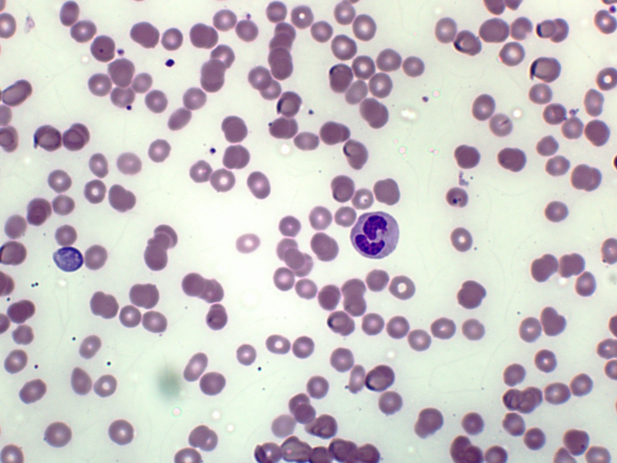 |
| Blast in PBS |  |
| Aspirate smear showing vacoules in the RBC precursors | 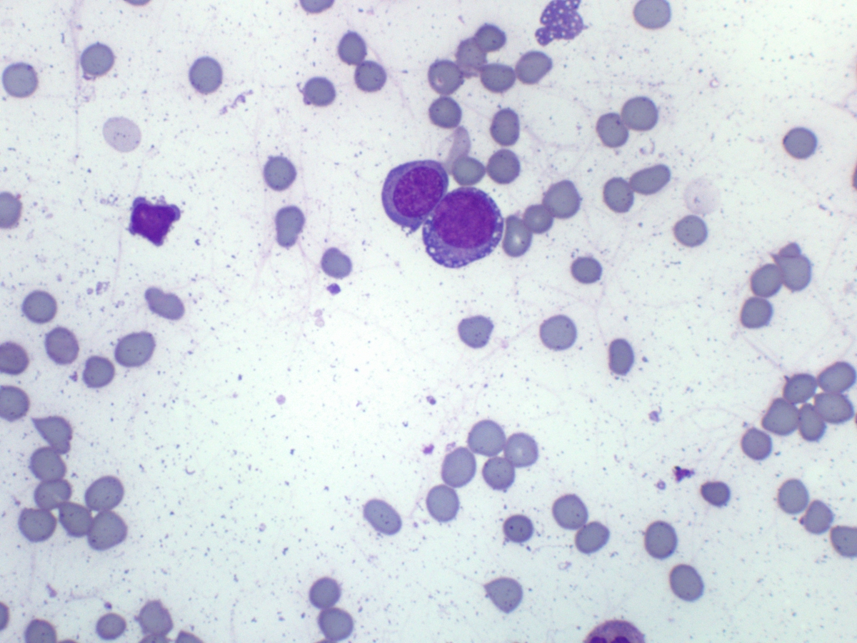 |
| Aspirate smear showing dyserythropoiesis |  |
| Hypolobated megakaryocytes |  |
| bone marrow biopsy |  |
| Hemoglobin A stain | 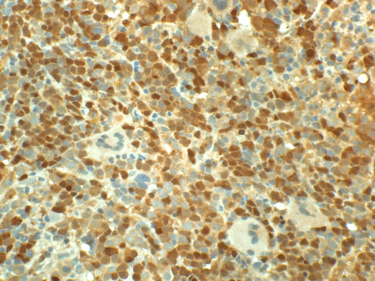 |
| Fish, deletion of chromosome 17 (TP53) | 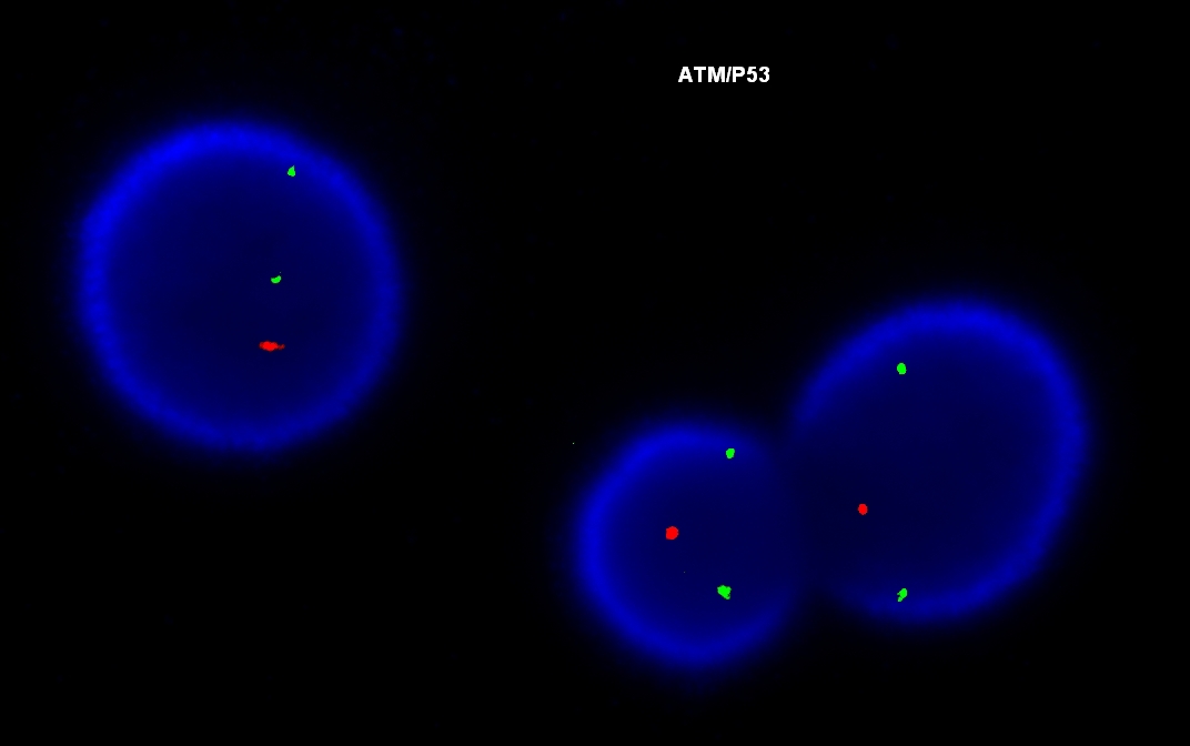 |
| FISH, deletion of chromosome 13 (D13S319) | 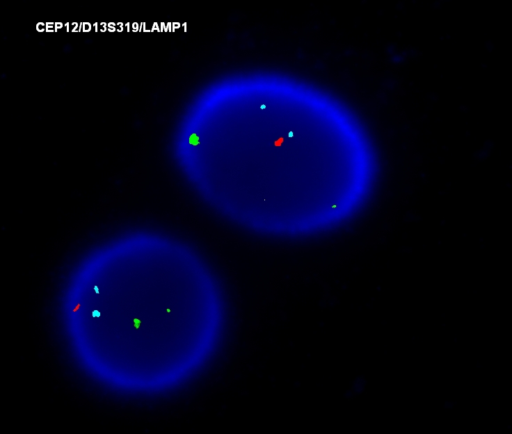 |