Institution: Moffitt Cancer Center
Additional authors:Peter Papenhausen, PhD and Ling Zhang, MD
Session: Myeloid and lymphoid neoplasms with eosinophilia and abnormalities of PDGFRA, PDGFRB, or FGFR1
HISTORY
70 year old man who was originally diagnosed with acute myeloid leukemia 4 years ago when he presented with weakness and fatigue. Cytogenetics showed a rearrangement involving 4q12. He received induction chemotherapy with mitoxantrone and cytarabine. He had residual disease on a post-therapy biopsy. He received reinduction therapy with CLAG and imatinib with a complete response. Subsequently he received consolidation with cytarabine followed by maintenance azacitidine for 1 year. Three years after diagnosis he had evidence of dyspoiesis on bone marrow biopsy. After 1 additional cycle of azacitidine he was found to have increased blasts in the peripheral blood. A bone marrow biopsy confirmed relapsed acute myeloid leukemia. Again he received induction chemotherapy with CLAG and imatinib with a partial response. Imatinib dosage was titrated up for an increasing white blood cell count. Again he had a response with a decrease in blasts and he subsequently remained on imatinib. Recently he was found to have low level blasts on a follow-up bone marrow biopsy. A repeat bone marrow biopsy 3 weeks later showed acute myeloid leukemia in relapse.
DETAILS
Over the course of therapy, the patient has undergone several bone marrow biopsies from the posterior iliac crests. Aspirate smears were prepared at the time of biopsy and stained with Wright Giemsa. The clot sections and core biopsies were fixed in B plus Fix (BBC) fixative. The most recent bone marrow is variably cellular, averaging 30%. Dysplastic changes are noted in the megakaryocytes. Eosinophils are not increased in the bone marrow or peripheral blood. Blasts approach 20% of the intact cellularity on the core biopsy.
IMMUNOHISTOCHEMISTRY AND FLOW CYTOMETRY
By immunohistochemical stains on the core biopsy, the blasts express CD34 and variable CD117. Flow cytometry identified a population of blasts expressing CD34, CD117 and CD56. Previous flow cytometry performed during the previous relapse showed the blasts to express CD7, CD33, CD34, CD56, CD117 and HLA-DR.
CYTOGENETIC FINDINGS
At the time of initial diagnosis cytogenetics reported 46,XY,t(2;4)(p11.2;q12)[18]/46,XY[2]. Three years after diagnosis FISH and cytogenetics again revealed a rearrangement involving PDGFRA in a marrow with dyspoiesis but no significant increase in blasts. On relapse of his acute myeloid leukemia cytogenetics showed an evolved clone with 46,XY,der(2)inv(2)(p21q11.2)t(2;4)(p11.2;p11.2),der(4)t(2;4)(p11.2;p11.2)[4]/46,XY[16]. This clone is again present in the most recent specimen in 8 or 20 cells. In the interim between the two episodes of relapse, the patient had normal cytogenetics on bone marrow biopsy.
MOLECULAR FINDINGS
FISH for PDGFRA was not performed on the initial bone marrow. Subsequent marrows have shown PDGFRA translocation by FISH confirming cytogenetic findings. FISH on the most recent marrow showed persistence of PDGFRA translocation while FISH for PDGFRB and FGFR were negative.
INTERESTING FEATURES
While some of the post therapy specimens showed a mild increased eosinophils in the marrow, there has not be a significant sustained peripheral eosinophilia to date in this patient. There were a couple of isolated mild increases in eosinophils (0.5 k/uL) on CBC over the last 4 years. PDGFR alpha rearrangements are classically associated with peripheral eosinophilia with degranulated eosinophils.
Neoplasms with PDGFRA rearrangement can present initially as acute myeloid leukemia. Neoplasms with PDGDRA rearrangements have been shown to have sensitivity to imatinib , including those in blast phase. FISH for PDGFRA should be considered in cases with increased atypical eosinophils or if cytogenetics demonstrates alteration involving 4q12.PROPOSED DIAGNOSIS
Acute Myeloid Leukemia with PDGFRA rearrangement.
CONSENSUS DIAGNOSIS
Acute myeloid leukemia with PDGFRA rearrangement, t(2;4)(p11.2;q12)
| Aspirate smear (WG) showing an area with increased blasts - 100X |  |
| CD34 immunohistochemical stained section of core biopsy showing area of increased blasts. |  |
| FISH using tricolor probes for SCFD2/FIP1L1, LNX/CHIC2 and PDGFRA demonstrated a population of nuclei with a translocation of the PDGFRA gene from chromosome 4q12. An additional LNX/CHIC2 signal was also observed. | 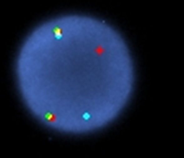 |
| H&E stained core biopsy section. |  |