Institution: Baylor College of Medicine
Additional authors:M. Tarek Elghetany; Choladda V. Curry
Session: Acute leukemias of ambiguous lineage
HISTORY
An 18 month old boy first presented in 2005 with acute myeloid leukemia with MLL gene rearrangement treated with St. Jude’s AML 2002 protocol . He then had a first relapse of disease in 2006 as B-acute lymphoblastic leukemia with MLL gene rearrangement. He was treated with individualized induction protocol based on COG AALL0331, achieved remission, and underwent a matched unrelated bone marrow transplant. He was in remission until in 2012 at 9 years of age, when he presented with leg and hip pain, and urinary and fecal incontinence. MRI scan of the spine revealed an intraspinal extradural enhancing soft tissue mass resulting in compression of the distal spinal cord from level of T11 through L2. Laminectomy and biopsy of the mass were performed. After the diagnosis of a second relapse was made, he was treated with an individualized protocol based on COG AALL0433 and is alive 7 months after a second relapse.
DETAILS
Biopsy Fixation Details:
10% neutral buffered formalinDetails of Microscopic Findings: Bone marrow 2005 (Figs. 1 and 2): The bone marrow biopsy shows extensive infiltration by blasts. Bone marrow aspirate smears show blasts (>90%) with features of myeloid differentiation, demonstrating medium to large sized blasts with high nuclear to cytoplasmic ratio, fine, lacy chromatin with occasional prominent nucleoli and a thin rim of pale blue cytoplasm.Bone marrow 2006 (Figs. 6 and 7): The biopsy shows extensive infiltration by blasts. The aspirate demonstrates a population of blasts (>90%) with an appearance of lymphoblasts. The blasts are predominantly small with some variation; having a high nuclear to cytoplasmic ratio, moderately fine nuclear chromatin, scant amounts of basophilic cytoplasm, and inconspicuous nucleoli. Extradural vertebral mass in 2012 (Fig. 11): The biopsy demonstrates a sheet of monomorphic blasts with irregular nuclear contours, and a small amount of cytoplasm. Scattered mitotic figures are seen.IMMUNOHISTOCHEMISTRY AND FLOW CYTOMETRY
Immunophenotyping by Immunohistochemistry and/ or Flow Cytometry:
Bone marrow 2005 (Fig. 3): Initial flow cytometric findings show a population of 74% blasts that are positive for CD33, CD15, CD4, CD16, CD56, HLA-DR, and CD11b. The blasts are negative for CD3, CD5, CD7, CD8, CD19, CD10, CD20, CD22, kappa and lambda, CD13 and CD14. Bone marrow 2006 (Fig. 8): Results of flow studies in the subsequent relapse bone marrow show a population of 91% blasts that mark as leukemia of early B lineage. The blasts are positive for CD19, CD22, and HLA-DR. No expression of CD20, surface light chains, CD10, CD3, CD4, CD5, CD7, CD8, CD71, CD138, CD16, CD56, CD34, CD117, CD33, CD15, CD11b, CD14 or CD13 is seen.Extradural vertebral mass 2012 (Fig.12): The extramedullary mass is composed of a population of 65% blasts that express CD45 (dim), CD19 (partial), CD11b (partial), CD79a (partial), CD38 and HLA-DR. The blasts do not express CD3 (surface and cytoplasmic), CD2, CD5, CD7, CD4, CD8, CD10, CD20, CD22 (surface and cytoplasmic), kappa and lambda, CD13, CD14, CD33, CD23, CD15, CD16, CD56, CD117, CD36, CD64, TdT and MPO.CYTOGENETIC FINDINGS
Bone marrow 2005 (Figs. 4 and 5):46,XY,add(12)(p11.2)[9].ish ins(9;11)(p22;q23q23)(5'MLL+,9cen+,WCP11-; MLL+, WCP11+) /46,XX[15] nuc ish 11q23(MLLx2)(5'MLL spx1)[124]/11q23(MLLx2)[76]
Interpretation: cryptic translocation/insertion of MLL gene into 9p22 leading to MLLT3-MLL fusion.Bone marrow 2006 (Figs. 9 and 10): 46,XY,t(14;19)(q32;p13).ish t(14;19)(q32;p13)(3’IGH+,5’IGH con 3’IGH+)[7]/46,XY[23] nuc ish (5’MLL x3,3’MLL x2)(5’MLL con 3’MLL x2)[45]/(MLL x2)[155]/12p13(TELx2), 21q22(AML1x2)[200]Interpretation: Abnormal cytogenetic and FISH results showing evidence of the previous cryptic MLL rearranged clone with a new clone involving IGH gene rearrangement.Extradural vertebral mass 2012 (Figs. 13 and 14): 46,XY,inv(3)(p21q27),inv(12)(q13q24.3),t(14;19)(q32;p13)[8]/46,XY[1].ish ins(9;11)(p22;q23q23)(5'MLL+;MLL+) nuc ish(5'MLLx3,3'MLLx2)(5'MLL con 3'MLLx2)[134/200]/(ETV6x2)[200]/(5'IGHx2,3'IGHx3)(5'IGH con 3'IGHx2)[136/200]Interpretation: Abnormal chromosome and FISH analyses showing previously reported crypticvariant MLLT3-MLL fusion and IGH gene rearrangements.INTERESTING FEATURES
Our patient initially presented with acute myeloid leukemia with recurrent genetic abnormality involving the MLL gene [t(9;11)(p22;q23); MLLT3-MLL]. Features of myeloid lineage included morphologic findings consisting of blasts of medium to large size with occasional prominent nucleoli and a rim of pale blue cytoplasm. Flow cytometric studies demonstrated that the blasts were of myeloid lineage without expression of any lymphoid markers. The patient subsequently had a bone marrow relapse with blasts showing different morphology and immunophenotyping. These blasts were small in sized with scant cytoplasm and inconspicuous nucleoli and expressed early B lymphoid markers without myeloid surface markers. Cytogenetics studies showed a clone with persistent MLL gene rearrangement in addition to another clone with IGH gene involvement. The patient later had a second relapse, which was extramedullary. The blasts were of ambiguous lineage but maintained the same cytogenetics abnormalities as the first relapse, including MLL gene rearrangement. While the WHO 2008 classification recognized acute myeloid leukemia with t(9;11)(p22;q23); MLLT3-MLL, this case demonstrates that t(9;11), like other MLL gene rearrangement with other partners, is not only involved in leukemogenesis but also in providing the cells with a potential for lineage switch through an alternative differentiation pathway to survive the initial chemotherapy and relapse in another cell line that requires different therapeutic strategies.
PROPOSED DIAGNOSIS
Acute leukemia with MLL gene rearrangement with cryptic variant t(9;11)(p22;q23) MLTT3-MLL fusion with evolving new clone of IGH rearrangement presenting as 1) acute myeloid leukemia in 2005, 2) first relapsed as B-acute lymphoblastic leukemia in 2006, and 3) second relapsed as extramedullary (vertebral mass) involvement by acute leukemia of ambiguous lineage in 2012.
CONSENSUS GROUP: ADDITIONAL INFORMATION/STUDIES
Additional immunostaining performed on the epidural mass by the conference consensus group:
MPO: Negative
CD68: Negative
Lysozyme: Negative
CD19: Weakly positive (70%)
CD79a: Positive (90%)
PAX5: Positive (90%)
CONSENSUS DIAGNOSIS
Acute leukemia with MLL gene rearrangement with cryptic variant t(9;11)(p22;q23) MLTT3-MLL fusion with evolving new clone of IGH rearrangement presenting as 1) acute myeloid leukemia in 2005, 2) first relapse as B-acute lymphoblastic leukemia in 2006, and (3) second relapse as extramedullary (vertebral mass) involvement by B-acute lymphoblastic leukemia
| Figure 1 |  |
| Figure 2 |  |
| Figure 3 |  |
| Figure 4 |  |
| Figure 5 | 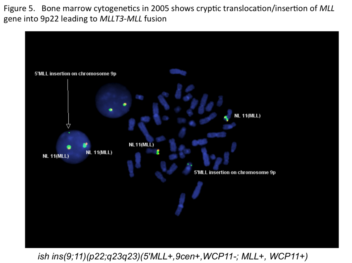 |
| Figure 6 |  |
| Figure 7 |  |
| Figure 8 | 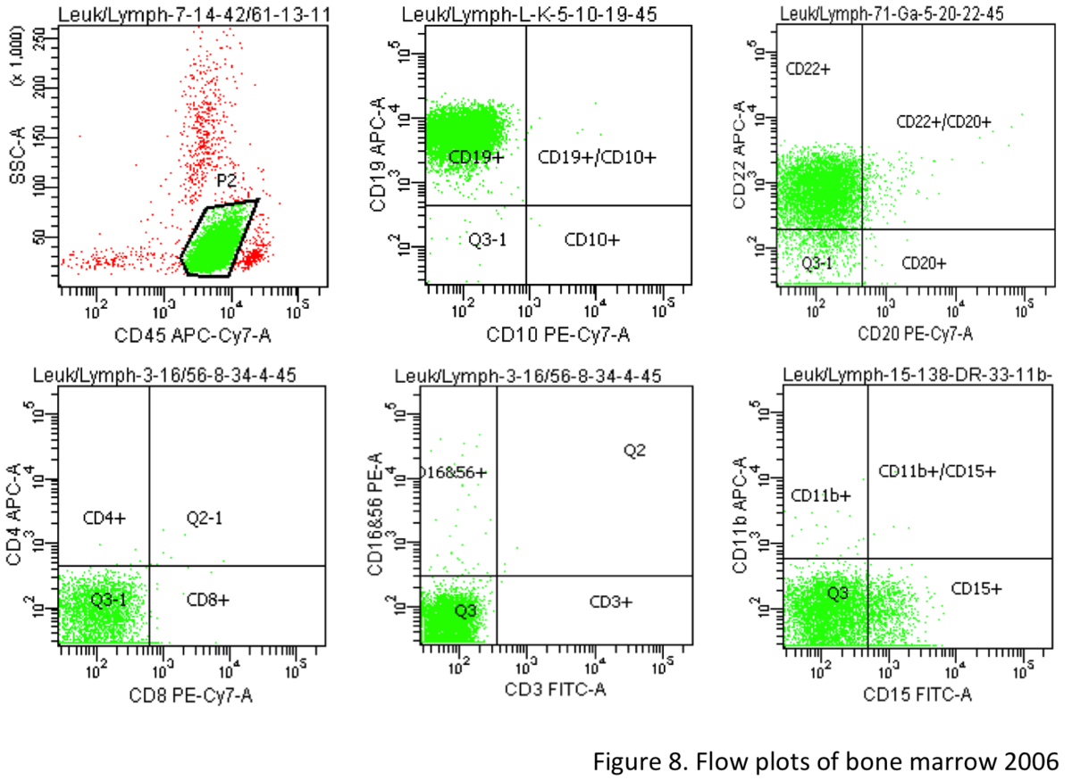 |
| Figure 9 |  |
| Figure 10 |  |
| Figure 11 |  |
| Figure 12 | 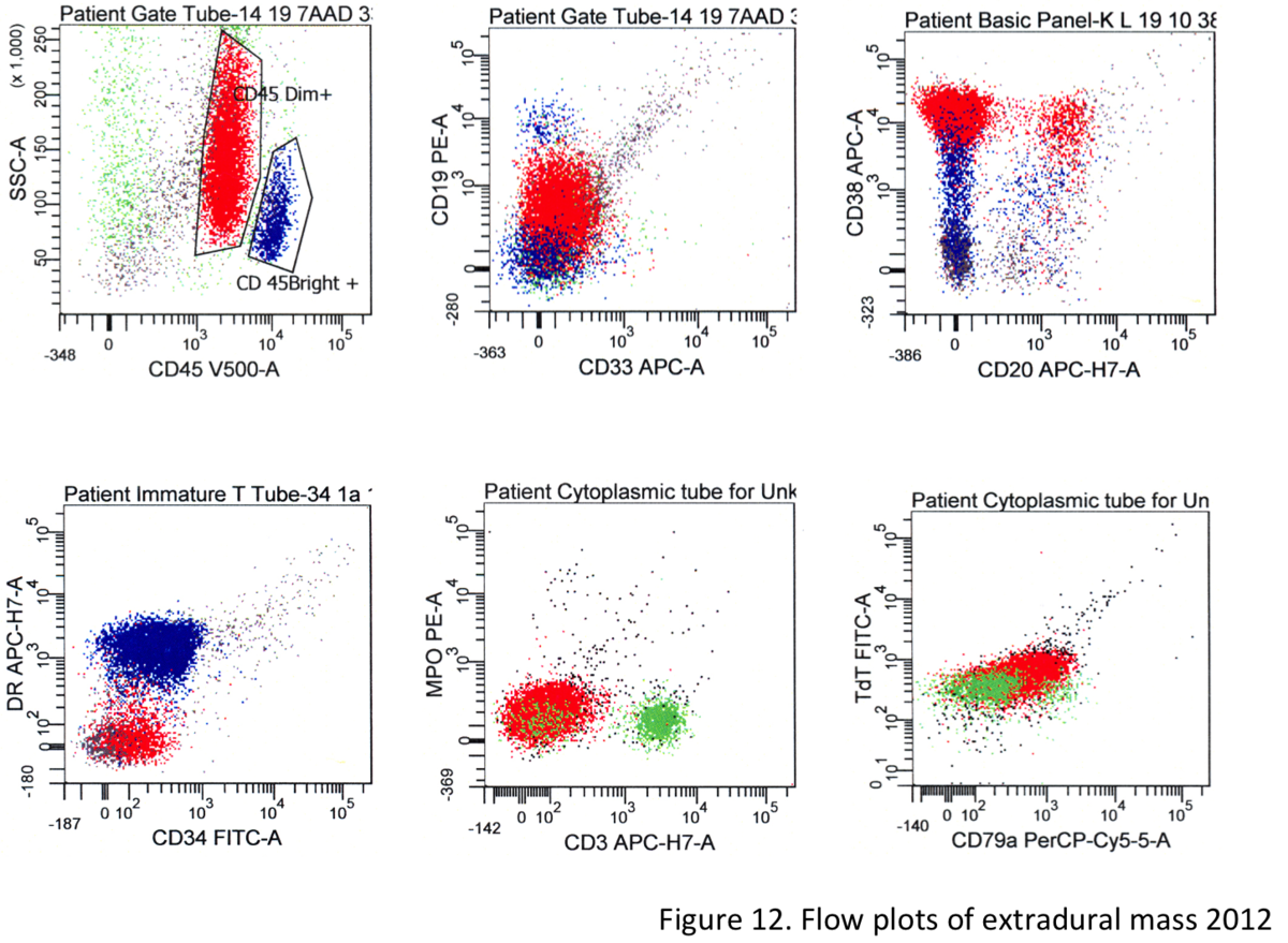 |
| Figure 13 |  |
| Figure 14 | 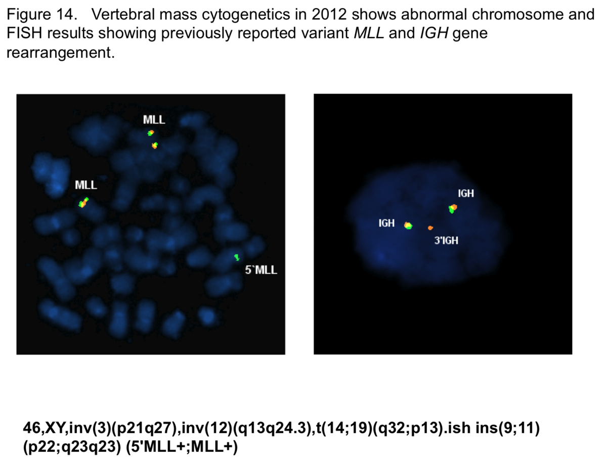 |