Institution: University of New Mexico
Additional authors:Fred Elder, Ph.D., Alexei Bakhirev, M.D., Qian-Yun Zhang, M.D., Ph.D.
Session: AML with myelodysplasia-related changes
HISTORY
The patient is a 65 year old male.
DETAILS
Peripheral blood and bone marrow aspiration and biopsy were submitted for examination. CBC shows WBC 35.6 x 10E3/mm3, Hb 11.2 gm/dL, plt 38 x 10E3/mm3, 24% blasts. The circulating blasts are intermediate to large cells with moderate to scant cytoplasm, high N/C ratio, fine chromatin and prominent nucleoli. A subset of blasts also have a few fine cytoplasmic granules. The maturing granulocytes show significant dysplastic changes including hypogranularity and nuclear hypolobation (Image 1). Bone marrow aspirate reveals an increased M:E ratio, 22% blasts, frequent hypolobated maturing granulocytes, and mild dyserythropoiesis. Megakaryocytes are exceedingly rare (Image 2). Bone marrow core biopsy and clot section show 100% cellularity. The marrow is predominantly comprised of left shifted myeloid precursors. Hypolobation is the dominant feature within the maturing cells. Scattered erythroid precursors are seen. Rare megakaryocytes demonstrate mononucleation or hypolobulation (Image 3).
IMMUNOHISTOCHEMISTRY AND FLOW CYTOMETRY
Flow cytometric analysis demonstrates a population of myeloblasts which express CD34, CD117, CD13, HLA-DR, subset CD33, subset CD4, and CD45 (dim). The blasts are negative for monocytic markers (Image 4).
CYTOGENETIC FINDINGS
Karyotype:
45,XY,der(16)t(16;17)(q12~13;q11.2),-17[3]/
46,sl,+11[10]/
47,sdl1,+9,+11[2]
47,sdl2,+11,+13[2]
46,XY[3]
FISH: FISH for t(8;21) and rearrangement of CBFB on chromosome 16(p13q22) are negative, though there is a loss of CBFB gene region in 87% of cells (Image 5).
FISH for p53 gene confirms the deletion of one copy (Image 6).
MOLECULAR FINDINGS
INTERESTING FEATURES
17p deletion is most often seen in the presence of complex cytogenetic abnormalities and results in poor clinical outcome. Nonrandom chromosomal abnormalities resulting in 17p deletion as sole abnormality are seen in 1-2% of AML cases. The prognostic significance of AML with loss of 17p as sole abnormality has now been linked to in vitro drug resistance and short overall survival. AML with sole 17p loss typically exhibits dysgranulopoiesis including hypolobulation and pseudo-Pelger-Huet forms. Here we present a case with t(16;17)(q12~13;q11.2) resulting in loss of p53 and CBFB genes. This case demonstrates the typical morphologic findings in association with p53 gene deletion. The presence of multiple side lines is also indicative of progression of the disease and predicts a poor clinical outcome. The significance of deletion of CBFB is less clear.
References:
1. Haferlach C, Dicker F, Herholz H et al. Mutations of the TP53 gene in acute myeloid leukemia are strongly associated with a complex aberrant karyotype. Leukemia 2008; 22: 1539–1541.
2. Grimwade D, Hills RK, Moorman AV, et al. National Cancer Research Institute Adult Leukaemia Working Group. Refinement of cytogenetic classification in acute myeloid leukemia: determination of prognostic significance of rare recurring chromosomal abnormalities among 5876 younger adult patients treated in the United Kingdom Medical Research Council trials. Blood. 2010 Jul 22;116(3):354-65.
3. Seifert H, Mohr B, Thiede C, et al. Study Alliance Leukemia (SAL). The prognostic impact of 17p (p53) deletion in 2272 adults with acute myeloid leukemia. Leukemia. 2009 Apr;23(4):656-63.
4. Nahi H, Lehmann S, Bengtzen S, et al. Chromosomal aberrations in 17p predict in vitro drug resistance and short overall survival in acute myeloid leukemia. Leuk Lymphoma 2008; 49: 508–516.
5. Soenen V, Preudhomme C, Roumier C, et al. 17p Deletion in acute myeloid leukemia and myelodysplastic syndrome. Analysis of breakpoints and deleted segments by fluorescence in situ. Blood. 1998 Feb 1;91(3):1008-15.
6. Lai JL, Preudhomme C, Zandecki M, et al. Myelodysplastic syndromes and acute myeloid leukemia with 17p deletion. An entity characterized by specific dysgranulopoïesis and a high incidence of P53 mutations. Leukemia. 1995 Mar;9(3):370-81.
7. Fenaux P, Jonveaux P, Quiquandon I, et al. P53 gene mutations in acute myeloid leukemia with 17p monosomy. Blood. 1991 Oct 1;78(7):1652-7.
8. Jary L, Mossafa H, Fourcade C, et al. The 17p-syndrome: a distinct myelodysplastic syndrome entity? Leuk Lymphoma. 1997 Mar;25(1-2):163-8.
PROPOSED DIAGNOSIS
Acute myeloid leukemia with myelodysplasia-related changes and deletion of 17p
CONSENSUS DIAGNOSIS
Acute myeloid leukemia with myelodysplasia-related changes (by complex karyotype and morphology), with TP53 deletion
| Image 1. Peripheral blood smear demonstrating circulating blasts (arrow) and dysgranulopoiesis including hypogranulation, hypolobation, and pseudo-Pelger-Huet forms (arrow heads). | 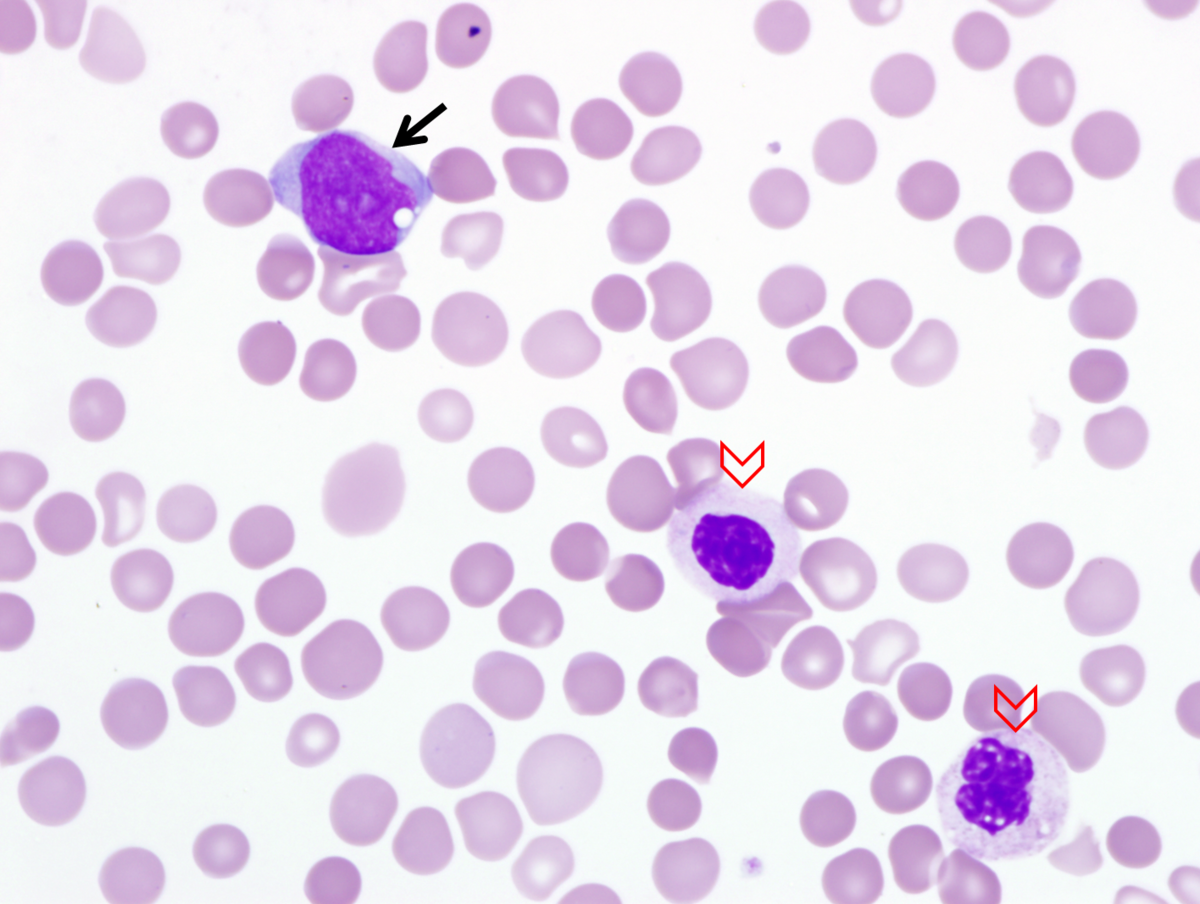 |
| Image 2. Bone marrow aspirate reveals increased blasts (arrows), maturing granulocytic precursors with hypolobulated nuclei including pseudo-Pelger-Huet form (arrow heads). Erythroid precursors exhibit dysplastic changes including binucleation, abnormal hemoglobinization, and basophilic stippling (double arrows). | 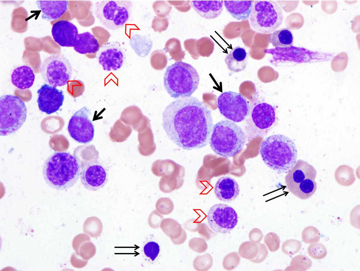 |
| Image 3. Clot section demonstrates 100% cellularity with predominantly immature myeloid precursors. The maturing granulocytes exhibit prominent nuclear hypolobulation. Erythroid and megakaryocytes are markedly reduced. | 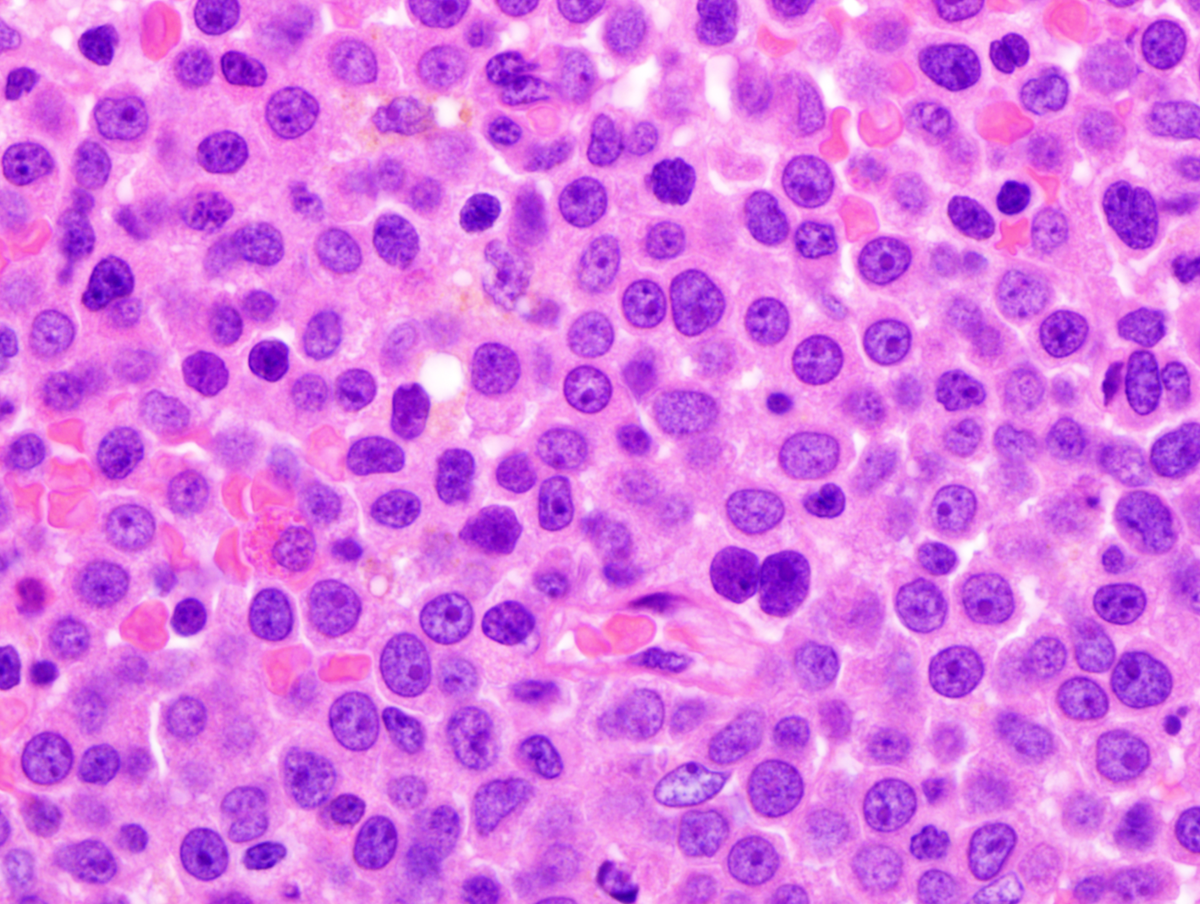 |
| Image 4. Flow cytometric analysis identified a population of blasts with expression of CD34, CD117, HLA-DR, CD13, subset CD4 (not shown), subset CD33. The blasts are negative for CD45 and monocytic markers. | 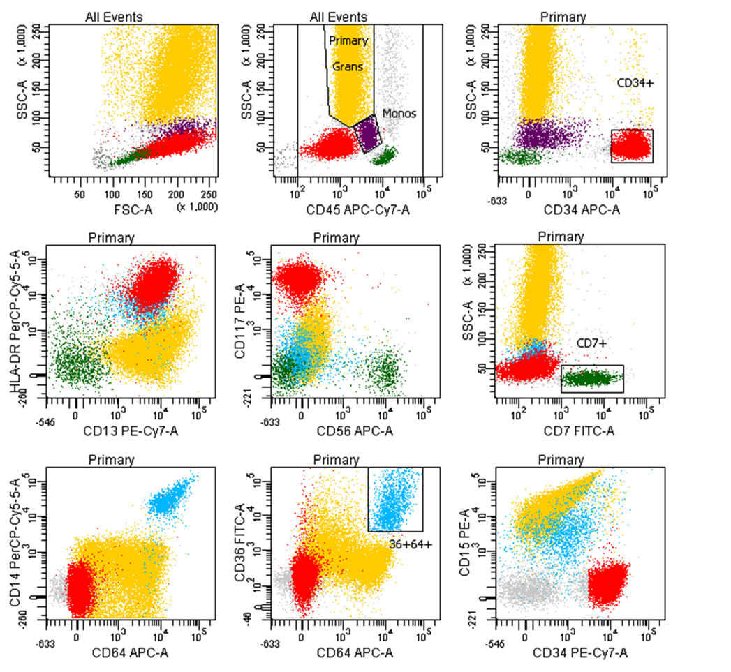 |
| Image 5. Chromosomal study reveals a stem line and multiple side lines. Karyotype: 45,XY,der(16)t(16;17)(q12~13;q11.2),-17[3]/ 46,sl,+11[10]/ 47,sdl1,+9,+11[2] 47,sdl2,+11,+13[2] 46,XY[3] | ![Image 5. Chromosomal study reveals a stem line and multiple side lines. Karyotype: 45,XY,der(16)t(16;17)(q12~13;q11.2),-17[3]/ 46,sl,+11[10]/ 47,sdl1,+9,+11[2] 47,sdl2,+11,+13[2] 46,XY[3]](cases/374/images/SH2013 kry.png) |
| Image 6. FISH for CBFB gene demonstrates deletion of one copy of CBFB gene in 86% of cells. A rearrangement was not detected within the remaining copy observed in this microphotograph. | 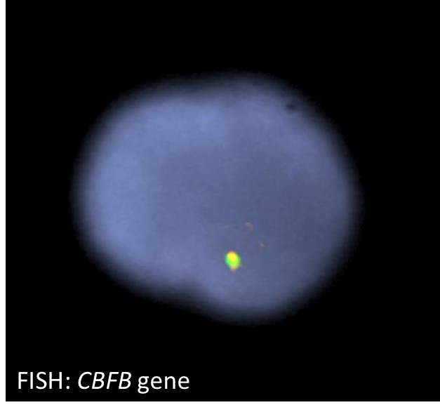 |
| Image 7. FISH for p53 gene confirms deletion of one copy within the majority of cells. | 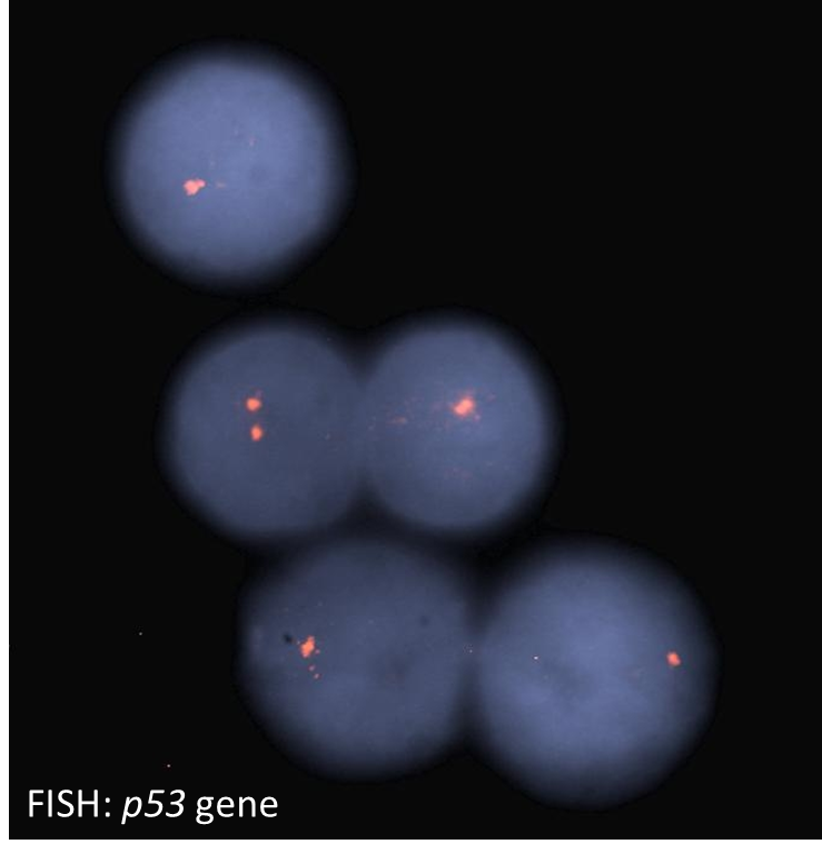 |