Institution: University of Pittsburgh
Session: Extramedullary manifestations of myeloid neoplasms
HISTORY
The patient is an 81 year old male who presented with a 6 month history of weakness, fatigue, skin lesions and decreased peripheral blood counts. Worsening peripheral blood counts and the appearance of blasts in the peripheral blood smear prompted bone marrow evaluation. The skin lesions were also subsequently biopsied.The patient was treated with chemotherapy with initial improvement in peripheral blood counts, but relapse occurred despite therapy with blasts then detected also in the cerebrospinal fluid.
DETAILS
Bone marrow aspirate and biopsy from left posterior iliac crest (fixed in B+ fixative) and peripheral blood smear.Peripheral Blood Counts:WBC 7.4 x10E+9/L ( 3.8 - 10.6)RBC 2.10 x10E+12/L ( 4.13 - 5.57)Hgb 7.7 g/dl ( 12.9 - 16.9)Hct 23.0 % ( 38.0 - 48.8)MCV 109.6 fL ( 82.6 - 97.4)RDW 19.0 % ( 11.8 - 15.2)PLT 52 x10E+9/L ( 156 - 369)Neutrophils 21 % 1.6 x10E+9/L ( 2.24 - 7.68)Metamyelocytes 1% 0.1 x10E+9/LBlasts 30 % 2.2 x10E+9/L Lymphocytes 41 % 3.0 x10E+9/L ( 0.80 - 3.65)Monocytes 6 % 0.4 x10E+9/L ( 0.30 - 0.90)Eosinophils 1 % 0.1 x10E+9/L ( 0.00 - 0.40)The peripheral blood smear shows blasts with large, oval to irregular nuclei containing fine chromatin and some prominent nucleoli. The blastic cells have variable but generally small amounts of agranular cytoplasm. Some hypogranular and hyposegmented neutrophils are seen as well as a mild left shift in neutrophilic maturation. Red blood cells show macrocytosis and mild-moderate anisocytosis.Bone Marrow Morphology: The biopsy shows hypercellular marrow for age with approximately 95% cellularity overall. Most of the marrow space is occupied by sheets of immature-appearing cells with high nuclear:cytoplasmic ratio; some with prominent nucleoli. Megakaryocytes are decreased, and few maturing myeloid or erythroid precursors are evident in the biopsy.In the bone marrow aspirate smears there is a predominance of blasts, which resemble those seen in the peripheral blood. Differential cell counts show approximately 88% blasts with very few maturing myeloid and only rare erythroid precursors found.
IMMUNOHISTOCHEMISTRY AND FLOW CYTOMETRY
Immunophenotype: Flow cytometric evaluation of the bone marrow aspirate shows approximately 80% dim CD45 positive cells with low side scatter that are positive for CD4, CD56, CD123, HLA-DR, and CD7 with partial CD2 and CD36 expression. This population is negative for: CD34, CD117, CD13, CD14, CD16, CD33, CD64, CD11b, CD15, CD3 (surface and cytoplasmic), CD1a, CD5, CD8, CD57, CD19, CD22, CD20, CD41, CD61, glycophorin A, TdT and myeloperoxidase.Immunohistochemical studies of the bone marrow biopsy confirmed expression of CD123, CD56 and CD4 by the blast cell infiltrate, and the absence of CD3. The blasts were also shown to be positive for CD43 and CD68 and negative for CD79a.
CYTOGENETIC FINDINGS
46,XY,del(12)(p11.2p12)[4]/46,sl,del(6)(q21q25)[12]/46,sdl,add(11)(q23)[5]Fluorescence in situ hybridization (FISH) was negative for MLL rearrangement, monosomy 7, trisomy 8 and deletions of 5q31, 7q31 and 20q12.MOLECULAR FINDINGS
Not performed.INTERESTING FEATURES
This case represents an unusual blastic neoplasm presenting as an apparent acute leukemia in which the blasts were negative for multiple lineage-associated antigens (CD3, CD19, CD22, CD79a, myeloperoxidase, CD13, CD33, CD14, CD64, TdT, CD117, CD41, CD61, glycophorin A), though showing some positivity for other, less lineage-specific myeloid and T-cell markers.
Further immunophenotypic evaluation showed the blasts to express CD4, CD56, and CD123 (as well as HLA-DR, CD68, CD43, and CD7). These positive results in combination with the negative findings for other markers analyzed, permitted diagnosis as a blastic plasmacytoid dendritic cell neoplasm.The diagnosis was also suggested by the presence of cutaneous lesions, which were subsequently shown to consist of similar CD4, CD56 and CD123 positive cells. The disease showed a typically aggressive clinical course with development of central nervous system involvement at relapse.PROPOSED DIAGNOSIS
Blastic Plasmacytoid Dendritic Cell Neoplasm
CONSENSUS GROUP: ADDITIONAL INFORMATION/STUDIES
Additional immunohistochemical stains performed by the consensus group:
TCL1: Positive
CONSENSUS DIAGNOSIS
Blastic plasmacytoid dendritic cell neoplasm
| Bone marrow aspirate, Wright-Giemsa. | 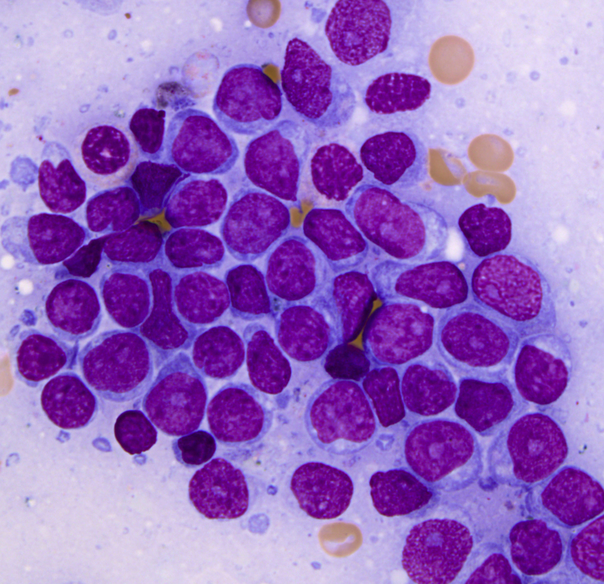 |
| Bone marrow biopsy, H&E, low magnification. |  |
| Bone marrow biopsy, H&E. | 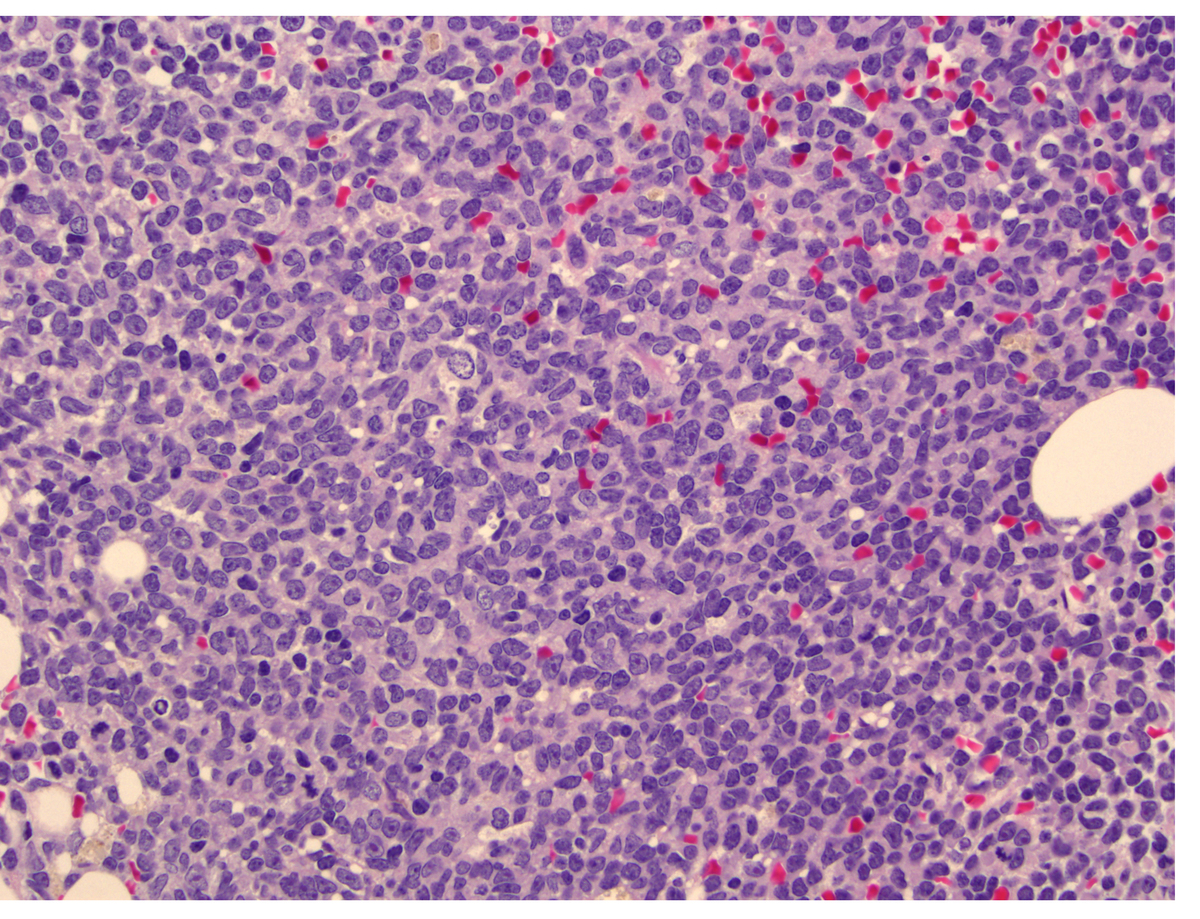 |
| Bone marrow biopsy, H&E. |  |
| Bone marrow biopsy, anti-myeloperoxidase immunostain. |  |
| Bone marrow biopsy, anti-CD4 immunostain. | 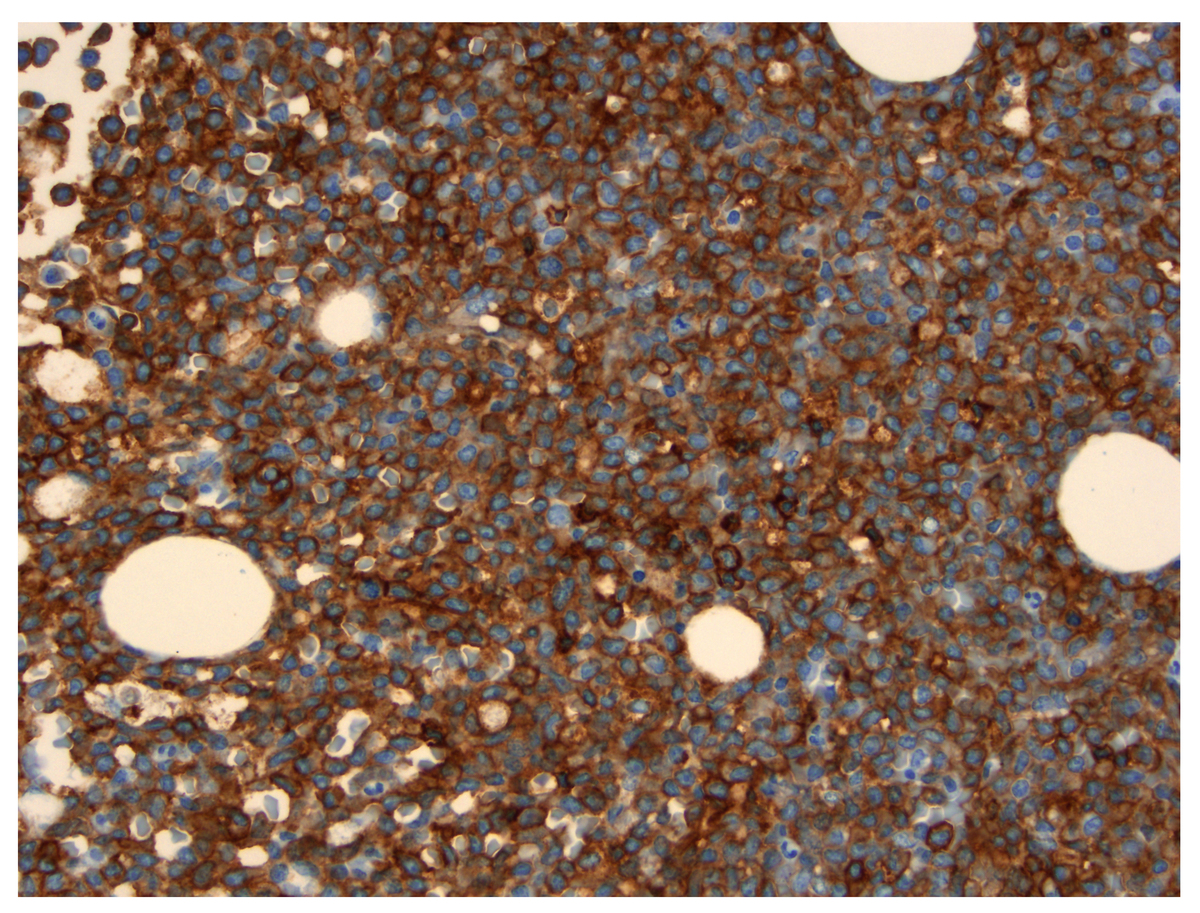 |
| Bone marrow biopsy, anti-CD56 immunostain. |  |
| Bone marrow biopsy, anti-CD123 immunostain. |  |
| Additional figure 1: TCL1 IHC, 400x |  |