Institution: Roswell Park Cancer Institution
Additional authors:George Deeb MD, Eunice S. Wang MD, Sheila N. Jani Sait PhD
Session: AML secondary to myeloproliferative neoplasms and other types of disease progression in MPN
HISTORY
The patient is a 59-year-old male who was diagnosed with essential thrombocythemia 10 years ago when an incidental platelet count showed 1,000, 000/ dL. As he was otherwise asymptomatic, he was managed expectantly on aspirin. In July 2011 he developed thrombocytopenia (~77K/dL), anemia (10.3g/dL) and massive splenomegaly. A bone marrow biopsy was performed in Aug 2011 and showed marked reticulin myleofibrosis, hypercellularity with maturing trilineage elements and dyspoietic megakaryocytic hyperplasia. He was started on hydroxyurea, and referred to Roswell Park Cancer Institute (RPCI) for follow up care. Molecular studies performed on the bone marrow at RPCI were negative for t(9;22) and positive for JAK2 (V617F). Cytogenetic studies showed a male karyotype with 18/20 cells with an interstitial deletion in the long arm of chromosome 13. 15/18 cells had additional deletion of part of the long arm of chromosome #20. 2/18 cells had in addition to the del(13q), trisomy of chromosome 8.
To follow up for persistent and worsening pancytopenia, he underwent a bone marrow biopsy in June 2012.DETAILS
A left iliac trephine core biopsy, aspirate smears, touch imprints and a peripheral blood smear were received. The aspirate smears were markedly hemodilute and non-diagnostic. The touch imprints showed blasts without auer rods and azurophilic granulation. H&E stained sections from the core biopsy showed marrow fibrosis with hypercellularity, and subtotal marrow replacement by blasts. A small focus of chronic myleoproliferative neoplasm (5%-10% of marrow space) was noted. Morphologic findings were consistent with acute leukemia. Peripheral blood smear showed 1% circulating blasts, normocytic anemia and thrombocytopenia
IMMUNOHISTOCHEMISTRY AND FLOW CYTOMETRY
The blasts were diffusely positive for PAX-5, TdT, CD20, CD79a, CD22, CD10, CD45, CD43 and Ki-67 (>95% proliferative index). The blasts were negative for CD34, CD117, MPO, lysozyme, hemoglobin, E-cadherin, CD163, CD3, CD68, CD56, CD123, CD33, CD15, Factor VIII, CD42b, and other T-cell markers tetsted by immunohistochemistry.
Flow cytometry studies reported a 2% CD34 positive myeloblast population and 1% atypical B-lineage population expressing CD19, CD10 (bright), and CD38 (bright), and lacking CD34 and surface immunoglobulin expression (other marker expression was difficult to characterize due to sparse evaluable events seen).Reticulin stain showed marked and diffuse reticulin fibrosis.CYTOGENETIC FINDINGS
August 2011 marrow with MPN (ET):
46,XY,del(13)(q13q21),del(20)(q11.2q13.3)[14]47,XY,+8,del(13)(q13q21)[2]46,XY,del(13)(q13q21)[1]June 2012 marrow with B-ALL:46,XY,del(13)(q13q21),del(20)(q11.2q13.3)[9]MOLECULAR FINDINGS
The marrow with acute B-lymphoblastic leukemia was positive for JAK2 (V617F) mutation and negative for t(9;22) [e1a2, e13a2, and e14a2 BCR-ABL1 transcripts].
INTERESTING FEATURES
1: Uncommon transformation of JAK2 positive and t(9;22) negative essential thrombocythemia to acute B-lymphoblastic leukemia.
2: Discordant flow cytometry results as compared to the bone marrow morphology and immunohistochemistry. 3: The cytogenetic studies showed overall similar chromsomal abnormalities, mainly del13 and del20, in both samples submitted as bone marrow from the chronic disease and the blast transformation.4: Follow up: The patient is status post chemotherapy for B-ALL and the bone marrow from January 2013 was reported as negative for B-lymphoblasts. Karyotype in the marrow continues to be abnormal as noted in the original diagnostic myeloproliferative neoplasm.PROPOSED DIAGNOSIS
Acute B-lymphoblastic leukemia arising from previously diagnosed essential thrombocythemia
CONSENSUS DIAGNOSIS
B-acute lymphoblastic leukemia arising from prior JAK V617F mutation positive myeloproliferative neoplasm (essential thrombocythemia by report)
| Sheets of blasts and scattered atypical megakaryocytes | 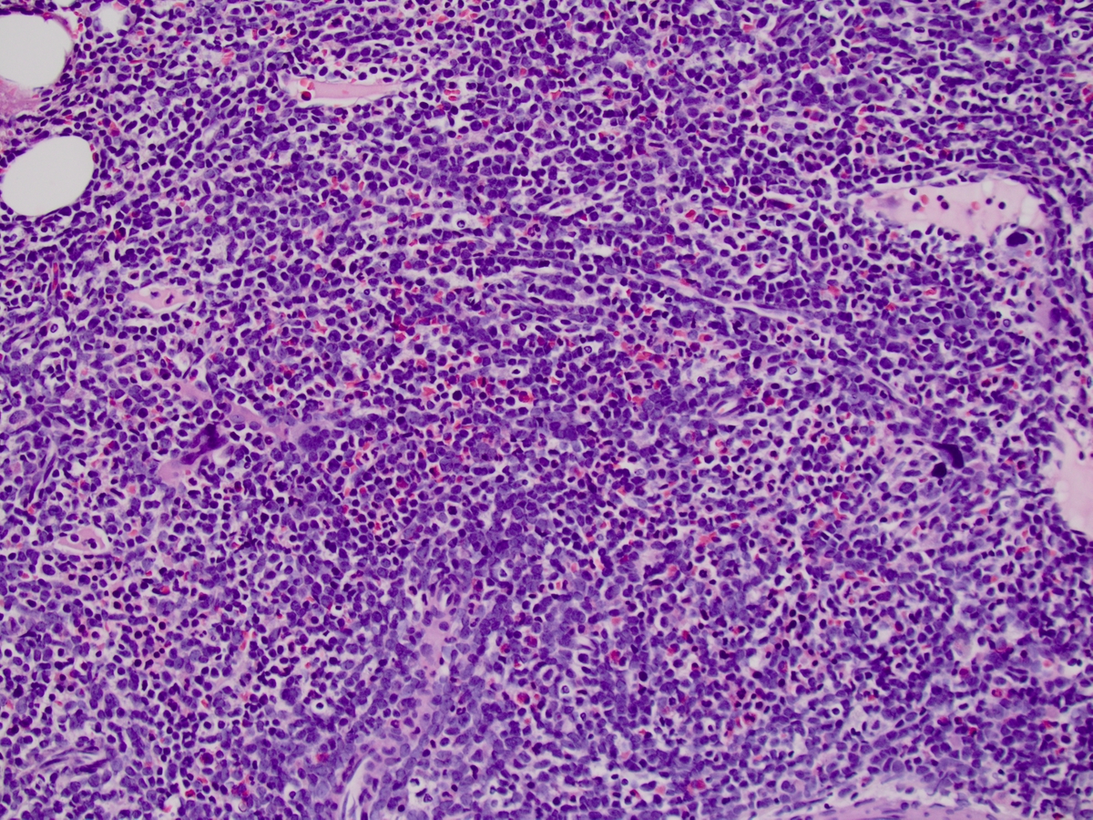 |
| Focus of Myeloproliferative neoplasm | 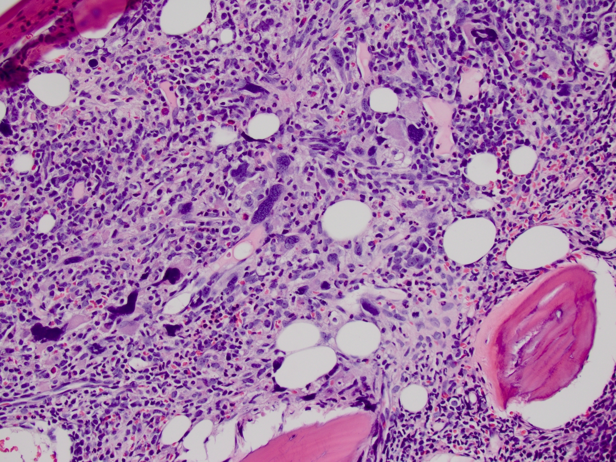 |
| CD20 stain | 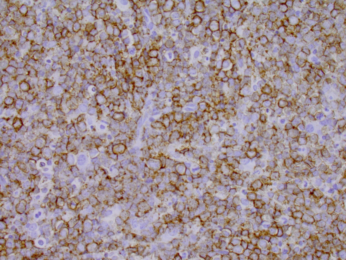 |
| TdT stain | 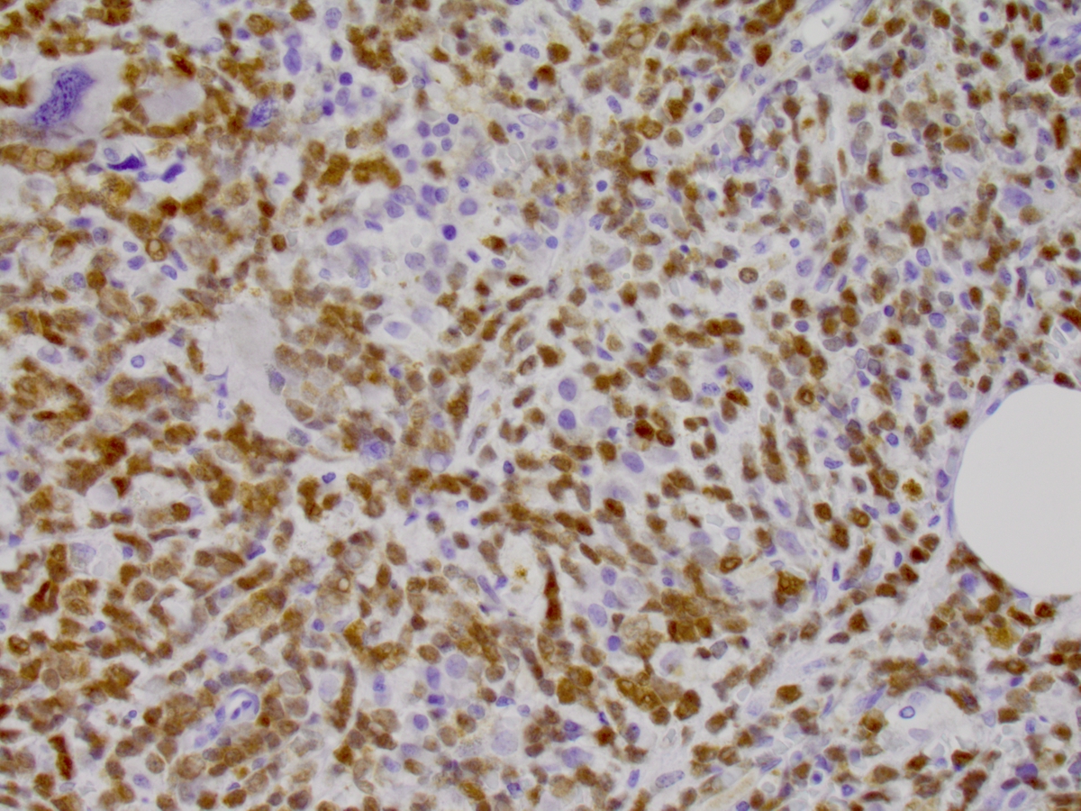 |
| Reticulin with marked fibrosis | 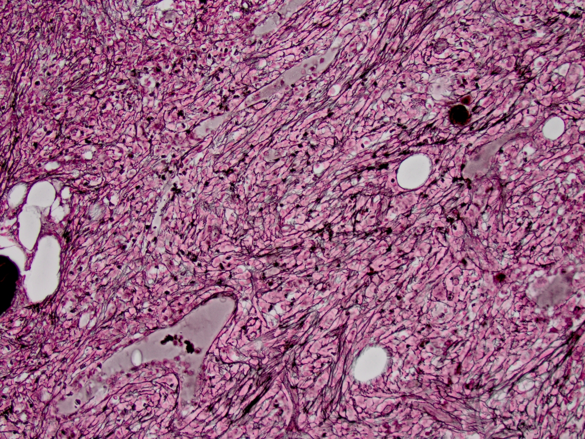 |
| Blasts positive for PAX5 | 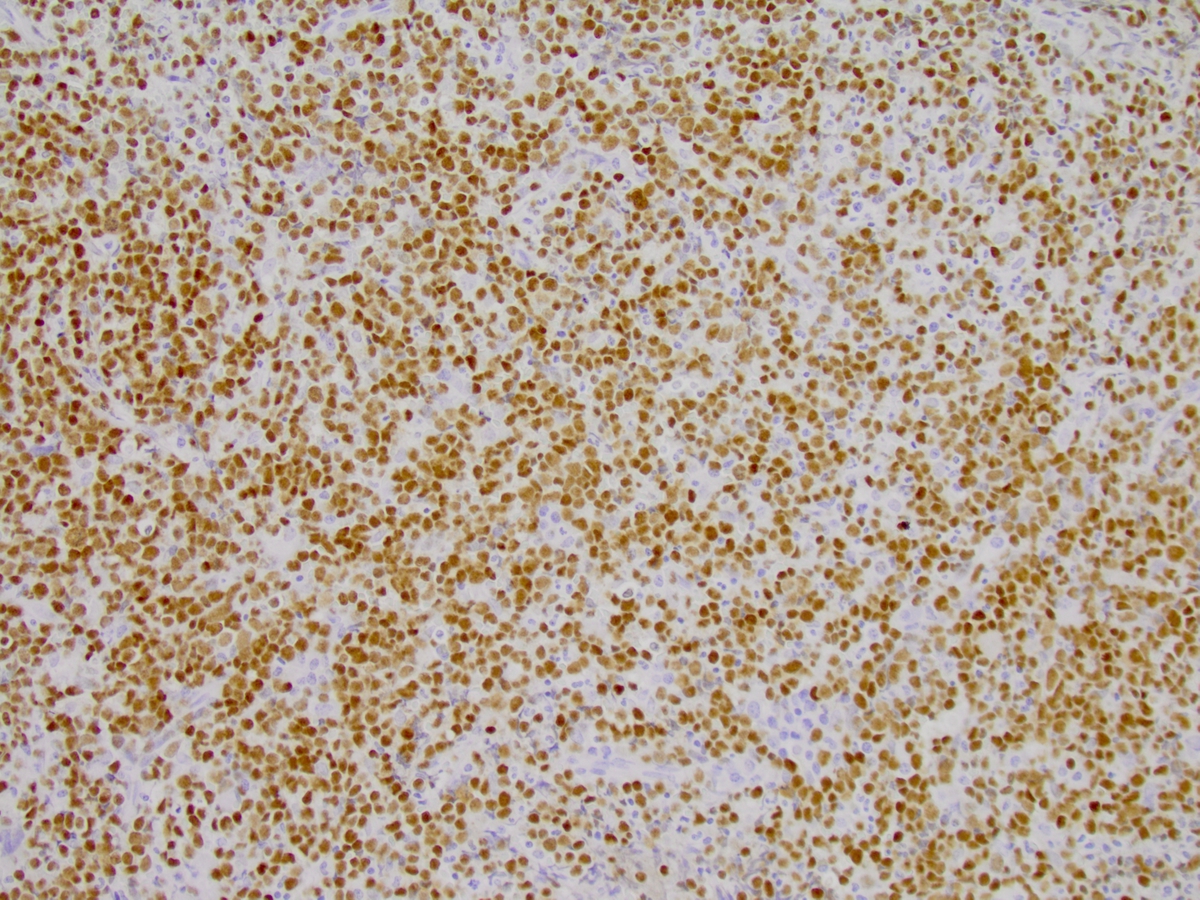 |