Institution: The University of Texas MD Anderson Cancer Center
Additional authors:Stefan H Faderl, MD, Carlos E Bueso-Ramos, MD, PhD, Marina Konopleva, MD, PhD, and Sergej Konoplev, MD, PhD
Session: AML with recurrent genetic mutations Part II
HISTORY
The patient is a 79-year-old gentleman who has been taking methotrexate for several years for treatment of psoriasis. He was noted to be pancytopenic at least since June 2011 with a white blood cell count of 3.8 and hematocrit of 30.3. In addition, the patient complained of more fatigue and dyspnea on exertion. Bone marrow (BM) biopsy performed at outside facility on 10/03/11 revealed Acute myeloid leukemia. Bone marrow (BM) biopsy performed at our institution on 11/08/2011 is submitted as specimen 1. On 11/12/2011, the patient started induction chemotherapy consisting of an investigational combination of omacetaxine and low-dose cytarabine. However, repeat BM biopsy on 12/13/2011 (specimen 2) showed persistent disease. The patient was then started on an investigational drug PR-104 as a monotherapy. PR-104 circulates in the blood in inactive form and is converted under hypoxic conditions by hypoxia-selective reductases into active nitrogen mustard. The patient received the first course on 12/16/2011 and achieved completed remission. Follow-up BM biopsy performed on 01/05/2012 is submitted as specimen 3. The patient underwent two more consolidation courses with PR-104 on 01/30/2012 and 02/29/2012, respectively. The patient has remained in complete remission over a year at the time of case submission (February 2013).
DETAILS
Specimen 1:
BM biopsy was markedly hypercellular (figure 1) and infiltrated by sheets of immature cells with finely dispersed chromatin and distinct nucleoli. A subset of immature cells showed nuclear folding and indentation (figure 2). Blast count on BM aspirate smear (figure 3) was 48%. The blasts were strongly positive for myeloperoxidase (MPO, figure 4);25% of blasts were positive for butyrate esterase (figure 5). Specimen 2:BM biopsy showed 20% cellularity (figure 6) and infiltration by increased immature cells (figure 7). BM aspirate smear revealed 25% blasts and increased monocytes including promonocytes.Specimen 3:BM biopsy showed patchy 5-10% cellularity, usual number of megakaryocytes with unremarkable morphology, and a small non-paratrabecular lymphoid aggregate (figure 8). Immunohistochemical stains of the biopsy specimen revealed no increase in CD34+ blasts (figure 9) or CD117+ immature cells (figure 10).IMMUNOHISTOCHEMISTRY AND FLOW CYTOMETRY
Specimen 1:
Flow cytometric immunophenotyping of BM aspirate detected increased blasts (53% of total cells) expressing CD13 (dim, partial), CD33, CD38 (partial), CD45 (dim to moderate), CD64 (subset), CD117, CD123, HLA-DR (partial) and MPO. The neoplastic cells were negative for CD2, cytoplasmic CD3, CD5, CD7, CD10, CD14, CD19, CD34, CD41, CD56 and TdT expression. Specimen 2:Flow cytometric immunophenotyping of BM aspirate showed an aberrant immature myelomonocytic population. The blasts were positive for CD13 (partial), CD14 (dim/partial), CD33 (bright), CD38, CD64 (bright), CD123 and HLA-DR. They were negative for CD2, CD5, CD7, CD19, CD34, and CD117.CYTOGENETIC FINDINGS
Specimen 1
Conventional cytogenetic study revealed diploid male karyotype 46, XY in all 20 metaphases analyzed.MOLECULAR FINDINGS
Specimen 1
A mutation in exon 12 of NPM1 gene and a mutation in codon 140 (CGG to CAG) in exon 4 of the IDH2 gene (R140Q) were identified.No CEBPA mutation, no mutation in any of the tested codons of the KIT gene on exon 17; no mutation in codon 87 to 138 in exon 4 of the IDH1 gene, no mutations in codons 12, 13, and 61 of the KRAS and NRAS genes, and neither internal tandem duplication (ITD) nor codon 835/836 mutation in the FLT3 gene were detected. In addition, no fusion transcripts were detected by Luminex, which included testing for b3a2, b2a2 and e1a2 transcripts of bcr-abl/ t(9;22)(q34;q11), the short and long forms of PML-RARA/t(15;17)(q22;q21), the A and D forms of CBFb-MYH11/inv(16), AML1-ETO/t(8;21)(q22;q22), E2A-PBX-1/t(1;19)(q23;p13), MLL-AF4/t(4;11)(q12;q23) and TEL-AML1/ t(12;21)(p12;q22). Specimen 2A mutation in exon 12 of NPM1 gene was detected. No additional testing was performed.INTERESTING FEATURES
This case demonstrates anti-leukemic activity of PR-104 as a single agent in a patient with refractory AML. It has been demonstrated that hypoxic conditions of bone marrow give leukemic stem cells growth advantage and additional protection from chemotherapeutic drugs. Targeting hypoxia in patients with acute leukemia has been recently proposed as a novel strategy to improve patient response and survival.
PROPOSED DIAGNOSIS
Acute myeloid leukemia with mutated NPM1, which responded to monotherapy with hypoxia-activating agent PR-104
CONSENSUS GROUP: ADDITIONAL INFORMATION/STUDIES
Additional immunostains performed by consensus group:
NPM1: Nuclear-restricted nucleophosmin
CONSENSUS DIAGNOSIS
Acute myeloid leukemia, with NPM1 mutation and IDH2 mutation
| Specimen 1, BM core biopsy, H&E x 40 | 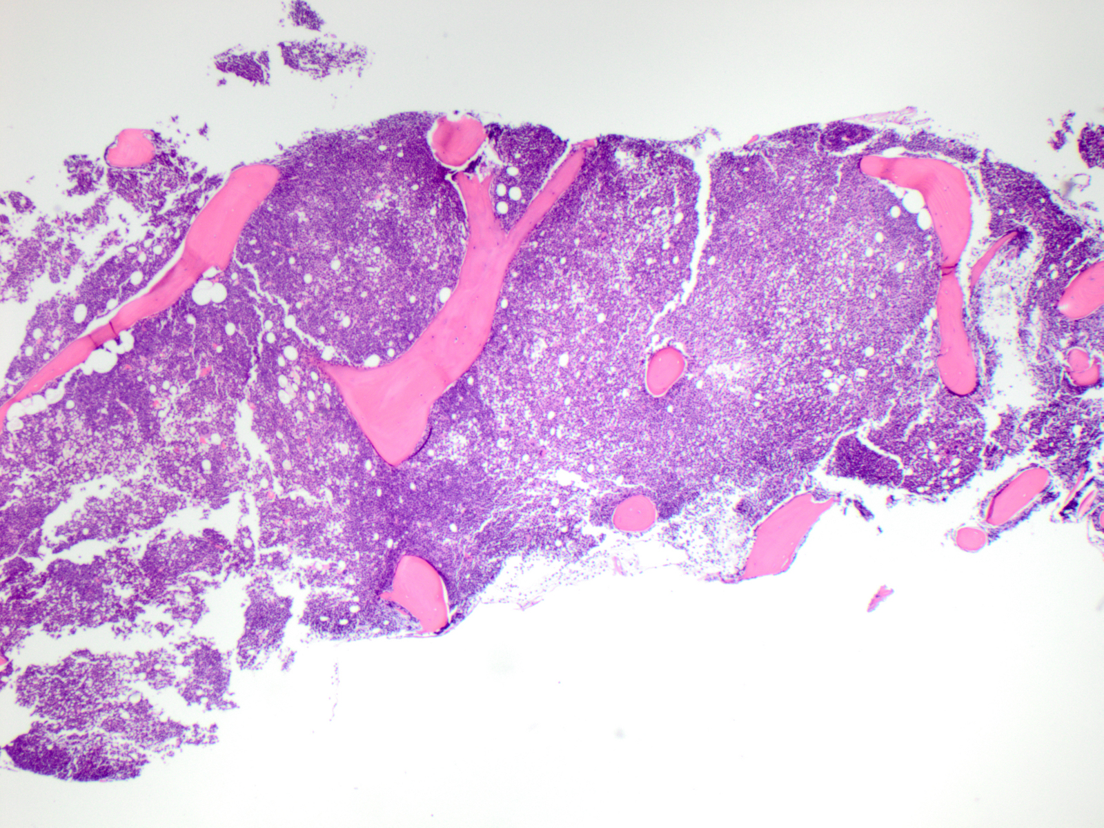 |
| Specimen 1, BM core biopsy, H&E x 400 |  |
| Specimen 1, BM aspirate smear, Wright Giemsa x 500 |  |
| Specimen 1, BM aspirate smear, MPO x 500 |  |
| Specimen 1, BM aspirate smear, non-specific esterase x 500 | 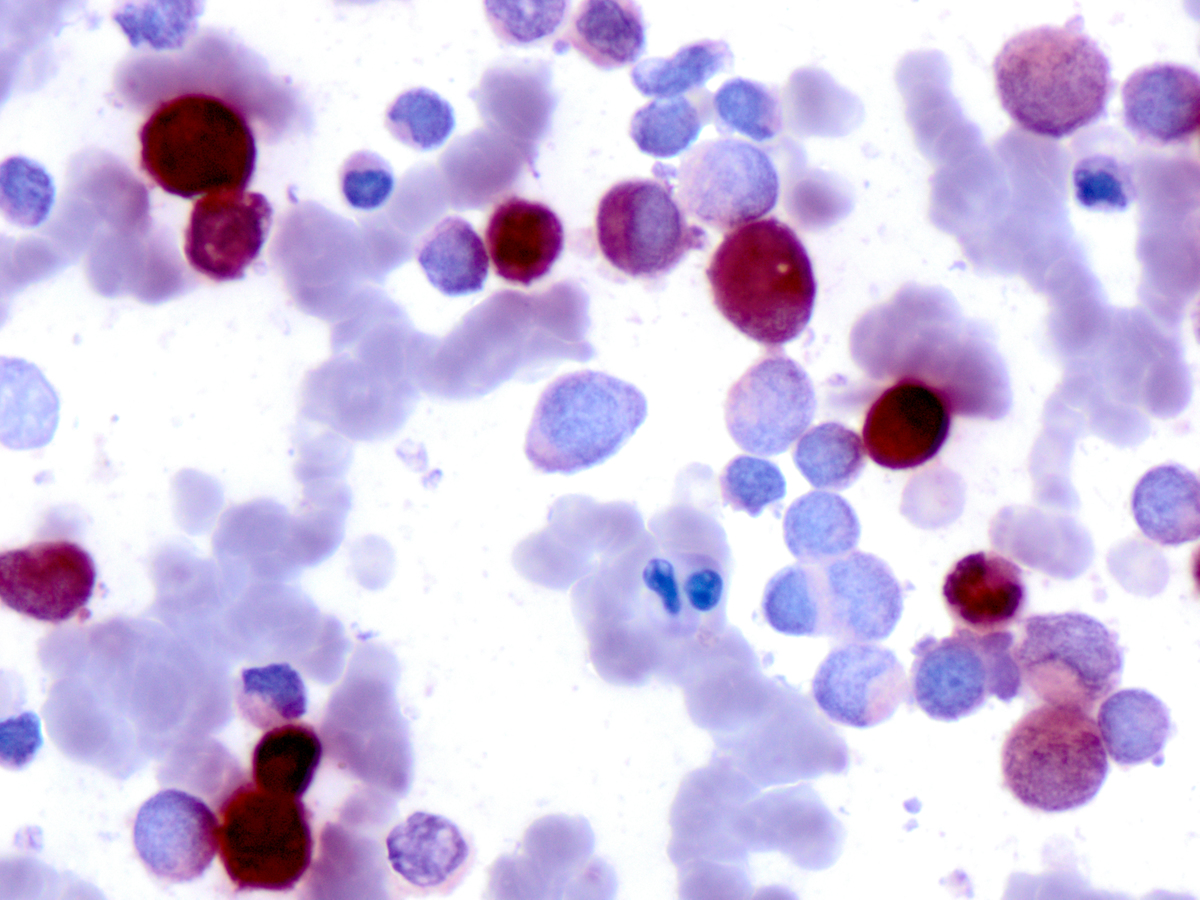 |
| Specimen 2, BM core biopsy, H&E x 40 | 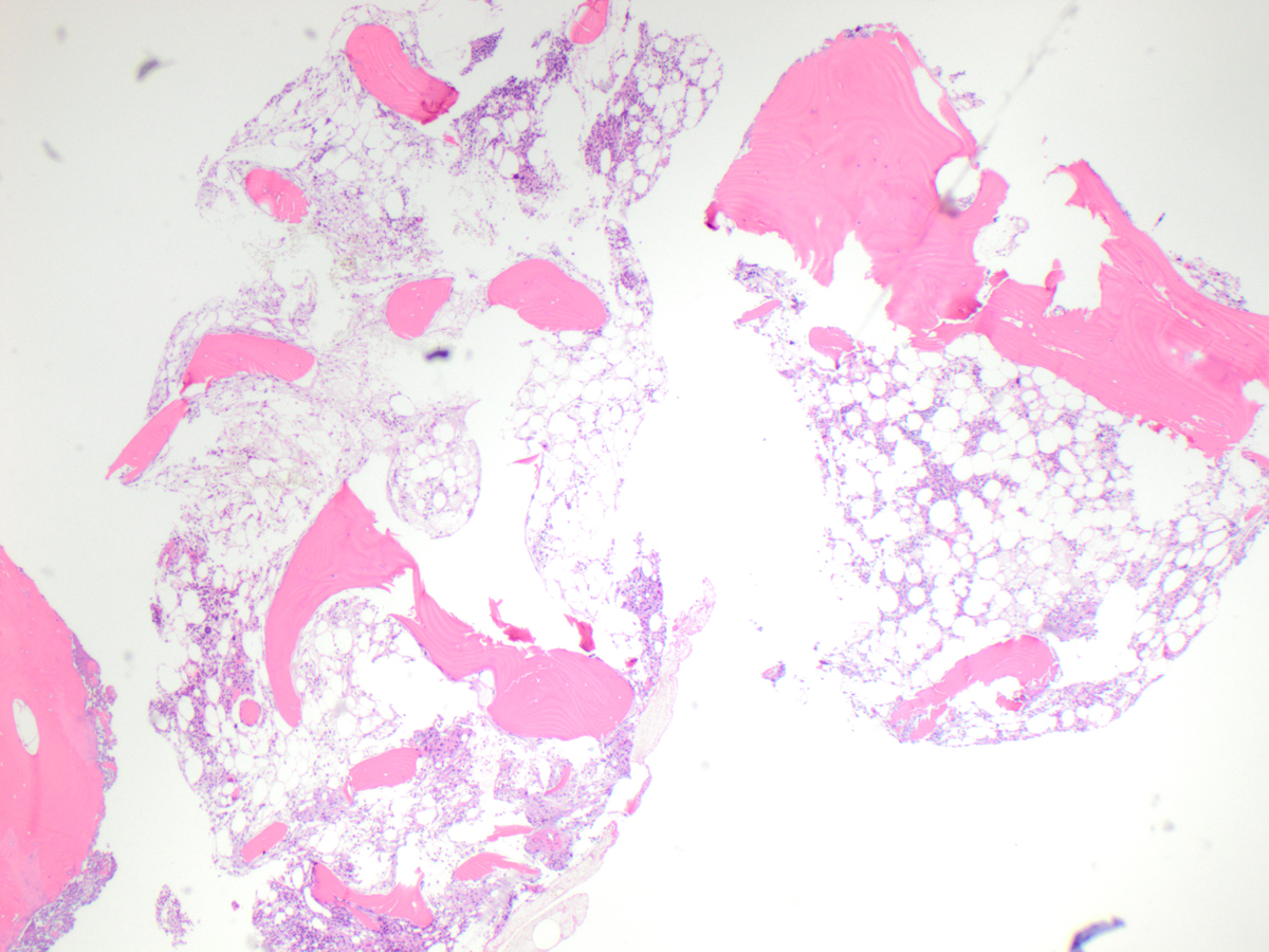 |
| Specimen 2, BM core biopsy, H&E x 100 |  |
| Specimen 3, BM core biopsy, H&E x 40 |  |
| Specimen 3, BM core biopsy, H&E x 100 | 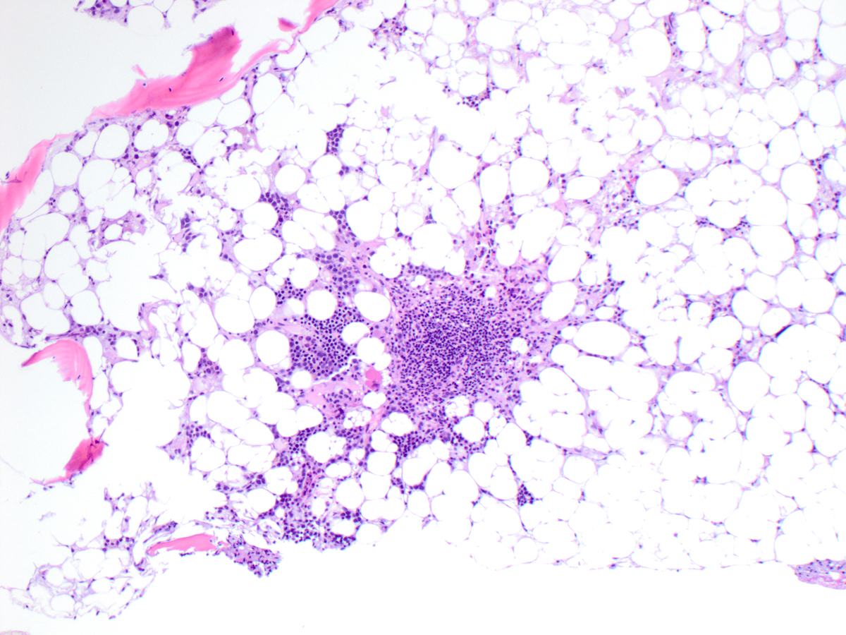 |
| Specimen 3, BM core biopsy, CD34 x 200 | 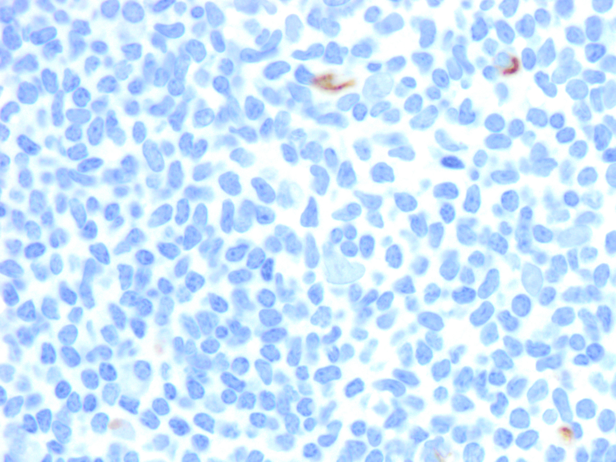 |
| Specimen 3, BM core biopsy, CD117 x 200 | 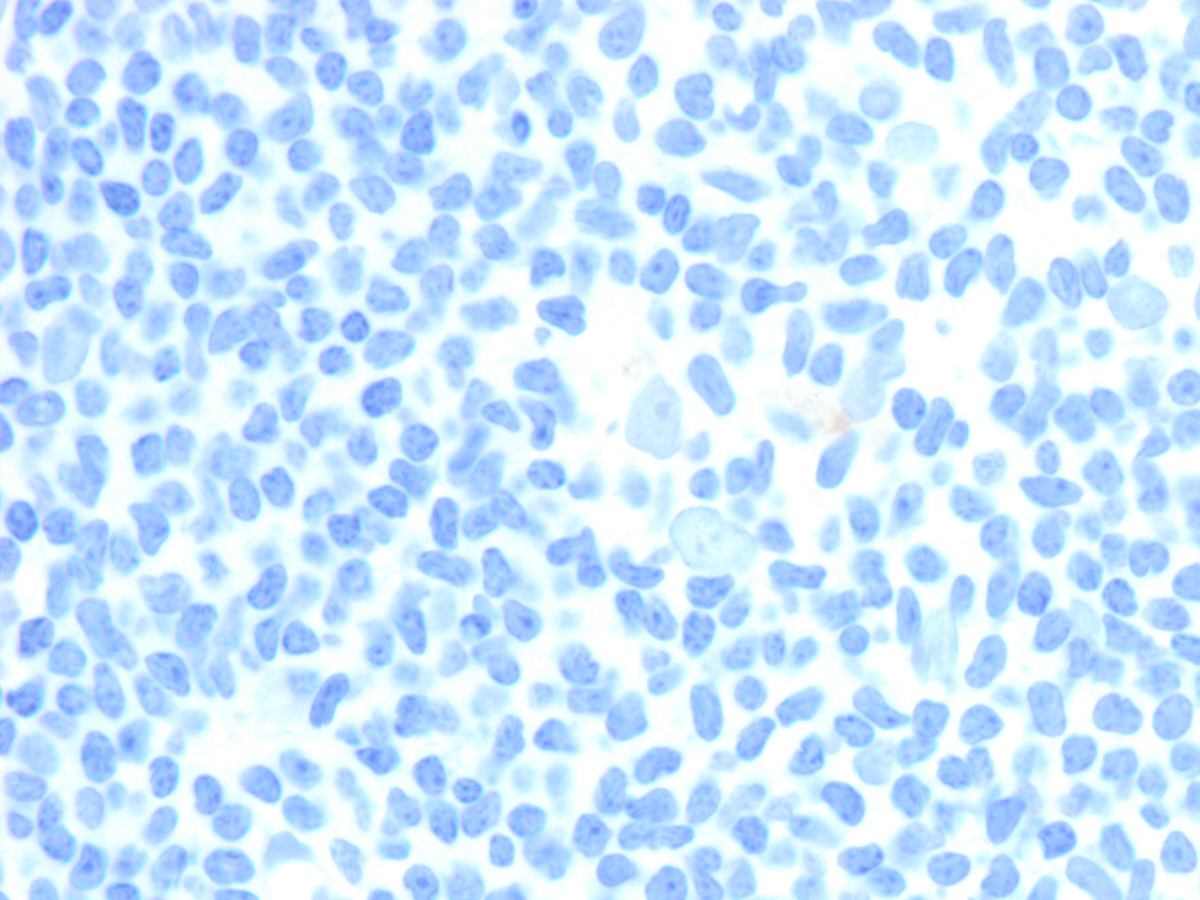 |