Institution: UT MD Anderson Cancer Center
Additional authors:Roberto Miranda, MD
Session: AML with recurrent genetic abnormalities Part I
HISTORY
Patient is a 72-year-old female with history of sarcoidosis (late 1980s), in 2007 treated with prednisone and subsequently with methotrexate (for 1.5 years) stopped in 10/2009, due to platelet count of 100,000. She developed superficial vein thrombosis in her arm and was placed on aspirin. She had no evidence of active bleeding; however, over the next month, platelets dropped progressively to 17. Patient developed spontaneous bruising and petechiae, and a new hematoma of the left thumb. She was given high-dose steroids 60 mg a day in an attempt to treat ITP, but had relatively little response. It was also noted the patient had an elevated LDH. Bone marrow work-up was performed at outside institution and reported as myelodysplastic syndrome with multilineage dysplasia (FISH and cytogenetic studies pending).
Patient was referred to UTMCACC. At admission:WBC: 3.8 k/ul; hemoglobin 10.7 g/dl; MCV 86 fl, platelet 22 k/ul; nucleated RBC 1.7%; 25% promyelocytes, 33% neutrophils and bands, 12% metamyelocytes, 24% lymphocytes, 6% monocytes; Protime-pt. 17.9 h sec (12.7- 15.0), activated PTT 33.1 sec (24.7- 35.9), fibrinogen 71 l mg/dl (202- 450), D-dimer automated 14.89 mcg/ml (0.00- 0.40)Bone marrow work up was performed in UTMDACC.DETAILS
Bone marrow aspiration and biopsy Site: posterior iliac crest Procedure - Formalin fixed biopsy after decalcification
Core biopsy: Sheets of immature cells, comprising 80% of marrow cellularity; cells are large, with oval to irregular nuclear, contours, finely dispersed chromatin, variably distinct nucleoli, and abundant amounts of eosinophilic granular cytoplasm, occasional mitotic figures present Aspirate: 70% promyelocytes; large, with folded or bilobed nuclei, finely dispersed chromatin, one to a few distinct nucleoli, moderate to abundant amounts of granular cytoplasm; no Auer rods identified Myeloperoxidase: strongly positive in most promyelocytesIMMUNOHISTOCHEMISTRY AND FLOW CYTOMETRY
Flow cytometry: blasts POSTIVE for CD13, HLA-DR, CD33(bright), CD117; NEGATIVE for CD34, B-cell and T-cell markers.
CYTOGENETIC FINDINGS
Karyotype:
46,XX,t(11;17)(q23;q21)[5] 46,XX[15]FISH: NEGATIVE for PML-RARA rearrangement; but POSITIVE for an extra RARA signal (81% of interphases), consistent with RARA rearrangement; NEGATIVE for MLL (11q23) rearrangement.Additional studies: LSI RARA dual color, break apart rearrangement probe that hybridizes to band 17q21 demonstrated 89.5% cells to have rearranged chromosome 17 within the RARA gene breakpoint.MOLECULAR FINDINGS
Molecular studies:
LUMINEX Multiplex leukemia translocation assay: NEGATIVE for PML-RARA/t(15;17)(q22;q21)NEGATIVE for FLT3, NPM1, RAS , CEBPA and KIT mutationsINTERESTING FEATURES
Patient was treated with Mylotarg, Arsenic and ATRA off protocol; treatment was complicated by ATRA differentiation syndrome. She achieved complete remission; and received one cycle of consolidation chemotherapy with ATRA and arsenic (x1). She then underwent autologous stem cell transplantation (04/10) with Cytoxan/busulfan conditioning regimen. Posttransplant, the patient had persistent thrombocytopenia; and she was started back on and ATRA and arsenic for maintenance therapy; doses reduced for the former, and the latter discontinued due to pericardial effusion.At last follow up (12/2012), she was in complete remission as assessed by karyotype and FISH.
This is a challenging case with atypical morphology associated with a variant translocation i.e. t(11;17)(q23;q21). Numerous neoplastic promyelocytes have well-developed secondary fine eosinophilic granules. Occasional neutrophils with dysplastic features are identified. In addition, no Auer rods are identified. This morphology has been reported to be associated with the t(11;17). The case would be classified as acute promyelocytic leukemia, FAB: AML M3. In spite of the WHO 2008 terminology, the term “acute promyelocytic leukemia” is still meaningful from a morphologic standpoint since it directs the pathologist’s attention to a related set of AMLs with different RARA translocations, and would lead to a consideration of a variant translocation irrespective of the blast count. Karyotypic and FISH studies could be expedited by the screening pathologist, and the clinician alerted to the possibility. In this case, the patient had labs compatible with disseminated intravascular coagulation; which could have been fatal if correct diagnosis had not been made in time. We propose to retain the original term “acute promyelocytic leukemia” in future terminology with variant translations as subtypes. FISH studies with a RARA dual color break apart probe should be considered where the level of suspicion for a RARA translocation is high and results are negative with the dual color fusion probe. This was done in this case.FISH is the only modality for monitoring minimal residual disease in absence of generally available molecular studies for this translocation.Prognosis is poorer than in the t(15;17) cases; hence autologous stem cell transplant and subsequent maintenance therapy was performed in this patient.PROPOSED DIAGNOSIS
Diagnosis submitted:
Acute myeloid leukemia with variant RARA translocationCONSENSUS DIAGNOSIS
Acute myeloid leukemia with variant t(11;17)(q23;q21); ZBTB16-RARA
| Bone marrow core biopsy | 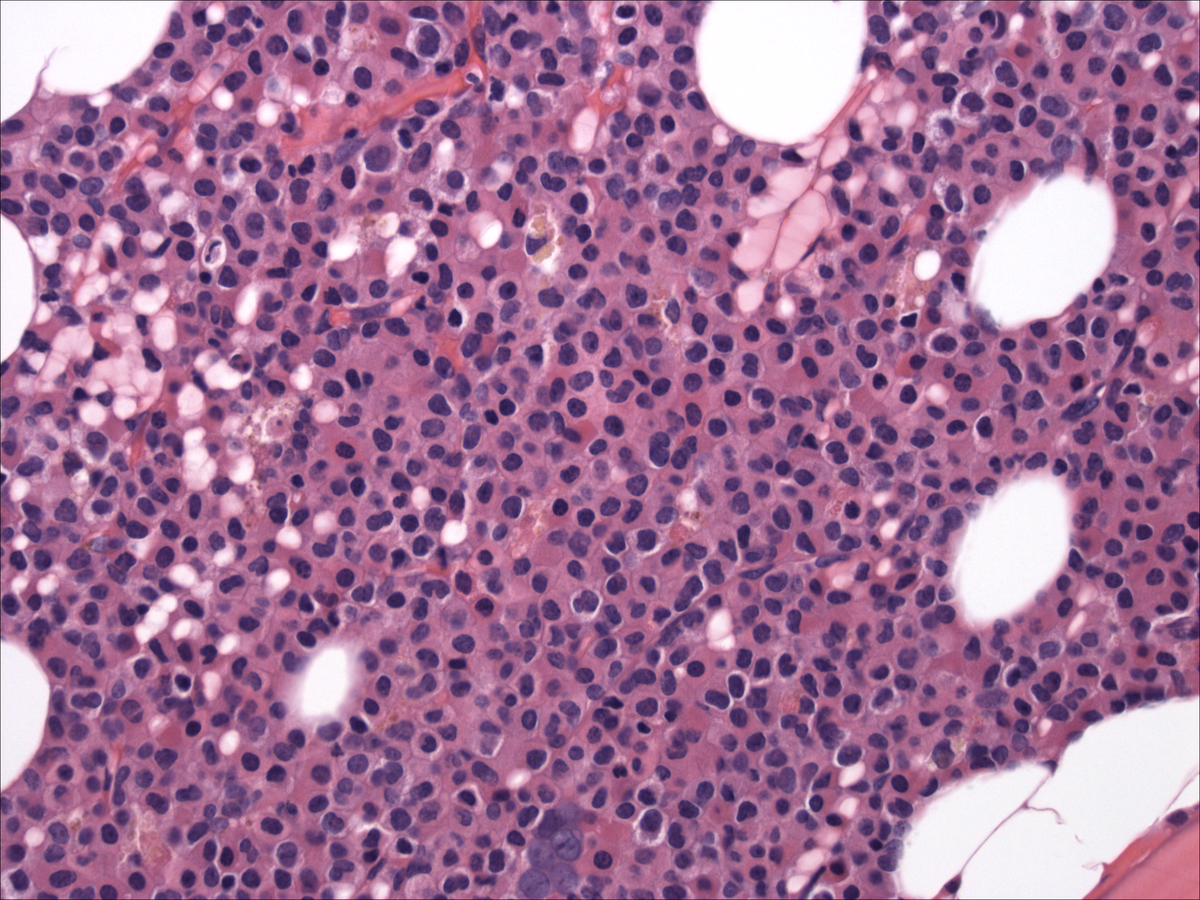 |
| Bone marrow aspirate |  |
| Aspirate_myeloperoxidase |  |
| Aspirate smear_myeloperoxidase_counterstain | 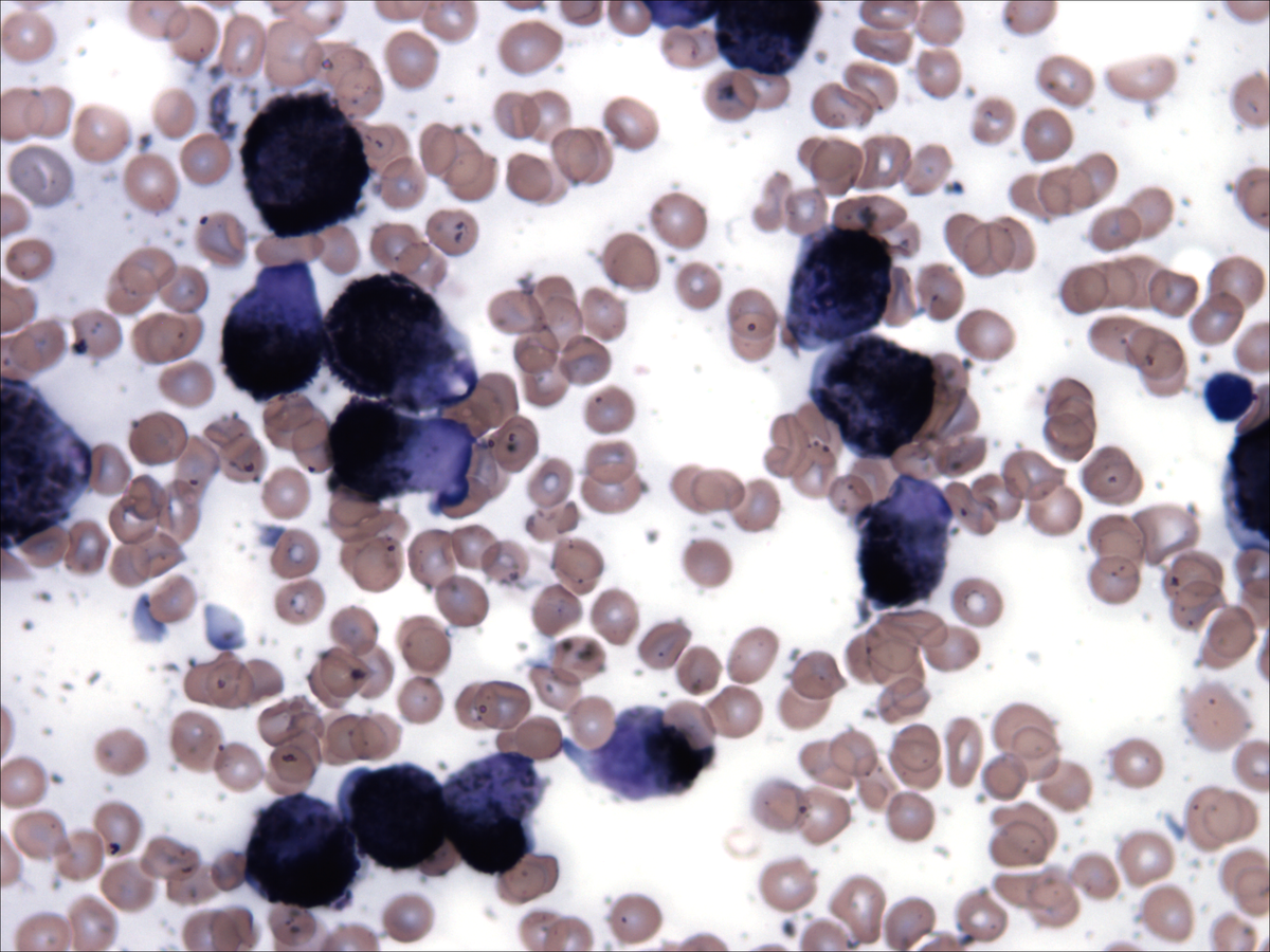 |