Institution: University of Texas Health Science Center San Antonio
Additional authors:Edward A. Medina, M.D., Ph.D., Russell A. Higgins, M.D.
Session: AML with recurrent genetic abnormalities Part I
HISTORY
A 58 year old man with past medical history of asthma was admitted for evaluation of fatigue, shortness of breath, and a 10 pound weight loss. He was found to have a leukocytosis (WBC of 54.1 K/uL) with 93% blasts, anemia (hemoglobin of 7.9 g/dL), and thrombocytopenia (82 K/uL). Bone marrow biopsy demonstrated acute myeloid leukemia (AML) with recurrent cytogenetic abnormality, t(3;3)(q21;q26.2). The patient underwent conventional 7+3 induction chemotherapy, and a day 14 post-induction bone marrow biopsy demonstrated residual AML with 45% myeloblasts. The patient was then reinduced with FLAG-IDA. A day 31 post-reinduction bone marrow biopsy demonstrated no morphologic, immunophenotypic, or cytogenetic evidence of disease; however, there was persistent unexplained anemia and thrombocytopenia. Approximately two months later, he was readmitted for proptosis and vision changes which were clinically and radiologically attributed to myeloid sarcoma/AML. The patient had a prolonged hospital course, which included Clostridium difficile colitis secondary to antibiotic coverage for neutropenia, one leukophoresis procedure for pulmonary leukostasis (WBC of up to 165.4 K/uL), and pneumonia. Approximately 5 ½ months after initial diagnosis, the patient passed away. Autopsy findings included relapsed acute myelogenous leukemia with myeloid sarcomas involving the spleen, lymph nodes, lungs, small intestine, right testis, and periorbital soft tissue (the latter by clinical and radiographic history). Additional contributory factors to the cause of death included pseudomembranous colitis and multilobar pneumonia due to invasive pulmonary aspergillosis.
DETAILS
Biopsy, bone marrow, right posterior iliac crest, B5 fixative. A peripheral blood smear demonstrated leukocytosis with two populations of blasts. One blast population (66% of cells) had medium-size cells with fine nuclear chromatin, one or more nucleoli, and scant cytoplasm. No Auer rods were observed. The second blast population of blasts (23% of cells) consisted of larger monoblasts/promonocytes with variably round to indented nuclei with delicate chromatin, one or more nucleoli, and abundant cytoplasm with pseudopod formation. Bone marrow biopsy: The bone marrow was hypercellular due to sheets of blasts (90% of cells) with a variable amount of cytoplasm. The granulocytic and erythroid precursors were morphologically unremarkable, although assessment was limited due to there relative paucity (<5% of marrow cellularity). Numerous small, monolobate or bilobate megakaryocytes were present.
IMMUNOHISTOCHEMISTRY AND FLOW CYTOMETRY
Flow cytometric immunophenotyping demonstrated two abnormal populations:
Myeloblasts (65% of cells) express CD34, HLA-DR (partial), CD117, CD33 (dim, partial), CD15, CD4 (dim), CD2 (dim,partial), CD56 (partial), and lack cMPO, cCD3, CD19, and CD10.
Monoblasts/promonocytes (25% of cells) express HLA-DR, CD33 (bright), CD4, CD11c, CD14 (partial), CD13 (moderate to bright), CD11b (moderate to dim), CD2 (dim), and CD56 (partial).
CYTOGENETIC FINDINGS
45,X,-Y,t(3;3)(q21;q26.2)[19]/46,XY[1]
MOLECULAR FINDINGS
Not performed.
INTERESTING FEATURES
Studies reporting the immunophenotypic features of AML with inv(3)(q21;q26.2) or t(3;3)(q21;q26.2) are limited to 27 cases. Prior studies have not reported the distinct immunophenotypes of separate myeloblast and monoblast/promonocyte populations as seen in this case.
PROPOSED DIAGNOSIS
Acute myeloid leukemia with recurrent cytogenetic abnormality [t(3;3)(q21;q26.2)], with myelomonocytic differentiation.
CONSENSUS DIAGNOSIS
Acute myeloid leukemia with t(3;3)(q21;q26.2); RPN1-EVI1, and -Y
| Peripheral Blood 600x Myeloblasts |  |
| Peripheral Blood 600x Promonocytes |  |
| Peripheral Blood 600x Spectrum of myeloblast, monoblast, promonocytes |  |
| Peripheral Blood 600x Spectrum of myeloblast, monoblast, promonocyte |  |
| Bone Marrow Biopsy 100x |  |
| Bone Marrow Biopsy 600x Myeloblasts and Monoblasts |  |
| Bone Marrow Biopsy 600x Hypolobate Micromegakaryoctes | 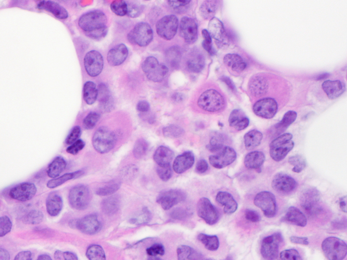 |
| Bone Marrow Biopsy 600x Hypolobate Micromegakaryoctes |  |
| Diagnostic Flow Cytomety Composite, Myeloblast population in Black, Monoblast/Promonocyte population in Red, Others (ie Lymphocytes) in Gray |  flow composite edit 1.JPG) |
| Diagnostic Karyotype with 45,X,-Y,t(3;3)(q21;q26.2)[19]/46,XY[1] | ![Diagnostic Karyotype with 45,X,-Y,t(3;3)(q21;q26.2)[19]/46,XY[1]](cases/394/images/AML t(3;3) karyotype.JPG) |
| Splenic involvement by AML at autopsy 600x |  |
| Testicular myeloid sarcoma, involvement at autopsy 100x |  |