Institution: Stanford University Medical Center
Additional authors:Yaso Natkunam MD, PhD
Session: Extramedullary manifestations of myeloid neoplasms
HISTORY
1-year-old Mayan female with fever, weight loss, and abdominal distension without splenomegaly or peripheral lymphadenopathy. Case received in consultation from Guatemala.
DETAILS
Formalin-fixed retroperitoneal mass. Microscopic findings: Sheets and nests of intermediate-sized cells with round nuclei, immature chromatin, and scant to moderate amounts of faintly eosinophilic cytoplasm. Numerous mitotic figures and areas with karyorrhectic debris are identified, with some areas showing a “starry sky” appearance.
IMMUNOHISTOCHEMISTRY AND FLOW CYTOMETRY
Positive: Glycophorin C, CD43 (subset), CD38 (weak), INI intact.
Negative: Cytokeratin, synaptophysin, NB84, WT1, desmin, myogenin, CD99, S100, SALL4, CD45, CD34, CD117, MPO, CD10, TdT, CD3, CD4, CD79a, CD20, bcl-2, CD30, ALK, EBV RNA, CD61, CD68, CD123, and CD138.
CYTOGENETIC FINDINGS
Not performed.
MOLECULAR FINDINGS
Not performed.
INTERESTING FEATURES
• Distinct erythroid differentiation demonstrated by positive glycophorin C immunostaining
• Differential diagnostic overlap with Burkitt lymphoma
• CD43 (subset positive) and CD38 (weakly positive) are only other positive markers of differentiation
PROPOSED DIAGNOSIS
Myeloid sarcoma with erythroid differentiation
CONSENSUS GROUP: ADDITIONAL INFORMATION/STUDIES
Additional immunohistochemical stains performed by the consensus group:
E-cadherin: negative
CD43: positive
CONSENSUS DIAGNOSIS
Myeloid sarcoma (erythroid differentiation) involving retroperitoneum
| Retroperitoneal mass, low power | 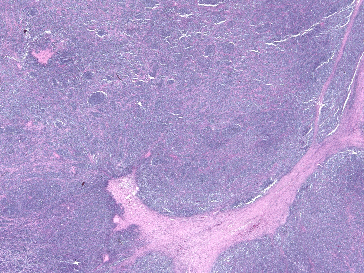 |
| Retroperitoneal mass, high power | 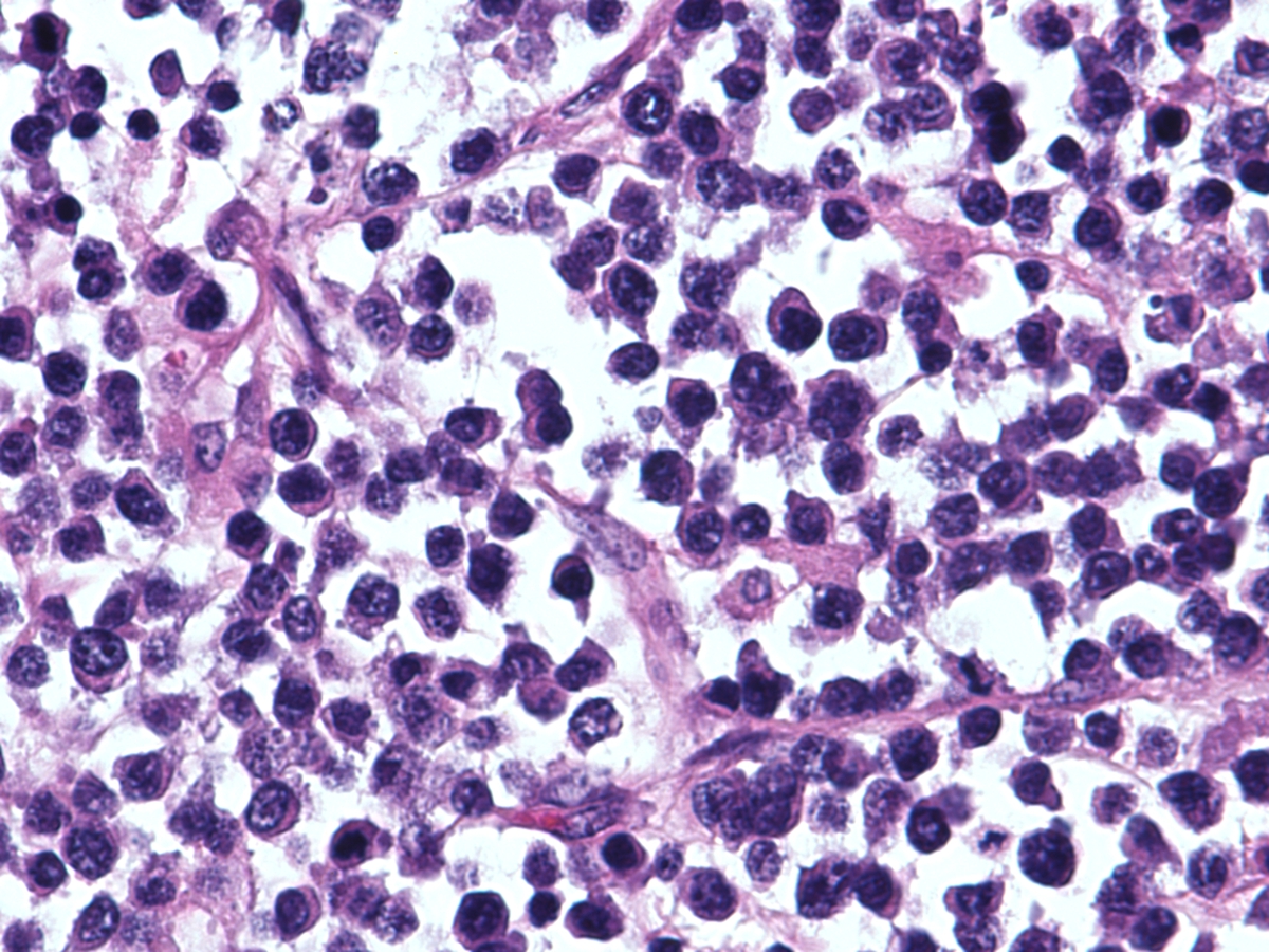 |
| Glycophorin C immunohistochemistry | 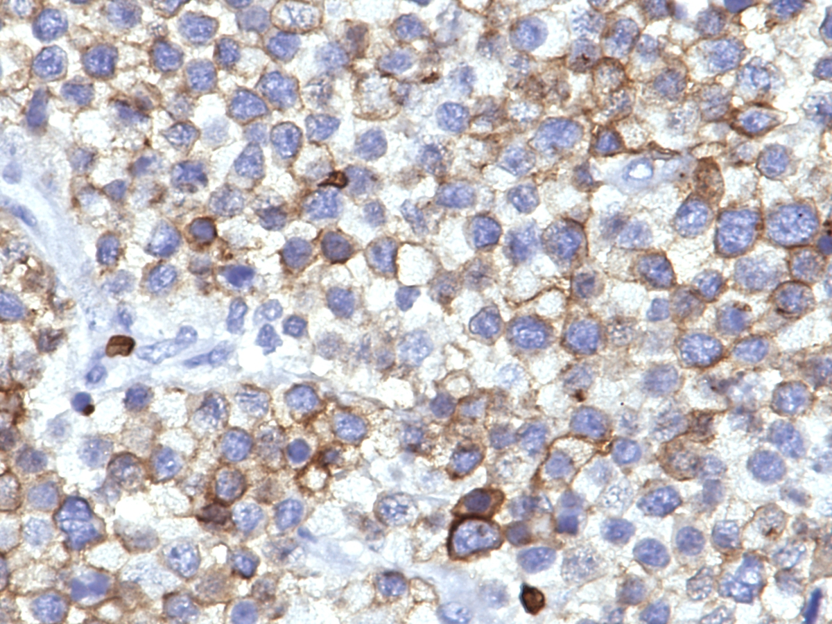 |
| Additional figure 1: CD43 IHC, 400x | 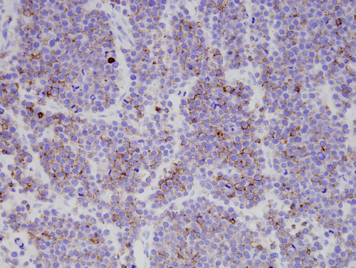 |
| Additional figure 2: E-cadherin IHC, 400x | 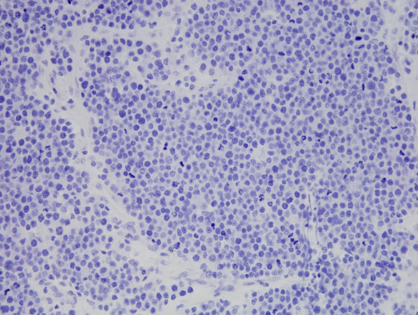 |