Institution: SUNY Upstate Medical University
Additional authors:Constance K. Stein, Ph.D., Theresa C. Gentile, M.D.
Session: AML with recurrent genetic mutations Part II
HISTORY
The patient is a 48 year old female who presented to her doctor with symptoms consistent with a sinus infection with head congestion, shortness of breath and cervical lymphadenopathy which did not improve with antibiotics. She was found to have a white blood cell count of 20,000 /ul with peripheral blasts, anemia and thrombocytopenia.
DETAILS
Blood: WBC=20.2 K/ul with 85.5% blasts and 14.5% lymphocytes; Hgb=7.5 g/dl, RBC=2.24 M/ul, MCV=101.8 fl, plt=61 K/ul. Blasts showed high N/C ratio, diffuse chromatin with 1-2 nucleoli and occasional Auer rods.
Aspirate: Hypercellular marrow with 50.8% blasts, 41.4% erythroid precursors with moderate megaloblastic/dysplastic features, 6.8% lymphocytes and 1.0% plasma cells. Auer rods were frequent and sometimes multiple. There were occasional megakaryocytes. Biopsy/clot sections: Hypercellular marrow with predominance of blasts and with admixed erythroid islands.IMMUNOHISTOCHEMISTRY AND FLOW CYTOMETRY
Flow cytometry: Blasts were positive for CD38, CD117, CD13, CD33, MPO (98%), CD34 and CD15 (dim/partial).
Blasts were negative for CD14, CD64, cCD3, cCD79a, cCD22 and Tdt.CYTOGENETIC FINDINGS
Karyotype: 47,XX,+4(9); 46,XX(11)
FISH: chr. 4: nuc ish (D4Z1x3)(64/100)Two cell lines were identified:1) Female chromosome complement with an additional copy of chromosome 4 (trisomy 4). 64% of cells showed this pattern.2) Female chromosome complement.MOLECULAR FINDINGS
Positive for NPM (exon 12) mutation, 40% mutant total
Negative for FLT3 length mutation (including internal tandem repeats) as well as point mutations at D835 and I836 with the FLT3 tyrosine kinase domain.INTERESTING FEATURES
AML with trisomy 4 as the sole cytogenetic abnormality is a rare to uncommon disease that has been described but not classified among AML with recurrent genetic abnormalities in the WHO Classification. It often shows morphology of AML without maturation or with minimal differentiation (some cases described with varied differentiation), with high blast proportions and with a generally poor prognosis.
AML with NPM1 mutation is a provisional entity in the WHO Classification that has a relatively good prognosis, usually occurs in patients with normal karyotype with monocytic or myelomonocytic differentiation, occasionally with erythroid differentiation or multilineage dysplasia, also has high blast proportions and is often considered to be a primary abnormality. In the few cases described of AML with trisomy 4, NPM1 mutation occurs at a similar frequency as in AML with normal cytogenetics.The simultaneous occurrence of trisomy 4 and NPM1 in this case raises questions of whether either is the primary event or if they are independent genetic abnormalities occurring coincidentally to produce a unique disease. The case suggests that in this situation the "worse" genetic feature, trisomy 4, holds sway. This patient initially achieved remission but relapsed after 6 months and is currently awaiting bone marrow transplantation.PROPOSED DIAGNOSIS
Acute myeloid leukemia with recurrent genetic abnormality
Current terminology; AML with mutated NPM1, and trisomy 4.Possible future terminology; AML with trisomy 4, and NPM1 mutation.CONSENSUS DIAGNOSIS
Acute myeloid leukemia, with NPM1 mutation and trisomy 4
| Blasts with Auer rods, BM aspirate 1000X | 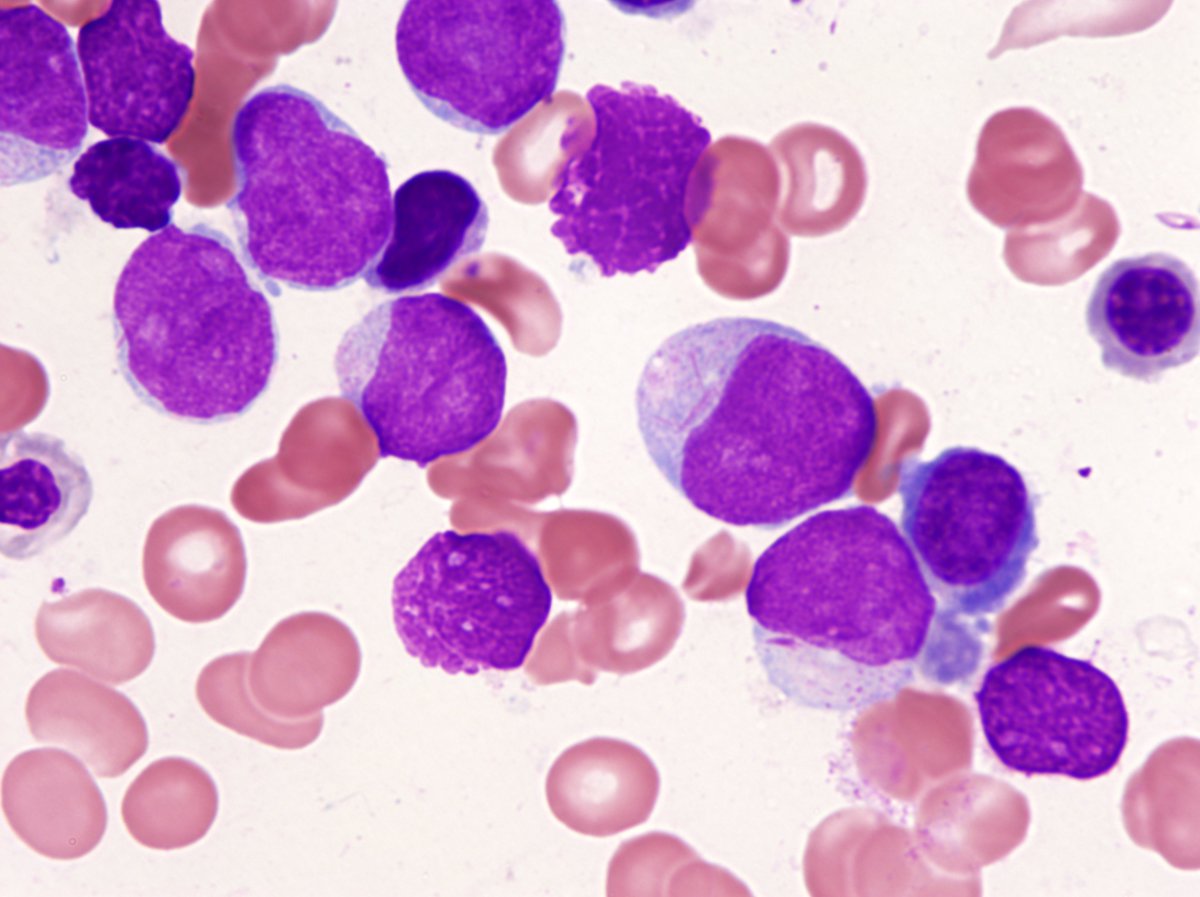 |
| Blasts and erythroid dysplasia, BM aspirate 1000X |  |
| BM biopsy 500X | 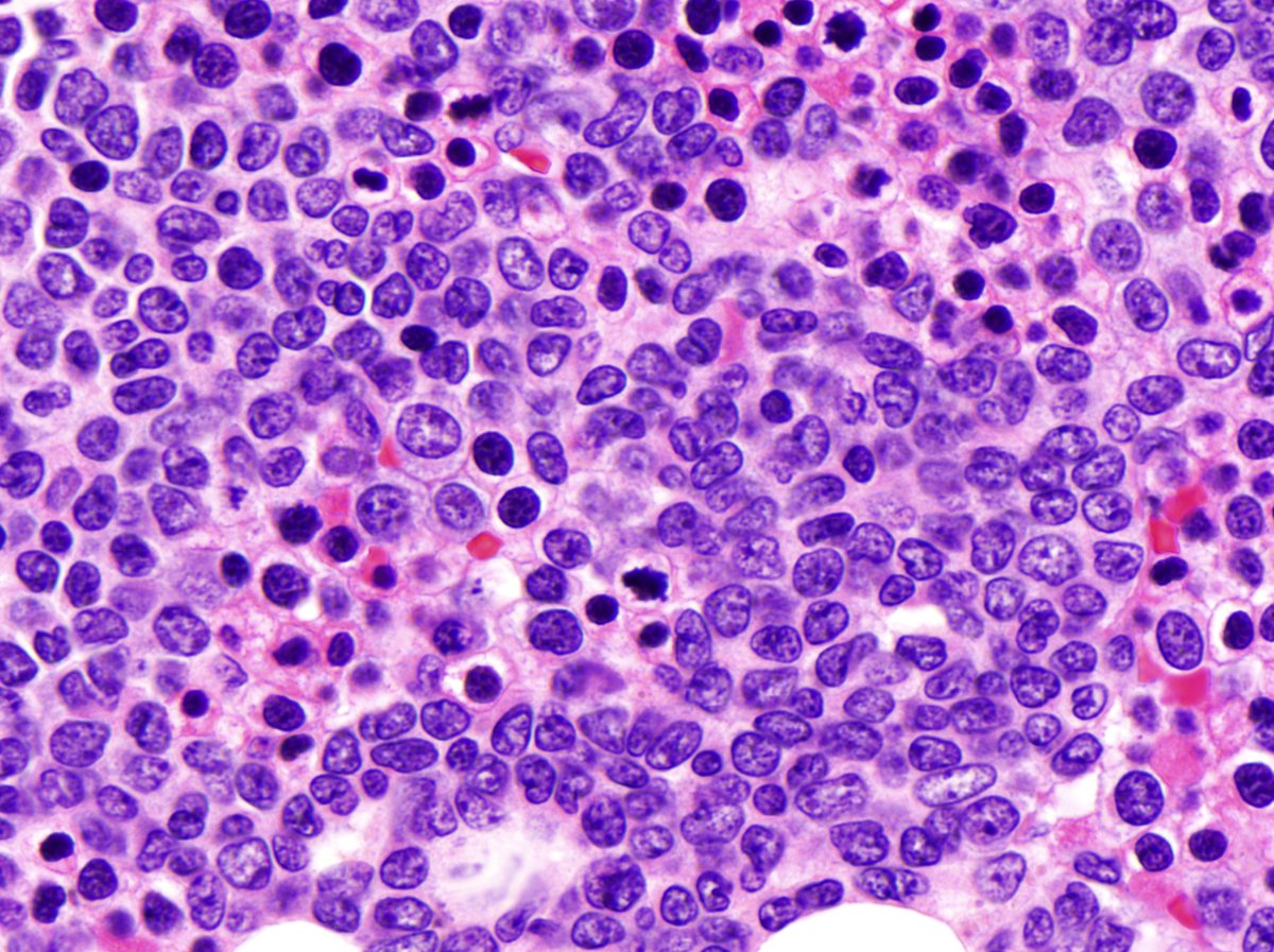 |
| Karyotype showing trisomy 4 | 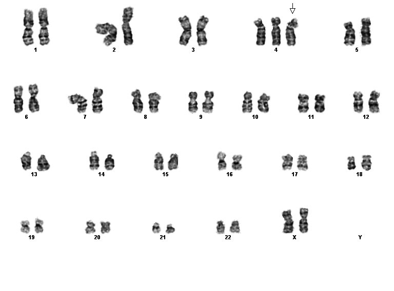 |
| FISH, Trisomy 4 | 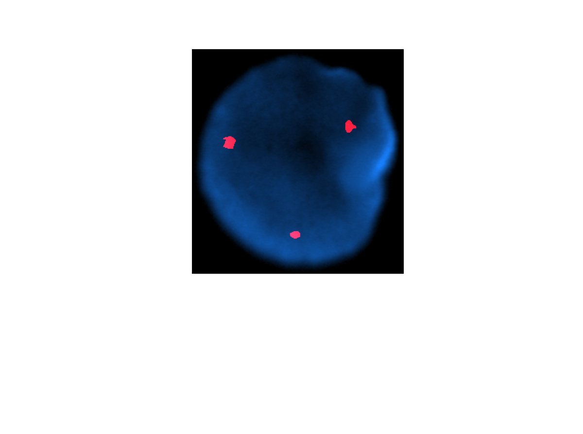 |