Institution: Fox Chase Cancer Center
Session: AML with recurrent genetic mutations Part II
HISTORY
The patient is a 73-year old woman with a 1.5 year history of myelodysplastic syndrome (MDS). She had a bone marrow biopsy at that time that showed a hypercellular marrow (>90%) with granulocytic hyperplasia and approximately 4-5% myeloblasts. Her karyotype and FISH studies for MDS were reported as normal.
She was now presenting with recurrent respiratory infections. On a CBC she was found to be anemic, thrombocytopenic and have an increased white count. On the peripheral blood smear numerous blasts were identified.DETAILS
The decalcified bone marrow trephine biopsy and aspirate clot of the posterior left iliac crest were fixed in AZF and stained with H&E. The peripheral blood and aspirate smears were stained with Wright-Giemsa.
The peripheral blood smear shows numerous blasts that are large in size with indented nuclei, powdery chromatin, several nucleoli and scant basophilic cytoplasm. Several blasts have granules and rare Auer rods. The neutrophils are hypogranular and circulating nucleated red blood cells are noted.The bone marrow smear shows sheets of blasts with similar morphology as described above. The neutrophils and precursors appear hypogranular and the very few megakaryocytes have hypolobulated nuclei.Sections of the aspirate clot and trephine biopsy show marrow cellularity of >95% with the normal bone marrow elements replaced by blasts.IMMUNOHISTOCHEMISTRY AND FLOW CYTOMETRY
Flow cytometric analysis performed on the peripheral blood show that the blasts express dim CD11c, CD13, CD33, CD34, CD38, dim CD45, CD117, HLA-DR and MPO.
CYTOGENETIC FINDINGS
Karyotype:
- Normal female karyotype: 46,XX[20]FISH:- Negative for monosomy 5 and a partial deletion of the long arm of chromosome 5 including EGR1.- Negative for monosomy 7 and a partial deletion of the long arm of chromosome 7 including D7S522.- Negative for trisomy 8.- Negative for deletion of D20S108 on chromosome 20 at q12.- Negative for both a rearrangement and deletion of MLLMOLECULAR FINDINGS
- CEBPA mutation analysis:
CEBPA mutation was identified: Single mutation (heterozygous) Nucleotide change: 968 G>C, predicted amino acid change: R322P- FLT3 mutation analysis:No evidence of internal tandem duplication (ITD) or exon 20 point mutation in the FLT3 gene- NPM1 mutation analysis:No NPM1 mutation was identifiedINTERESTING FEATURES
CEBPA (transcription factor CCAAT/enhancer binding protein α) is a critical regulator of myeloid development. It is found to be dysregulated in more than half of patients with acute myeloid leukemia (AML) as a result of genetic and epigenetic alterations such as AML1-ETO, BCR-ABL, FLT3-ITD or CEBPA promoter methylation (1). In addition 6-15% of de novo AML have mutations of the CEBPA gene. This last category when associated with a normal karyotype has been designated as a provisional disease entity under the 2008 WHO classification (2).
Usually AML with mutated CEBPA presents de novo and the patients with this mutation have a favorable prognosis. There are very few reports of CEBPA mutations in myelodysplastic/ myeoloproliferative neoplasms and the CEBPA mutations are significantly absent in cases of AML with myelodysplasia related changes (3). This case is interesting because of the associated history of MDS with normal cytogenetics and normal myelodysplasia FISH panel. Unfortunately, it is unknown if the CEBPA mutation was present at the time of her MDS diagnosis. Interestingly the identified CEBPA mutation in this case was single (heterozygous). Recent data suggest that the favorable outcome is mainly observed in AML patients with double mutation CEBPA and not single mutation CEBPA. Moreover, concurrent mutations were significantly less frequent in double mutation CEBPA compared with single mutation CEBPA AML (4). Another study found that most CEBPA-positive cases of MDS/AML, therapy-related AML or MDS patients had single mutation CEBPA (5). Our case might fit under this category. Thus, there have been proposals in the literature to separate the AML with a single mutation CEBPA from those with a double mutation CEBPA (3). The patient underwent induction chemotherapy and achieved remission with no evidence of disease on her day 14 marrow biopsy. Unfortunately she relapsed 3 months latter and now she is palliatively treated with decitabine with stable disease.References:1. Paz-Priel I, et al. Crit Rev Oncog. 2011;16(1-2):932. Swerdlow SH, et al, eds. WHO Classification of Tumours of Haematopoietic and Lymphoid Tissues. 2008 IARC Press3. Weinberg OK, et al. Blood 2009;113(9):19064. Taskesen E, et al. Blood 2011;117(8):24695. Kato N, et al. Blood 2011;117(1):221PROPOSED DIAGNOSIS
Acute myeloid leukemia with CEBPA mutation (single), possible history of myelodysplasia or other myeloid neoplasm
CONSENSUS DIAGNOSIS
Acute myeloid leukemia with heterozygous CEBPA mutation
| Peripheral blood smear with blasts and Auer rod | 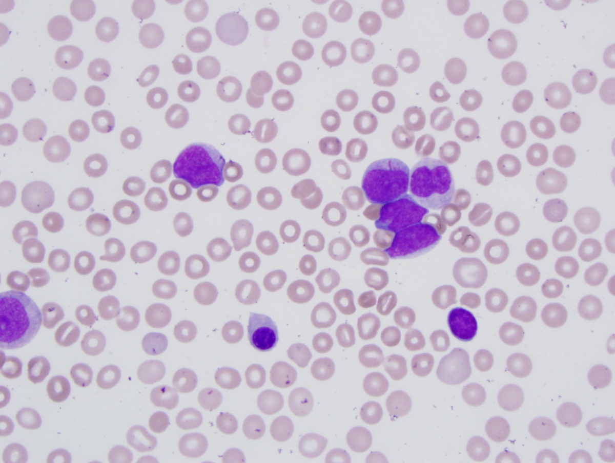 |
| Peripheral blood smear with blasts and a hypogranular neutrophil | 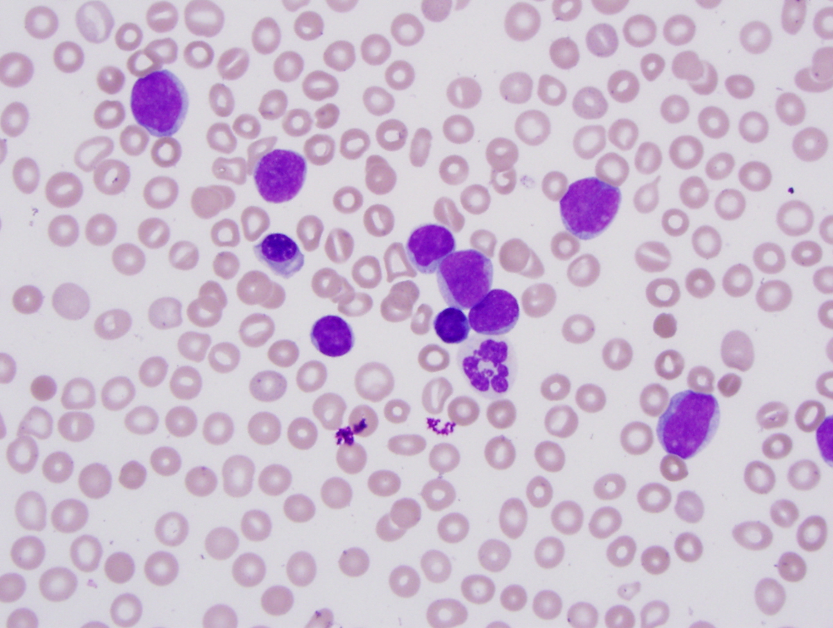 |
| Bone marrow aspirate | 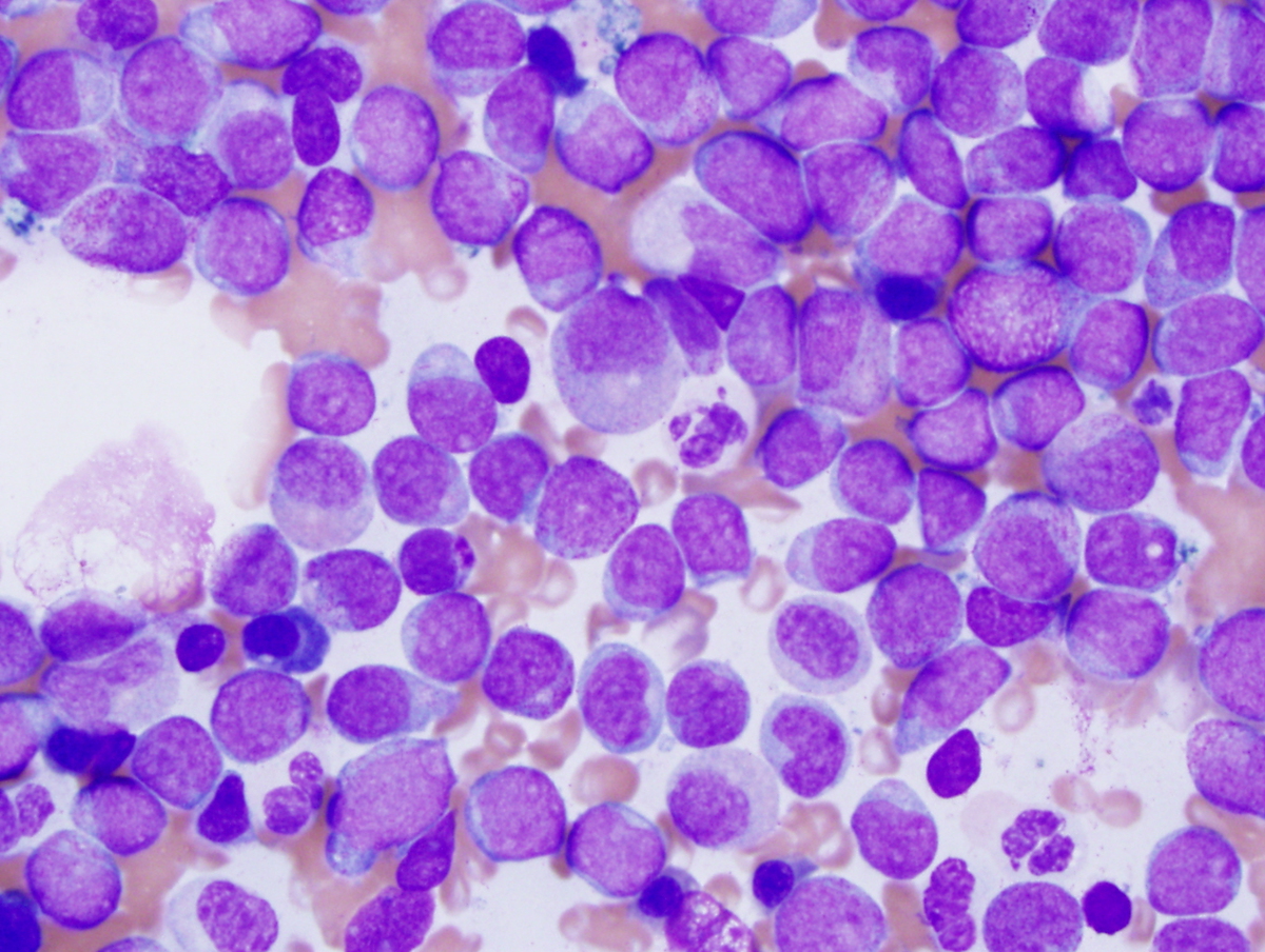 |
| Clot section showing sheets of blasts and dysplastic megakaryocytes | 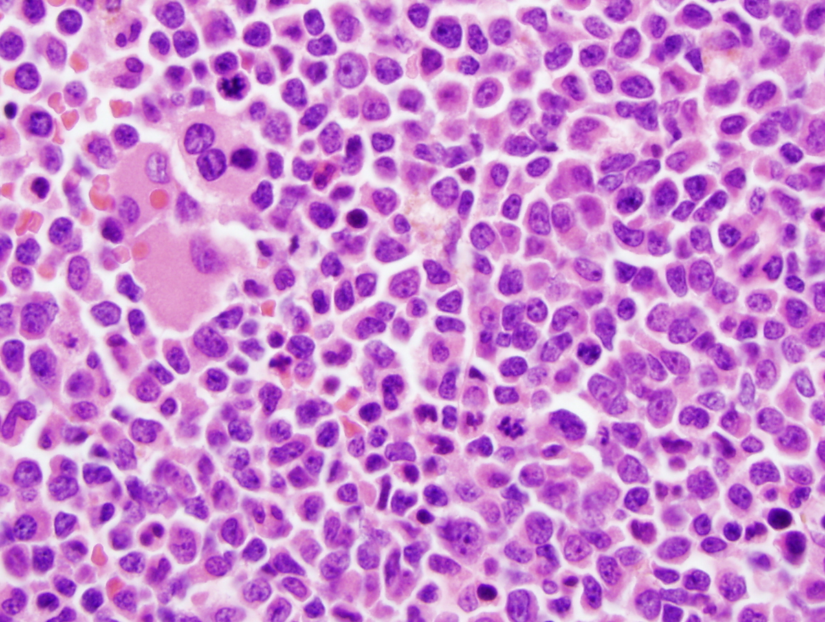 |
| Peripheral blood flow cytometry |  |