Institution: The University of Chicago
Additional authors:Andres Morales de la Madrid, M. Kamran Mirza, Gordana Raca, Jennifer McNee John Anastasi
Session: T Lymphoblastic Leukemia/Lymphoma
HISTORY
The patient is a 4-year-old girl who initially presented to her pediatrician with 2-week history of persistent fever, pallor, fatigue and earache, which was presumed to be acute otitis and was treated with antibiotics (previously published case, Morales La Madrid, A. et al. Leukemia & Lymphoma. 2013, 54(2):403-405). Showing no improvement, she shortly thereafter presented to our hospital with marked hyperleukocytosis, anemia, thrombocytopenia and hepatosplenomegaly but no lymphadenopathy. Laboratory values upon this presentation were as follows:
WBC 731x10^9/L, Hb 100g/L, Platelets 52x10^9/LAbsolute (x10^9/L) & relative (%) WBC differential count:Malignant lymphoid cells 629 (86%), myelocytes 7.3 (1%), neutrophils 37 (5%), basophils 7.3 (1%), eosinophils 7.3 (1%), lymphocytes 37 (5%), monocytes 7.3 (1%) HCT 35.6%, RBC 2.88x10^12/L, LDH 3310IU/L, uric acid 11.8 mg/dLPeripheral blood smear:The peripheral blood smear showed a marked leukocytosis in keeping with the automated WBC count. The circulating cells were almost entirely malignant-appearing lymphoid cells. These cells showed a range of morphology from cells with intermediate-sized nuclei, moderately fine chromatin, variable nucleoli and scant cytoplasm to cells exhibiting more abundant, blue cytoplasm often with blebs. Background small, more mature appearing lymphoid cells were also noted. The remainder of the leukocytes consisted of left-shifted granulocytes, eosinophils and basophils that, together, comprised <10% of WBCs.DETAILS
Bone marrow (BM) biopsy (Right posterior iliac crest, B-5 fixative):
The bone marrow biopsy consisted of a single core demonstrating a cellularity close to 100%, comprised of predominantly sheets of intermediate-sized malignant lymphoid cells. Admixed residual hematopoietic elements were noted but were much reduced with relative sparing of megakaryocytes.Bone marrow aspirate: The bone marrow aspirate smears shows no bony spicules but markedly increased cellularity. Malignant appearing lymphoid cells accounted for the majority of the total nucleated elements on the differential. On these preparations, the cells appeared somewhat ‘blastoid’ with fine nuclear chromatin, variable but mostly absent nucleoli and moderate to scant blue cytoplasm. The cells exhibited morphologic variation similar to that described in the peripheral blood. Trilineage hematopoiesis was markedly reduced.IMMUNOHISTOCHEMISTRY AND FLOW CYTOMETRY
Flow cytometric analysis (peripheral blood):
Flow cytometry was performed on the peripheral blood and showed a dominant population of cells (92% of total events) in the bright CD45 vs low side scatter region (typically ‘mature lymphoid region’) with the following phenotype: CD34-, HLADR-, TdT-, CD1a-, sCD3+, CD2+, CD5+, CD7+, CD4-, CD8-, TCR gamma/delta+. All B-cell and myelomonocytic antigens tested were negative.Immunohistochemical, in situ hybridization and special stains:Immunohistochemical staining using the MYC antibody (Epitomics) revealed >90% of tumor nuclei to be MYC positive with moderate to strong intensity staining. Stains for cytotoxic markers perforin, granzyme B and TIA1 were weakly positive in the malignant cells. TdT and TCL1 were negative. In situ hybridization for Ebstein-Barr virus encoded RNA (EBER) was negative. A reticulin stain revealed no increase in reticulin fibrosis.CYTOGENETIC FINDINGS
Cytogenetic analysis was performed on cells from the aspirate and revealed two related abnormal clones characterized by t(8;14)(q24;q11) involving the MYC gene at 8q24 and the T-cell receptor (TCR) alpha/delta gene at 14q11:
47, XX, t(8;14)(q24.1;q11.2), +17[85%]46,idem,-9,i(9)(q10)[15%]Fluorescence in situ hybridization (FISH) analysis was positive for a MYC rearrangement (8q24.1) in 75% of analyzed cells and negative for abnormalities of chromosome 7 and the BCR-ABL1 fusion.INTERESTING FEATURES
Course:
Rare malignant lymphoid cells were present in the cerebrospinal fluid but with a normal WBC count (clinically considered CNS2 status). The patient was started on typical pediatric induction chemotherapy for high-risk acute lymphoblastic leukemia (ALL). In the first week of treatment, the WBC count dropped to <100 x10^9/L with tumor lysis syndrome that responded well to medical management. In week 3 of induction, there was persistent leukocytosis with 70% circulating malignant cells indicating a less than optimal response. After suggesting that the process shared some features of Burkitt Lymphoma (BL), treatment was altered to a clofarabine-based rescue regimen, including high dose cytarabine and high dose methotrexate, both of which are essential components of current successful regimens for treatment of BL. At the end of this course, BM aspirate revealed complete morphologic remission and normal hematopoiesis with minimal residual disease of <0.01% by flow analysis. FISH was negative for the MYC rearrangement. The patient then received two consolidation cycles and subsequently underwent allogeneic stem cell transplantation (HSCT) using BM from matched sibling donor as well as stem cell source. She is currently 2-3 years post HSCT with no morphologic or molecular evidence of relapse as well as no signs of chronic graft-versus-host disease.Interesting features of case:This case shares some features of the mature B-cell leukemia/lymphoma, Burkitt lymphoma (BL). Similar to BL, there is dysregulation of MYC from the t(8;14)(q24;q11) leading to oncogenesis, and similar to Burkitt, the process appears to be occurring in a mature cell, initially mistaken as a blast. A small number of pediatric cases with this cytogenetic abnormality have been previously reported (Lange, BJ et al. Leukemia. 1992, 6:613-618) which were characterized typically by high WBC count, CNS infiltration and bulky extramedullary disease. These patients were treated with standard pediatric ALL regimens but with poor survival and dismal outcome. Specific immunophenotypes were generally not reported. Our case demonstrated brightly CD45 positive cells with surface CD3, absent of markers of immaturity (CD1a, CD34 and TdT), presence of cytotoxic markers and assembly of TCR gamma/delta. The Burkitt-like MYC translocation, the mature phenotype, together with the less than adequate response to standard high risk ALL therapy all raised the question as to whether this entity had a different cell of origin or biologic behavior compared to a precursor lymphoblastic malignancy. Notably, our patient attained complete remission with modification of the initial ALL therapy to a more BL-like approach of aggressive clofarabine-based regimen with high dose cytarabine and methotrexate. Consolidation cycles also consisted of some of the same components used in mature B-cell malignancies. With complete remission, the patient had the opportunity to proceed to HSCT and is currently disease-free.This case draws attention to the question of how this entity should be best classified, i.e., as a lymphoblastic versus a mature a T cell malignancy (“T-Burkitt”). The question has important therapeutic and prognostic significance given the consideration that a BL-like high intensity chemotherapy may be more appropriate and more efficacious.PROPOSED DIAGNOSIS
Gamma/delta T-cell leukemia/lymphoma with t(8;14)(q24;q11)/MYC-TCR (a possible “T-Burkitt”)
CONSENSUS DIAGNOSIS
Blastoid gamma/delta T-cell leukemia/lymphoma (mature phenotype) with t(8;14)(q24;q11)/MYC-TCRA/D
| Peripheral blood smear (Wright stain) showing marked leukocytosis of malignant lymphoid cells | 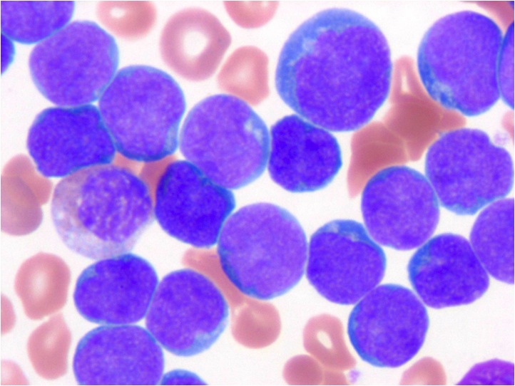 |
| Conventional cytogenetic analysis showing two related abnormal clones characterized by t(8;14)(q24;q11) involving the MYC gene at 8q24 and the T-cell receptor (TCR) alpha/delta gene at 14q11 |  |